Archive 21/2/20

Chitinase 3-like-1 could be a good Therapeutic Target for the new coronavirus (COVID-19)

A group from Brown University, etc. has reported that Chitinase 3-like-1 (CHI3L1/YKL-40) could be a good therapeutic target for the new coronavirus (COVID-19).
https://pubmed.ncbi.nlm.nih.gov/33442679/

In the case of COVID-19, majority is asymptomatic or mild, but 10-20% of patients require hospitalization. Especially, it is so interesting that the severity of COVID-19 is deeply related to aging and comorbid disorders (such as diabetes, hypertension, obesity and metabolic syndrome, cardiovascular disease and chronic lung diseases like COPD and asthma). It is know that CHI3L1 is secreted from a spectrum of cells in response to a variety of injury and inflammation, and regulates innate and adaptive immunity and also protects apoptosis. What’s even more interesting is that CHI3L1 increases with aging and also with risk factors of COVID-19 (i.e., presence of comorbid disorders). Taking these things into consideration, authors focused on the relationship between CHI3L1 and angiotensin-converting enzyme 2 (ACE2), transmembrane protease serine 2 (TMPRSS2), cathepsin L (CTSL).

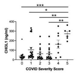
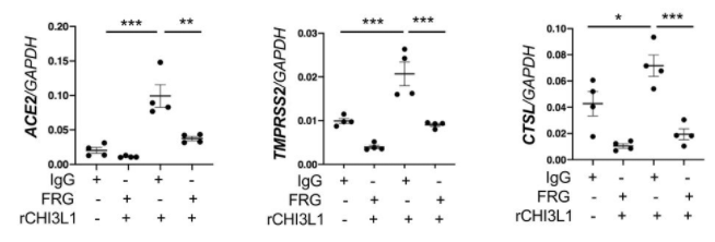
In an in vivo experiment using mice, it was found that CHI3L1 stimulates expression of pulmonary ACE2, TMPRSS2, and CTSL. In other experiments using Calu-3 lung epithelial cells, the expression of ACE2, TMPRSS2, CTSL increased monotonically with CHI3L1 dose, and also it was found that a monoclonal antibody for CHI3L1 (FRG) strongly inhibits the expression of ACE2, TMPRSS2, and CTSL. In actual COVID-19 patients hospitalized, it was also indicated that there is a significant correlation between severity and CHI3L1.

These evidences suggest strongly that CHI3L1 could be a good therapeutic target for COVID-19. Let’s anticipate the future research. 
IgM plays a major role unexpectedly in preventing the new coronavirus (SARS-CoV-2) infection

A group from University de Monterey, etc. has reported an evaluation result of SARS-CoV-2 neutralization activities of IgM, IgA, and IgG using plasma samples from 25 convalescent patients with the new coronavirus (COVID-19).
https://pubmed.ncbi.nlm.nih.gov/33596407/

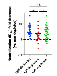
IgM, IgA, and IgG were selectively depleted using isotype-specific ligands immobilized on beads, respectively. A comparison of the SARS-CoV-2 pseudoviral inhibitory dilution (ID50) for each plasma is shown below. In the IgM-depleted plasma, neutralization activity was greatly reduced by 5.5 times, the secondary one was the IgG-depleted plasma with the reduction of 4.5 times, and the reduction of IgA-depleted plasma was 2.4 times. Considering that IgM is only 5% of total immunoglobulin, the neutralization activity of IgM is very curious.

Q677 mutations in clade 20G (lineage B.1.2) of the new coronavirus (SARS-CoV-2) are increasing rapidly since late 2020

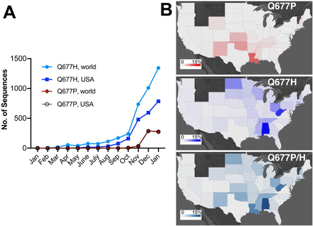
A group from University of Bern, Switzerland, etc. has reported a sharp increase of Q677 mutations (Q677H and Q677P) in clade 20G (lineage B.1.2) of the new coronavirus (SARS-CoV-2) in the USA. Since this mutation is present in the proximity of the furin cleavage site, it may have an effect on infectivity, but detailed research will be a future issue.
https://pubmed.ncbi.nlm.nih.gov/33594385/

Excessive complement activation occurs in kidneys of patients with the new coronavirus (COVID-19)

A group from Friedrich-Alexander-University (FAU) Erlangen-Nürnberg, etc. has reported excessive complement activation in kidneys of patients with the new coronavirus (COVID-19).
https://www.ncbi.nlm.nih.gov/pmc/articles/PMC7878379/

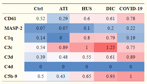
COVID-19 causes severe acute respiratory syndrome (ARDS), and the tissue damage is not restricted to lungs but also develop cardiac and kidney injury. In the case of kidneys, the glomerules and tubules seem to be damaged. Excessive activation of complements develops excessive membrane attack complex (MAC) resulting in damage of the glomerular and tubular tissues.
The table below shows a comparison of the level of complement expression among COVID-19 and typical kidney diseases comparing with a control.


ATI: acute tubular injury
HUS: hemolytic uremic syndrome
DIC: disseminated intravascular coagulation

The functions of glycans and lectins in myogenesis: Galectin-1 promotes muscle development

A group from University of Melbourne, etc. has investigated changes in glycan modification in myogenesis over time, while studying the functions of Galectins.
https://www.mcponline.org/article/S1535-9476(20)35144-6/fulltext

In myogenesis, there was a tendency that terminal di-Gal modification down regulated, α2-6Sia modification up regulated, α2-3Sia modification down regulated inversely, and paucimannose up regulated. These changes were thought to be related to intercellular signaling in myogenesis, but the specific signal paths were still unknown. On the other hand, as for galectins, the expression of Galectin-1 increased and the expression of Galectin-3 decreased.
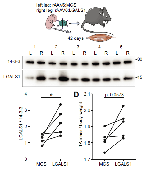
Using newborn mice, an empty multiple cloning site (MCS) was injected with AAV6 into the left foot (as a control), and a galectin-1 gene (LGALS1) was injected with AAV6 into the right foot, and the difference between them was compared after 42 days. it was found that muscle mass was significantly increased with the injection of LGALS1. So, it is clear that galectin-1 is able to promote muscle development.
In the figure below, 14-3-3 protein was referenced as a comparison of LGALS1. 14-3-3 protein is related to intercellular signaling, and recognizes Ser/Thr residues in a specific domain of target protein with a phosphorylation-dependent way, and extracts the physiological function of the phosphorylation state.

Efficacy of therapeutic agents targeting Galectin-3 for breast and ovarian cancers

A group from Memorial Sloan Kettering Cancer Center, New York etc. has reported on the development of therapeutic agents targeting Galectin-3 for breast and ovarian cancers.
https://www.nature.com/articles/s41598-021-82686-3

In ovarian and breast cancers, mucin named MUC16 with CA125 epitope is highly expressed. Mucins are strongly glycosylated with O-glycans, and the glycan chains are sometimes stretched by adding a poly LacNAc chain to the terminal. Galectin-3 (Gal-3) has affinity to the poly LacNAc, hence Gal-3 could bind to MUC16 via such glycans. Galectin family has various functions, and MUC16 is closely related to cancer cells and is considered to be related to cancer growth and metastasis.

Using a monoclonal antibody (14D11) against Gal-3, the authors evaluated the inhibitory effects of Gal-3 on ovarian and breast cancers in vitro and in vivo. The Kd value for Gal-3 and LacNAc binding was ~0.2mM, and the Kd value for Gal-3 and 14D11 was ~14.6nM. Therefore, it turns out that 14D11 has stronger affinity about 13,000 times.

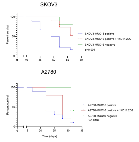
Two ovarian cancer cell lines (A2780, SKOV3) with high expression of MUC16 were transplanted into mice and the effects of 14D11 administration were compared with survival rates.

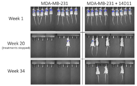
In addition, breast cancer cells (MDA-MB-231) were transplanted into mice and the effects of 14D11 administration were compared as below. These experimental results show that inhibition of Gal-3 can reduce cancer growth significantly.

ACE2-Fc fusion protein expressed in N. benthamiana as a therapeutic drug against the new coronavirus (SARS-CoV-2)

A group from UC Davis had proposed the idea of using ACE2-Fc fusion protein as a therapeutic agent for COVID-19 as follows,
https://journals.plos.org/plosone/article?id=10.1371/journal.pone.0237295

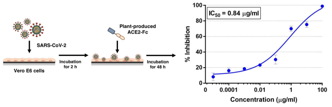
A group from Chulalongkorn University, Bangkok, Thailand etc. has  developed ACE2-Fc fusion proteins expressed in N. benthamiana leaf, and demonstrated the inhibitory effect of SARS-CoV-2 in vitro. Vero cells were infected with SARS-CoV-2, and then ACE2-Fc fusion protein was applied, 0.84 μg/ml (IC50) was obtained as the inhibitory effect. Why did authors use plants? The advantages of using plants are low cost, scalability of production, and no risks from pathogens of animal or human origin.
https://www.frontiersin.org/articles/10.3389/fpls.2020.604663/full

Strain determination of influenza virus using glycan array formed on an Arrayed Imaging Reflectometry (AIR) platform

A group from Univ. of Rochester’s group has developed and used sensor chips with various glycans immobilized on the Arrayed Imaging Reflectometry (AIR) platform as a simple way to detect the hemagglutinin subtypes of influenza viruses and the subtypes of neuraminidase.
https://pubs.acs.org/doi/10.1021/acs.bioconjchem.0c00718

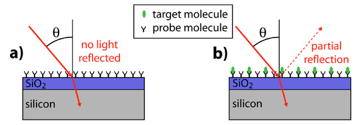
What is more curious is what is the Arrayed Imaging Reflectometry (AIR) platform rather than the application example itself. This platform seemed to be developed in Benjamin Miller Lab., Univ. of Rochester. The principle is physically very simple, growing a thin oxide on a Si substrate with the mirror surface, and using the interference effect of light reflected on the upper and lower surfaces of SiO2 (i.e., at the SiO2/Si interface). There could be a reflection condition that the surface becomes non-reflective as a result of the interference of the diagonally incident light onto the substrate, and molecular interaction between probes pre-fixed on the substrate and applied ligands happens, reflected light appears depending on degree of disruption of the light interference. Therefore, like SPR, AIR does not need fluorescence labeling to the sample (i.e., label-free). The question about this technology would be the sensitivity. However, the above example explained that it provides comparable or better sensitivity than SPR.
https://www.urmc.rochester.edu/labs/benjamin-miller/projects/arrayed-imaging-reflectometry.aspx

Prognostic markers for IgA nephropathy: Lectins detect changes in glycan modification

A group from Okayama University School of Medicine has reported on a study focusing on glycans and lectins as prognosis markers for IgA nephropathy.
https://www.nature.com/articles/s41598-020-77736-1

They have found excellent prognostic markers from urine as a diagnostic sample with using lectin microarrays for 157 IgA nephropathy patients who received a renal biopsy from 2010 to 2017 at Okayama University Hospital.
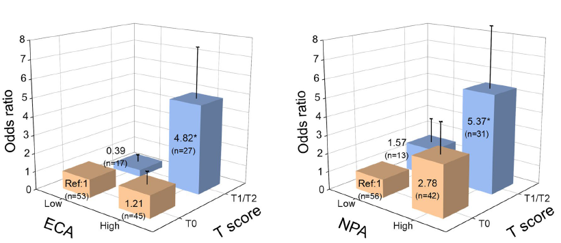
The prognosis outcome were based on a decrease in glomerular filter rate (eGFR) (> 4 mL/min/1.73 m2/year), or eGFR ≥ 30% decline from baseline. T Score was used for the statistical evaluation, and  it was found that ECA lectins (with glycan binding specificity to Galβ1-4GlcNAc) and NPA lectins (with glycan binding specificity to High Mannose) could be good prognostic markers.
ECA:(odds ratio[OR] 2.84, 95% confidence interval[CI] 1.11–7.28)
NPA:(OR 2.32, 95% CI 1.11–4.85)
T Score is calculated by multiplying the standardized percentile rank by 10 and adding 50. T0, T1, T2 indicate Oxford classification about tubular atrophy and interstitial fibrosis.

On the effects of O-glycan modification around furin cleavage site of the new coronavirus (SARS-CoV-2), the infectivity of UK variant (B.1.1.7)

A group of NIH has reported the effects of O-glycan modification around the furin cleavage site of the new coronavirus (SARS-CoV-2) to the increased infectivity of the UK variant (B.1.1.7).
https://www.ncbi.nlm.nih.gov/pmc/articles/PMC7872346/

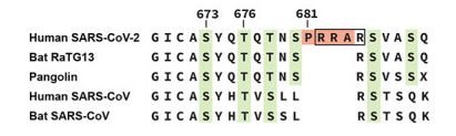
At the S1/S2 boundary of SARS-CoV-2 S-protein, an unique sequence of 4 amino acids (PRRA) is inserted forming the furin cleavage site as shown below.


S673, T676 are the positions where O-glycans are modified, as shown above. The presence of proline at 681 was deeply related to O-glycan modification at those positions. The authors also found that GALNT1 is the most active one glycosylating the furin proximal region among glycosyltransferase GALNT family. Furthermore, addition of O-glycans was shown to significantly suppress the cleavage at the furin cleavage site into S1 and S2. The UK variant (B.1.1.7) has a mutation called P681H, which replaces proline with histidine. Based on the above mentioned results, it is inferred that the non-presence of proline at 681 in UK variant suppresses O-glycan modification, resulting in increased cleavage efficiency and increased infectability.

Powered by WordPress |Copyright © 2020 Emukk. All rights reserved