Archive 22/4/26

Plant Natural Biopesticides: natural antimicrobial volatile organic compounds produced by entomopathogenic fungi

A group from Faculty of Science and Engineering, Swansea University, Singleton Park, Swansea SA2 8PP, Wales, UK, etc. has reported that natural antimicrobial volatile organic compounds (VOCs) produced by entomopathogenic fungi (EPF) have have the potential for use as plant natural biopesticides.
https://www.ncbi.nlm.nih.gov/pmc/articles/PMC9025432/

EPF form symbiotic relationships with plants, leading to improved plant growth and productivity. Plants treated with EPF species, such as Metarhizium brunneum and Beauveria bassiana, usually have more extensive root systems, greater biomass and higher crop yields than untreated plants.

In this study, antimicrobial properties of fungal VOCs produced by these EPF were investigated against the following soil microbes,
Gram-negative bacteria (Escherichia coli, Pantoea agglomerans, Pseudomonas aeruginosa),
Gram-positive bacteria (Micrococcus luteus, Staphylococcus aureus, Bacillus subtilis, B. megaterium, B. thuringiensis),
Yeasts (Candida albicans, Candida glabrata), and
Plant pathogenic fungi (Pythium ultimum, Botrytis cinerea, Fusarium graminearum).

The most potent antimicrobials were isovaleric acid and 1-octen-3-ol, which inhibited or killed bacteria, yeasts, filamentous fungi and the oomycete P. ultimum. In contrast, isoamyl formate, 3-octenone, cedrene and farnesene, although potent, were more restricted in their specificity. Some microbes were clearly more sensitive to M. brunneum VOCs than others. In order of sensitivity, F. graminearum was inhibited by far more compounds than any other test microbe, followed by B. cinerea, then P. ultimum. Of the test bacteria, B. megaterium was the most sensitive, followed by B. thuringiensis.

The natural antimicrobial VOCs identified in this study have the potential for use as plant natural biopesticides.

Difference in N-glycan modification of exosomes secreted from small-cell lung carcinomas and non-small-cell lung carcinomas

A group from Department of Pulmonary Medicine, Kagoshima University Graduate School of Medical and Dental Sciences, Kagoshima, Japan, etc. has reported about the difference in N-glycan modification of exosomes secreted from small-cell lung carcinomas and non-small-cell lung carcinomas.
https://www.jbc.org/article/S0021-9258(22)00390-8/fulltext

Lung cancer is a histologically complex disease that arises from different types of cells in the lungs. Small-cell lung carcinomas (SCLCs) and non-small-cell lung carcinomas (NSCLCs) are the two major lung cancer types classified based upon their histological properties. NSCLCs are further divided into three subtypes, lung adenocarcinomas (LUADs), squamous-cell lung carcinomas (SCCs), and large-cell lung carcinomas (LCCs), in descending order of frequency.

Compared lung cancers are as follows,
SCLC (H446 and SBC-3),
NSCLC-LUAD (HCC827),
NSCLC-SCC (H520, SK-MES-1, and LK-2), and
NSCLC-LCC (H1299).

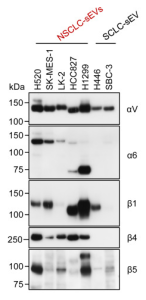
Interestingly, the core N-glycan structures showed striking differences between SCLC-small extracellular vesicles(sEVs) and NSCLC-sEVs. NSCLC-sEVs were commonly enriched in bi-antennary and tri-antennary N-glycans, the core fucosylated forms being the major glycan structures. In contrast, the N-glycan profiles of SCLC-sEVs showed considerable heterogeneity, with an enrichment of N-glycans with antennal fucose (73% of total) in H446-sEVs and those with LacdiNAc (40% of total) or bisecting GlcNAc (29% of total) in SBC-3-sEVs.

As almost all N-glycans were sialylated, glycoproteins from the detergent-solubilized sEVs with SSA-conjugated beads and WGA-conjugated beads were pulled down, and the proteins were subjected to shotgun proteomics. The analysis revealed that integrin αV was commonly expressed in sEVs of both cancer cell types, while the epithelium-specific integrin α6β4 heterodimer was selectively expressed in NSCLC-sEVs.

Why does the neutralizing antibody 35B5 show high neutralizing ability against the SARS-CoV-2 Omicron variant?

A group from Department of Gastroenterology of the Second Affiliated Hospital, School of Medicine and Life Sciences Institute, Zhejiang University, Hangzhou, China, etc. has reported that 35B5 potently neutralizes SARS-CoV-2 Omicron and other variants by causing significant conformational changes within a conserved N-glycan switch that controls the transition of RBD from the “down” state to the “up” state, which allows recognition of the host entry receptor ACE2.
https://www.ncbi.nlm.nih.gov/pmc/articles/PMC8960183/

Neutralization of SARS-CoV-2 by antibodies is carried out through mechanisms including ACE2 competition, ACE2 molecular mimicry, and Fc-receptor-mediated neutralization. N-linked glycosylation has important roles in viral pathology, including mediating protein folding and stability and shaping viral tropism. Glycosylation shields specific epitopes to facilitate viral immune evasion. Beyond the shield function, the glycans at N165 and N234 from the N-terminal domain (NTD) in SARS-CoV-2 act a molecular switch to control the conformational transition of the RBD from the “down” state to the “up” state, which is required for the receptor to bind to ACE2. N165 and N234 are conserved in SARS-CoV-1 and MERS-CoV, highlighting the common mechanism of RBD conformational transition in S proteins.

The Omicron S protein has significant antigenic shifts and structural changes, leading to immune escape of the Omicron variant from most mAbs. Previously, it had been shown that 35B5 has neutralizing activities against the WT and the Beta and Delta variants of SARS-CoV-2. 35B5 dissociates the S trimer and neutralizes SARS-CoV-2. In this study, it was found that 35B5 could neutralize the Omicron variant with a potent neutralizing efficacy much higher than that of many other neutralizing antibodies.
The actual reason why the S protein dissociates is that 35B5 displaces the conserved glycan switch from the RBD, leading to the unstable up states of the RBD and eventually causing the shedding of S1 from the S trimer. The glycan-displacement action of 35B5 represents an unprecedented neutralizing action of mAbs against SARS-CoV-2, which is different from the previously determined ACE2 competition and molecular mimicry mechanisms utilized by class 1 and 2 RBD mAbs and the Fc-receptor-mediated neutralizing mechanisms of class 3 and 4 RBD mAbs

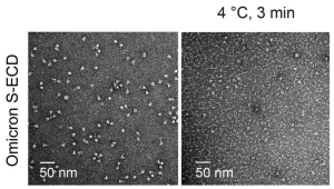
 This figure shows that in vitro incubation with 35B5 led to complete dissociation of the Omicron S trimer.

Salivary glycan profiles could be good markers for discriminating diabetic nephropathy from non-diabetic renal disease

A group from Department of Nephrology, The First Medical Centre, Chinese PLA General Hospital, Chinese PLA Institute of Nephrology, State Key Laboratory of Kidney Diseases, National Clinical Research Center of Kidney Diseases, Beijing Key Laboratory of Kidney Disease, Beijing, China, etc. has reported that salivary glycan profiles could be good markers for discriminating diabetic nephropathy (DN) from non-diabetic renal disease (NDRD).
https://www.ncbi.nlm.nih.gov/pmc/articles/PMC9009518/

Glycan profiles in the saliva could be a non-invasive tool for distinguishing between DN and NDRD.

The AAL, LEL, LCA, VVA, and NPA levels could reflect the severity of DN, and

the LEL and LCA levels could indicate the prognosis of DN.

A new β-trefoil lectin (R-type lectin) was found in protoctista, Salpingoeca rosetta

A group from Univ. Grenoble Alpes, CNRS, CERMAV, 38000 Grenoble, France, etc. has found a β-trefoil lectin (R-type lectin) in protoctista, Salpingoeca rosetta.
https://www.biorxiv.org/content/10.1101/2022.02.10.479907v1.full

Lectin domains are often associated with other functional proteins such as enzymes or toxins. Life-threatening examples are ricin or cholera toxin, in which the lectin domain is responsible for the specificity and adhesion to cell surface glycans, prior to the cellular uptake of the toxin that interferes with metabolism.

Authors have found a eukaryotic Mytilec domain in the genome of Salpingoeca rosetta for the first time using the TrefLec database, and named it as SaroL-1. It has been kbnown that the Mytilec domain functions as a β-trefoil lectin (R-Type lectin), and binds efficiently to globotriaosyl ceramide (Gb3). β-trefoil lectins (R-type lectins) are widely distributed in bacteria, fungi, plants and animals.

The binding of SaroL-1 to different galactosyl-ligands was assessed in solution by isothermal titration calorimetry. The monosaccharides N-acetylgalactosamine (GalNAc) and α-methyl galactoside (GalαOMe) displayed a similar millimolar affinity with a Kd of 2.2 and 2.8 mM, respectively. All tested αGal disaccharides and the p-nitrophenyl-α-D-galactopyranoside (PNPG) derivative bound with affinities twice as strong, with a Kd close to 1 mM, except for αGal1-4Gal, the terminal disaccharide of the globoside Gb3. Lactose that contains βGal displayed very weak binding, being 20 times less efficient than αGal1-4Gal, confirming the preference for the αGal epitope.

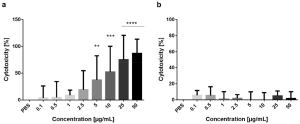
SaroL-1 could form pore-like structures upon membrane binding and work as pore-forming toxins. Actually, SaroL-1 showed strong cytotoxicity towards cancer cells (H1299 cells). The dose-dependent increase of cytotoxicity was confirmed with a standard cell proliferation assay as shown below (a), and cell viability was reduced by approximately 87% after stimulation with SaroL-1. Interestingly, the soluble sugar PNPG could inhibit SaroL-1 cytotoxicity as shown below (b).

This lectin might be a valuable tool for cancer therapy.

Leptospira Virulence-modifying (VM) proteins are novel R-type lectins containing tandem N-terminal ricin B-chain-like β-trefoil domains

A group from Section of Infectious Diseases, Department of Internal Medicine, Yale University School of Medicine, New Haven, CT, USA, etc. has reported that virulence-modifying (VM) proteins are novel R-type lectins containing tandem N-terminal ricin B-chain-like β-trefoil domains
https://www.ncbi.nlm.nih.gov/pmc/articles/PMC9002632/

The so-called virulence-modifying (VM) proteins contributes to the pathogenesis of leptospirosis. Leptospirosis is a globally important neglected zoonotic disease.

From the homology searches of virulence-modifying (VM) proteins (PF07598), tandem ricin B-like lectin subdomains were identified in the amino–terminal regions of that protein. If so, ricin B chain, leptospiral VM proteins could bind to terminal galactose and N-acetyl-galactosamine residues, such as asialofetuin, as a model protein.
Actually, it was confirmed experimentally that the recombinant VM protein expressed in E. coli bound to asialofetuin, showing similar carbohydrate-binding specificity to ricin B chain as determined by competitive asialofetuin-binding assays.

Leptospira VM proteins bound to, and are quickly internalized by, HeLa cells via an N-terminal R-type lectin domain with specificity for terminal galactosyl residues. Cell surface binding and internalization were shown to be rapid, occurring within 30 min after exposure, and induced pleiotropic effects on HeLa cells, including actin depolymerization, caspase-3 activation, nuclear fragmentation, and ultimately blebbing and cell death.
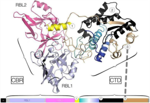
One mechanism of cell death appears to be originated with genomic DNA degradation, which occurs after nuclear localization of the VM protein. Corroborating in vitro experiments using purified HeLa cell genomic DNA, and supercoiled and linearized bacterial plasmid DNA indicates that VM proteins tested so far, possess DNase activities. It was found that CTD of VM protein possess conserved active site residues identical to bovine DNase I.

So, the combination of cell surface binding, internalization, nuclear translocation, and DNase functions indicate that VM proteins evolved as novel forms of the bacterial AB domain-containing toxin paradigm.

T-antigen (with or without sialylation) on MUC21 is required to elicit an anti-apoptotic effect

A group from Laboratory of Cancer Biology and Molecular Immunology, Graduate School of Pharmaceutical Sciences, The University of Tokyo, Tokyo, Japan, etc. has reported that T-antigen (with or without sialylation) on MUC21 is required to elicit an anti-apoptotic effect.
https://pubmed.ncbi.nlm.nih.gov/35410995/

Mucins are highly O-glycosylated proteins thought to protect epithelial surfaces from physical, chemical, and biological insults, and attention to mucins has been paid in the fields of cancer diagnosis and cancer therapy. Mucin 21 (MUC21) is a unique transmembrane-type mucin, found in an endeavor to identify mouse epiglycanin, a mammary carcinoma-associated mucin.

It was first reported that expression of MUC21 by transfection makes epithelial cells resistant to apoptosis. For this effect to occur, MUC21’s O-glycans need to be extended to contain galactose (Gal) residues that may or may not be sialylated. In addition, we show that MUC21-dependent apoptosis resistance is not altered in the absence of galectin-3, suggesting that another galectin or carbohydrate-binding molecule is mediating the effect.

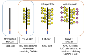
To investigate whether the extension of O-glycans with galactose is necessary for the apoptotic effect of MUC21, CHO-glycosylation variant ldlD cells were used. ldlD cells can synthesize O-glycans only after addition of GalNAc and/or galactose to the culture medium due to a deficiency in the enzyme UDP galactose and UDP-Gal/UDP-GalNAc 4-epimerase. ldlD–MUC21 transfectants grown in regular medium showed only one band at molecular weight around 160 kDa, indicating that ldlD cells expressed only nonglycosylated MUC21. When the medium was supplemented with GalNAc, bands at molecular weight around 200 kDa were seen when lysate was blotted with VVA lectin, suggesting that MUC21 carried Tn-antigen. When the medium was supplemented with GalNAc plus Gal, two bands appeared, one at approximately 200 kDa (no sialidase treatment) and one above 200 kDa (after sialidase treatment). The latter band was also seen when PNA lectin was used for blotting. These results indicate that GalNAc plus Gal supplementation led to the expression of sialyl T-antigen on MUC21.
Comparing these three kinds of samples, it was clearly shown that T-antigen (with or without sialylation) on MUC21 is required to elicit an anti-apoptotic effect.

Galectin-3 has been shown to have antiapoptotic effects in a variety of cell types. So, it was tested if galectin-3 is involved in the induction of MUC21-dependent apoptosis resistance by silencing galectin-3 in MUC21-transfectant HEK293 cells. However , it was found that galectin-3 silencing did not significantly alter the amount of apoptosis resistance in MUC21-transfectant cells.

What’s going on here?

Some urinary free-glycans were upregulated in some cancer patients

A group from Department of Molecular Biology, Osaka International Cancer Institute, Chuo-ku, Osaka, Japan, etc. has reported that some urinary free-glycans were upregulated in some cancer patients.
https://pubmed.ncbi.nlm.nih.gov/35413075/

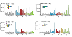
Free glycans in urine with α1,3-fucosylation, triply α2,6-sialylated tri-antennary structures and/or a (Man3)GlcNAc1-core were upregulated in cancer patients. Simple α2,3-sialylation and α1,6-core-fucosylation did not appear to contribute to the observed increase in the level of glycans. The figure below shows such examples.

G: gastric cancer, P: pancreatic cancer, B: cholangiocarcinoma and C: colorectal cancer

Distinct N-Linked IgG glycosylation profiles observed in Lymphatic filariasis

A group from Institute of Medical Microbiology, Immunology and Parasitology Institute for Medical Microbiology, University Hospital Bonn, Bonn, Germany, etc. has reported about distinct N-Linked IgG glycosylation profile observed in Lymphatic filariasis.
https://www.ncbi.nlm.nih.gov/pmc/articles/PMC8992374/

Lymphatic filariasis, commonly known as elephantiasis, is a tropical disease caused by vector-borne filarial parasites Wuchereria bancrofti, Brugia malayi, and Brugia timori. In endemic areas, most infected individuals remain asymptomatic, showing no external signs of the infection while bearing microfilaria (MF+) and contributing to the transmission of the parasite. In contrast, putatively immune individuals, also known as endemic normals (EN), remain infection-free despite continuous exposition to the vector. In some of the exposed individuals, the infection develops into chronic pathology (CP), manifesting as lymphoedema (tissue swelling), elephantiasis (skin/tissue thickening) of limbs, or hydrocele (scrotal swelling).

The clinical outcomes of the infection are tightly linked to the host’s immune reactivity. Typically, alongside classical parasite-induced Th2 immune responses, asymptomatic patients present a strong immune-regulated profile with high levels of regulatory cells, anti-inflammatory cytokines.

The Fc glycans in the IgG molecule are bi-antennary glycans with varying fucose content, bisecting N-acetylglucosamine (GlcNAc), galactose (Gal), and sialic acid; most IgG molecules are fucosylated. These glycan structures have profound impacts on antibody functions and thereby on health. Agalactosylated and asialylated IgG glycoforms, for example, were seen to be particularly abundant in chronic inflammatory diseases such as rheumatoid arthritis, systemic lupus erythematosus, inflammatory bowel disease (IBD), HIV, and mycobacterial infections.

Distinct N-Linked IgG glycosylation profiles observed are as follows:
CP patients presented significantly more glycan residues compared to MF+ and EN,
IgG from EN presented the highest numbers of agalactosylated residues,
IgG from CP patients presented the highest levels of both mono- and bigalactosylated residues,
IgG from MF+ patients presented lower Level of afucosylation, higher Level of sialylated residues, and bisecting GlcNAc.

This study has identified clear differences in IgG glycan profiles between CP, MF+ individuals, and EN and suggests that IgG glycome alterations might potentially be useful as biomarkers for disease severity prediction in lymphatic filariasis.

Relationship between HLA alleles and COVID-19: HLA-A*68 is protective and HLA-A*01 is not protective

A group from Department of Transplantation, Instituto Nacional de Ciencias Médicas y Nutrición Salvador Zubirán, Mexico City, Mexico, etc. has reported about protective HLA alleles against COVID-19.
https://www.ncbi.nlm.nih.gov/pmc/articles/PMC8982524/

In this pilot study in Tapachula, Chiapas México, the frequency of the allele HLA-A*68 is diminished in patients with severe disease and who had a fatal outcome. HLA-A*68 is an ancestral allele present in high frequencies in indigenous ancestral populations in Mexico, and the highest frequency is presented by the Teenek population, Yucatan Merida, and Yucatan rural inhabitants. The presence of protective alleles like HLA-A*68 could explain a little bit why this locality has been one of the least affected throughout Mexico, presenting low incidence rates of COVID-19.

On the other hand, the HLA-A*01 allele was associated with a fatal outcome.

Powered by WordPress |Copyright © 2020 Emukk. All rights reserved