Roles of PSGL-1 on HIV infection

A group from Department of Biological Sciences, University of Toronto Scarborough, 1265 Military Trail, Toronto, ON Canada, etc. has reported that P-selectin glycoprotein ligand-1 (PSGL-1/CD162) is expressed on HIV-1 envelops and can mediate virus capture and subsequent transfer to permissive cells (CD4+ T-cells etc).
https://www.ncbi.nlm.nih.gov/pmc/articles/PMC9123692/

P-selectin glycoprotein ligand-1 (PSGL-1/CD162) has been studied extensively for its role in mediating leukocyte rolling through interactions with its cognate receptor, P-selectin. Structurally, PSGL-1 is a highly glycosylated homodimeric transmembrane protein, with an extracellular domain (ECD) of 50–60 nm in length that extends far out from the cellular surface.

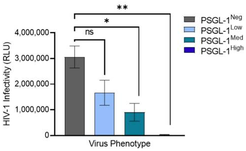
In reality, PSGL-1 plays diverse roles in the physiology of HIV-1 infection. It has been shown that the viruses with high levels of PSGL-1 are not infectious as show below.

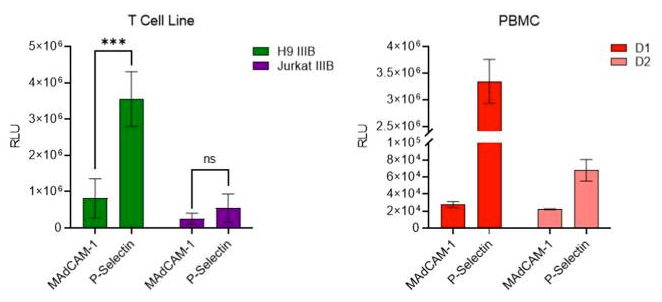
Since the primary target of HIV-1 infection, CD4+ T cells, are often found on activated endothelial tissues which display P-selectin in inflammatory conditions, it was interested in testing whether virions captured by P-selectin could be transferred to nearby permissive cells to elicit infection.
It was decided to test this model with T cell line and PBMC viruses, which contained lower levels of PSGL-1 and higher levels of gp120. We observed that viruses produced in T cell lines and primary PBMC were captured by P-selectin at levels that were markedly higher than the control. Most importantly, both viruses were effectively captured by P-selectin and transferred to HIV-1-permissive cells, suggesting that this mechanism of PSGL-1-mediated virus capture and transfer might also occur when HIV viruses encounter P-selectin on cell surfaces in vivo.

Burkholderia vietnamiensis B418 could be an effective biological nematicide for nematode management

A group from School of Bioengineering, Qilu University of Technology (Shandong Academy of Sciences), Jinan, 250353, China, etc. has reported that Burkholderia vietnamiensis B418 is an effective biological nematicide for nematode management.
https://www.nature.com/articles/s41598-022-12472-2

Root-knot nematodes (RKN), Meloidogyne spp., are highly polyphagous sedentary parasites capable of infesting a wide range of crops especially in greenhouse vegetable cultivation. During infestation, RKN can incite obvious knots or galls on plant roots, destroy the normal structure of the roots, compete with the host for water and nutrition, and make the host susceptible to secondary pathogens. Globally RKN damage is estimated to cause an annual economic loss of over $100 billion, accounting for about 12.6% of total crop losses.

The effects of different treatments on the reduction rate of nematode density and control efficacy against RKN are as follows.
All treatments (Burkholderia vietnamiensis B418 inoculation, fosthiszate, and avermectin) reduced nematode density to some extent compared with the negative control.
The highest reduction rate and control efficacy was observed with B. vietnamiensis B418 inoculation alone by 74.84% and 71.15%, respectively.
The introduction of B418 enhanced the inhibitory effects of chemical nematicide fosthiazate (from 38.92% to 62.71%) and biological nematicide avermectin (from 59.24% to 67.87%), which were still lower than that of B418 inoculated alone, indicating there was slight incompatibility within the combinations of B418 with fosthiazate and avermectin.

The application of B418 increased bacterial 16S rRNA sequences from 9.1% to 34.6%, and fungal ITS-2 rRNA sequences from 7.1% to 30.3%, which both exhibited more variation than the disease control treatments (9.1%).

The beneficial effects of PGPR on plant growth involve either direct mechanism such as biofertilization (facilitation of nutrient uptake including nitrogen and phosphorus primarily) and phytostimulation (production of plant growth promoting hormones), or indirect mechanism as biocontrol agents that antagonize the deleterious effects of phytopathogens by producing inhibitory substances or by inducing plant systemic resistance. The reason why inoculation of B. vietnamiensis B418 was effective in inhibiting RKN. However, it was found that the increment of Mortierella with B418 application was in accordance with the control effect against RKN. These variations in fungal community indicated that B418 inoculation resulted in the inhibition of pathogen-related species and the enhancement of plant-beneficial ones.

CK: control; FOST: fosthiazate; AVM: avermectin. (+ /−) stands for with and without B418 inoculation.

Novel serum glycan monosaccharide composite-based biomarkers for colorectal cancer (CRC) and colorectal adenoma (CRA)

A group from Department of Hepatobiliary and Pancreatic Surgery, The Affiliated Hospital of Qingdao University, Qingdao, China, etc. has developed novel serum glycan monosaccharide composite-based biomarkers for colorectal cancer (CRC) and colorectal adenoma (CRA).
https://www.ncbi.nlm.nih.gov/pmc/articles/PMC9099097/

HPAEC-PAD method was used to quantify the concentrations of two free monosaccharides (Glc-F and Man-F) and the six hydrolyzed monosaccharides (Fuc-H, GalN-H, GlcN-H, Gal-H, Glc-H, and Man-H) in the sera of healthy individuals, CRA, and CRC patients.
The concentrations of monosaccharides in serum were significantly higher in CRA and CRC patients than in healthy individuals. In concreat, concentrations of Fuc-H, GalN-H, GlcN-H, Gal-H, Glc-H, and Man-H in serum were significantly higher in CRC patients compared with healthy individuals. In addition, concentrations of Fuc-H, Gal-H, Glc-H, and Man-H in serum were significantly higher in CRA patients than healthy individuals

The logistic regression analysis was used to develop diagnostic models based on the above results. Tow types of the models were developed, one is MC1 and the other is MC2.
The logistic regression equation for MC1 was as follows: MC1 = -8.18 – 0.021 Fuc-H – 0.004 GlcN-H + 0.011 Man-H, where the Fuc-H, GlcN-H and Man-H were in μmol/L, and
the logistic regression equation for MC2 was as follows: MC2= -6.639 – 0.022 Fuc-H + 0.003 Gal-H + 0.003 Man-H, where the Fuc-H, Gal-H and Man-H were in μmol/L

MC1 was used to distinguish between healthy individuals and CRC, and MC2 was used to distinguish between healthy individuals and CRA. Area under receptor operating characteristic curve (AUC) of MC2 and MC1 was 0.8025 and 0.9403 respectively.
As a reference, the AUC of the existence marker CEA between healthy individuals and CRC was 0.7384.

Cocaprin (a β-trefoil fold lectin) from the mushroom Coprinopsis cinerea inhibits both cysteine and aspartic proteases

A group from Department of Biochemistry and Molecular and Structural Biology, Jožef Stefan Institute, 1000 Ljubljana, Slovenia, etc. has reported that a lectin with β-trefoil fold from the mushroom Coprinopsis cinerea, named cocaprins, inhibits both cysteine and aspartic proteases.
https://www.ncbi.nlm.nih.gov/pmc/articles/PMC9104457/

Sequence-based structural analysis about Coprinopsis cinerea predicted that the two encoded proteins, CCP1 and CCP2, respectively, and their paralog CCP3, containing a Ricin-type β-trefoil lectin-like domain. All three proteins lack a signal peptide for classical secretion and are, thus, predicted to be cytoplasmic.

It was found that cocaprins inhibit plant cysteine proteases belonging to the C1 family with Ki in the low micromolar range, and also inhibit aspartic protease pepsin with Ki in the low micromolar range.

It has shown that cocaprins have lectin activity in addition to protease inhibition. Glycan microarrays were used to analyze carbohydrate binding specificity of cocaprins. For CCP1, very weak binding was observed on a mammalian glycan array to structures including LacNAc or polyLacNAc and for CCP2 the binding was even weaker. This shows a potential for glycan-binding activity in cocaprins.

However, the biological function of cocaprins is unknown. Regarding a potential role in defense, it is noteworthy that CCP2 expression was induced upon challenge with a fungivorous nematode . However, no toxicity of the protein was detected against nematodes or dipteran insect larvae, although all of which have been shown to be targeted by other β-trefoil protease inhibitors and lectins. Strange isn’t it.

Diagnosis of IgA nephropathy (IgAN): A sandwich assay focusing on aberrant glycosylation of serum IgA1 with WFA lectin

A group from Reagent Engineering, Protein Technology Group, Sysmex Corporation, Kobe 651-2271, Hyogo, Japan, etc. has reported about evaluation of serum WFA+-IgA1 as IgA nephropathy (IgAN) diagnostic marker.
https://www.ncbi.nlm.nih.gov/pmc/articles/PMC9104065/

Authors have reported results of an automated sandwich immunoassay system for diagnosing IgA nephropathy (IgAN), using Wisteria floribunda agglutinin (WFA) lectin and anti-IgA1 monoclonal antibody focusing on aberrant glycosylation of IgA1.

The usefulness of WFA for the immunoassays was investigated by lectin microarray. A figure below shows the typical images of microarray results in HC and IgAN patients, and it was confirmed that the WFA signal increased significantly in IgA1 derived from IgAN patients compared with that in HCs.

Unfortunately, the results seemed to be disappointing from a view point of diagnostic assay, i.e., Sensitivity = 66% and Specificity = 62%.

The importance of booster vaccine doses for protection against all Omicron variants

A group from Center for Retrovirus Research, The Ohio State University, Columbus, OH 43210, USA, etc. has reported about the importance of booster vaccine doses for protection against all Omicron variants.
https://www.sciencedirect.com/science/article/pii/S1931312822002207?via%3Dihub

There are several Omicron sub-lineages with significant variations in their S proteins, including BA.1, BA.1.1, and BA.2. While the constellation of mutations varies between isolates, the BA.1.1 lineage is defined by the presence of a single R346K mutation that is absent from the BA.1 lineage, whereas the BA.2 lineage is defined by key S mutations T19I, L24S, Δ25/27, V213G, T376A, and R408S. Although BA.1 was the major variant during the Omicron wave of the pandemic, the BA.2 variant, and to a lesser extent BA.1.1, has begun to account for an increasing proportion of cases. In particular, the BA.2 variant exhibits enhanced transmissibility relative to BA.1 and can reinfect previously BA.1-infected individuals.

In this study, authors have demonstrated the followings:
Omicron BA.1.1 is effectively neutralized by Omicron patient sera, and Omicron sub-lineages are comparably neutralized by Omicron patient sera.
Omicron BA.1 and BA.2 are resistant to neutralization by two-dose mRNA vaccination but sensitive to neutralization after a booster dose.

Effects of antibiotic sulfonamides on soil microbial population and respiration in rhizospheric soil of wheat

A group from Molecular Plant Physiology, Institute of Botany, University of the Punjab, Lahore, Pakistan, etc. has reported about the effects of sulfonamides on soil microbial population and respiration in rhizospheric soil of wheat.
https://journals.plos.org/plosone/article?id=10.1371/journal.pone.0264476

Sulfonamide is widely used in livestock farming and has been used to treat a variety of bacterial diseases. Due to poor management, they are excreted into the soil after treatments and these are extremely hazardous. Sulfonamides may impair the growth of plants, leaves, and roots at concentrations of several hundred mg/L. Accumulations of different antibiotics, including sulfonamides harmed the function and activity of microorganism and reduce soil enzymatic activity. Antibiotics in soil can bring constant changes in organisms and plants and exert harmful impacts on soil microbes.

This study was conducted to infer impact of four newly synthesized sulfonamides on isolated native strains from rhizosphere of wheat cultivar ‘Chakwal-50’. Furthermore, present research focused on the susceptibility of soil microbes and microbial respiration in the rhizospheric soil of wheat.

Sulfonamides: 2-(phenylsulfonyl) hydrazine carbothioamide (TSBS-1), N, 2-bis phenyl hydrazine carbothioamide (TSBS-2), aminocarbonyl benzene sulfonamide (UBS-1), and N, N’-carbonyl dibenzene sulfonamide (UBS-2) were applied on five isolated bacterial strains, i.e., AC (Actinobacter spp), RS-3a (Bacillus spp.), RS-7a (Bacillus subtilis), RS-4a (Enterobacter spp.) and RS-5a (Enterobacter spp.) isolated from the wheat rhizosphere. All sulfonamide derivatives exhibited antibacterial activity against tested bacterial strains, except for TSBS-1. In comparison of all sulfonamide derivatives, UBS-1 exhibited the highest inhibition zone (11.47 ± 0.90 mm) against RS-4a at the highest concentration (4 mg/ml).

numbers in the figure are UBS-1 concentrations (mg/mL)

Thus, sulfonamides have a negative influence on the soil microbiome, and some soil microorganisms that are unable to resist such stressors, so it is difficult to retain soil fertility and plant development as well. Soil microbial respiration changes mediated by sulfonamides were dependent on length of exposure and concentration. It is needed that antibiotics should be carefully watched and their impact on plant growth should be tested in the future studies.

Multivalent 9-O-Acetylated-sialic acid glycoclusters could be potent inhibitors for SARS-CoV-2 infection

A group from Louvain Institute of Biomolecular Science and Technology, Université catholique de Louvain, Louvain-la-Neuve, Belgium, etc. has reported that multivalent 9-O-Acetylated-sialic acid glycoclusters could be potent inhibitors for SARS-CoV-2 infection.
https://www.nature.com/articles/s41467-022-30313-8

The S1-subunit of SARS-CoV-2 Spike protein can be divided into the N-terminal domain (NTD) containing the glycan-binding domain (GBD) and the C-terminal domain (CTD) accommodating the receptor-binding domain (RBD). The GBD engages glycoproteins and glycolipids in most CoVs8, whereas the RBD binds to the ACE2 receptor.

Here authors focused on 9-O-acetylated-SA (9-AcSA), that are of particular interest, since it has been shown that coronaviruses OC43 and HKU bind this glycan via a conserved receptor-binding site in their spike protein.

While monovalent 9-AcSA shows a high IC50 > 100 µM, we observed for all 9-AcSA-derived glycoclusters screened an IC50 in the range 1-10 µM confirming a multivalent effect for these molecules. Among these, 9-AcSA-porphyrin even shows an IC50 in the sub-micromolar range, both on purified 9-AcSA and in the cellular context, making it an excellent candidate as a therapeutic agent.

Activated lung interstitial macrophages are a prominent site of SARS-CoV-2 infection and focus of inflammation

A group from Department of Biochemistry, Stanford University School of Medicine, Stanford, CA, USA, etc. has identified activated interstitial macrophages as a prominent site of SARS-CoV-2 viral takeover and focus of inflammation.
https://www.biorxiv.org/content/10.1101/2022.05.10.491266v1.full

Here, an experimental model of SARS-CoV-2 infection was proposed to allow systematic interrogation of the early molecular events and pathogenic mechanism of COVID-19 at cellular resolution in native human lung tissue.
To define the early events of SARS-CoV-2 infection in human lung, thick sections (~300-500 µm “slices”) of fresh lung tissue were prepared from therapeutic surgical resections or organ donors, and placed the slices in culture medium containing DMEM/F12 and 10% FBS. Then those tissue slices were infected with SARS-CoV-2 (USA-WA1/2020) at a multiplicity of infection (MOI) of 1 for two hours, and the cultures continued for 24 or 72 hours to allow infection to proceed.

To characterize viral and host gene expression during SARS-CoV-2 infection, slices were dissociated and analyzed by single-cell RNA sequencing. Multiplexed single molecule fluorescence in situ hybridization (smFISH) of the infected lung slices were also performed to simultaneously detect positive strand viral RNA (S gene probe), negative strand viral RNA (replication intermediate, Orf1ab gene probe), the canonical viral receptor ACE2, and markers of the infected cell types detected in scRNA-seq.

The results indicate that the most susceptible lung target of SARS-CoV-2 and focus of inflammation is activated interstitial macrophages. In this newly characterized lung macrophage subtype, viral RNA amplification results in host cell takeover with viral transcripts comprising up to 60% of the total cellular transcriptome. During takeover, there is cell-autonomous induction of an interferon-dominated inflammatory response, including induction of chemokines that can recruit local innate immune cells expressing the cognate receptors (CCL8, CCL2, CCL13, CXCL10). Takeover also induces expression of cytokine IL6, the potent inflammatory molecule central to cytokine storm. Thus, SARS-CoV-2 infection and takeover of interstitial macrophages and interferon-dominated induction of this suite of chemokines and cytokines forms a focus of lung inflammation and immune infiltration, which we propose initiates the transition from COVID-19 pneumonia to ARDS.

To explore the mechanism of SARS-CoV-2 entry into human lung macrophages, a modified recombinant Spike-pseudotyped lentivirus system was applied. Treatment of purified lung macrophages with hydroxychloroquine, a lysosomal protease inhibitor, or cytochalasin D did not block infection by the lentivirus across a wide range of concentrations. This indicates that Spike-mediated entry into lung macrophages does not require phagocytosis. Then, neutralization assays using three potent anti-Spike monoclonal antibodies (mAbs) were evaluated. Although each of these mAbs robustly inhibited lentivirus infection of HeLa-ACE2/TMPRSS2 at nanomolar concentrations, none reduced lentivirus infection of purified lung macrophages. Thus, this means that spike-mediated entry into lung macrophages occurs by a potentially novel mechanism that does not require phagocytosis or the ACE2-interacting receptor binding motif of the SARS-CoV-2 Spike protein.

Chitinase-3-like protein-1 at hospital admission predicts COVID-19 outcome

A group from Division of Immunology, Transplantation and Infectious Diseases, IRCCS San Raffaele Scientific Institute, San Raffaele Via Olgettina 58, 20132, Milan, Italy, etc. has reported that Chitinase-3-like protein-1 at hospital admission predicts COVID-19 outcome.
https://www.nature.com/articles/s41598-022-11532-x

It was found hat high levels of CHI3L1 were associated with an increased risk of adverse outcome, including transfer to the ICU or mortality, independently of age, sex, comorbidities, degree of respiratory insufficiency and systemic inflammation at admission, all known to be associated with COVID-19 clinical outcome.

The ideal biomarkers should not just reflect the overall inflammatory burden but disclose the events responsible for adverse disease evolution, such as vascular inflammation and lung remodeling. Moreover, since the interplay of antigen-presenting cells and T cells is crucial determinant of COVID-19 outcome, molecules involved in this process could be suitable candidates. Chitinase-3 like-protein-1 (CHI3L1), a member of the glycoside hydrolase family, meets these requirements. CHI3L1 binds to chitin, although being devoid of the ability to cleave the protein. It also binds to other substrates such as hyaluronic acid and heparin. Various signals that are activated in the early phases of COVID-19, including extracellular matrix (ECM) alterations, cell and tissue injury and response to cytokines and growth factors, elicit its synthesis by tissue cells and inflammatory leukocytes. In turn, CHI3L1 stimulates the expression of ACE-2 and viral spike protein priming proteases in pulmonary epithelial and vascular cells.

Since CHI3L1 is a recognized biomarker of kidney injury, CHI3L1 levels may at least in part reflect kidney damage, but it must be studied further.