α2,6-Sialylation is upregulated in Severe COVID-19

A group from Department of Chemistry, University of Alberta, Edmonton, Alberta, Canada, etc. has reported that α2,6-Sialylation is upregulated in severe COVID-19.
https://pubmed.ncbi.nlm.nih.gov/35702159/

In influenza, severity of disease was found to be associated with levels of high mannose and the innate immune lectin MBL2. In SARS-CoV-2 infection, antibody glycosylation has been studied as a marker of severity. Antibodies to the spike protein were altered in severe patients, with lower fucosylation and sialylation observed. This has potential consequences for effector function. However, such studies have focused on a single protein type (IgG). To date there has been no work on the systemic glycomic response to SARS-CoV-2 infection in plasma and no analysis of infected tissues.

Herein, high-throughput analysis of plasma and autopsy sample glycosylation was performed from COVID-19 patients using a lectin microarray technology. It was revealed that plasma α2,6-sialic acid could be a marker of severity. This modification is known to increase the half-life of select proteins, including IgG. In plasma, it was found that the fraction of α2,6-sialylated C5 and C9 in severe COVID-19 patients is significantly upregulated. In line with this, it was observed that the staining for complement proteins C5 and C9 in COVID-19 autopsy samples got stronger.

However, the functional significance of sialylation on complement proteins remains poorly understood. Glycosylation can also play a role in controlling both serum half-life and resistance to proteolytic cleavage, which is of particular importance to this cascade. The α2,6-sialylation may be increasing half-life, prolonging the cell-mediated damage from the cascade. There may also be other effects of α2,6-sialylation on complement biology that have yet to be discovered. It will be needed to understand the functional impact of α2,6 sialylation and other glycans on complement.

N-GlcNAc has a plant growth promoting effect

A group from State Key Laboratory of Food Nutrition and Safety, College of Food Science and Engineering, Tianjin University of Science and Technology, Tianjin, China, etc. has demonstrated that N-GlcNAc has a plant growth promoting effect.
https://pubmed.ncbi.nlm.nih.gov/35665438/

Chitin is the second most abundant polysaccharide in living organisms, following cellulose. Chitin and chitosan are biodegradable and nontoxic, drawing increasing attention due to their ability to improve soil and substrate quality, plant growth, and plant resilience and to contribute extensively to the development of enhanced and sustainable crop production. The addition of chitin and its derivatives can improve the fresh yield weight of many crops. However, knowledge of their specific functions in plant growth promotion, cultivation, and agro-environmental sustainability remains limited, restricting their further contribution to yield increase, the predictable activation of plant defenses, the extension of harvest storage life, and the improvement of slow release of optimized nutrients in fertilizers for agricultural products and their microbiomes.
N-Acetyl-D-glucosamine (N-GlcNAc), the most abundant carbon-nitrogen bio compound on Earth, is a derivatized glucose monomer found in polymers of chitin, chitosan, and peptidoglycan, which are major constituents of arthropod exoskeletons, filamentous fungi, and bacterial cell walls.

Using Tomato as a model plant, a hypothesis that N-GlcNAc promotes plant growth was examined. As expected, N-GlcNAc-treated plants produced greater plant height, greater whole fresh weight, and greater stem weight in natural soil. The increase in plant height of N-GlcNAc-exposed plants was 1.29-fold comparing with the control. The whole fresh weight of N-GlcNAc-exposed plants was 1.33-fold that of plants comparing with the control.

The rhizosphere soil samples exposed to N-GlcNAc contained 142 unique OTUs that contained relatively abundant Proteobacteria, Actinobacteria, and Planctomycetes. The relative abundance of Proteobacteria and Actinobacteria in the rhizosphere soil samples of N-GlcNAc-exposed plants was increased by 3.89% and 45.82% comparing with the control, respectively.

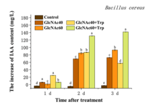
Interestingly, auxin indole-3-acetic acid (IAA) produced by B. cereus increased with N-GlcNAc treatment and reached 92.9 mg/L when cocultured with 60 mmol/L N-GlcNAc in LB medium. This ability of N-GlcNAc to activate IAA production was dependent on supplying the exogenous substrate tryptophan to the strains of P. mirabilis and P. putida. The addition of tryptophan also promoted the accumulation of IAA in strains of B. cereus and S. thermocarboxydus.

Thus, this study provides a new direction for understanding and utilizing the benefits and stability of PGPRs in the field and reveals a key microbial signaling molecule, N-GlcNAc, which shapes the microbial community structure and induces changes in metabolism of the rhizosphere microbiome, thereby simultaneously enhancing plant growth.

Trichoderma wheat seed dressing reduces Fusarium in the rhizosphere microbiome

A group from School of Bioengineering, Qilu University of Technology (Shandong Academy of Sciences), Jinan, China, etc. has reported about effects of inoculation of Trichoderma atroviride HB20111 onto wheat rhizosphere microbiome.
https://www.ncbi.nlm.nih.gov/pmc/articles/PMC9188553/

Fusarium crown rot and wheat sharp eyespot are major soil-borne diseases of wheat.
Biological agents Streptomyces, Bacillus subtilis, Pseudomonas, Actinomyces, Trichoderma, etc. can reduce the colonization of pathogenic fungi, produce antibiotics and organic compounds in the soil, and promote plant growth. Seed dressing is a cost-effective method that has the potential for large-scale application in the prevention and control of crop diseases. As a biocontrol agent, Trichoderma seed dressing has been increasingly used to control plant fungal diseases. Common pathogens controlled by Trichoderma include Bipolaris sorokiniana, F. pseudograminearum, F. oxysporum, R. solani, R. cerealis and many other plant pathogens.

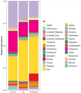
The copy number measured by qPCR of total fungi in T. atroviride HB20111 treatment was lower 47.2% than that of the control. Ascomycota and Olpidiomycota were changed significantly in the rhizosphere fungal community following Trichoderma seed dressing, compared with the control, among which, the content of Ascomycetes decreased by 54.2%, and the Olpidiomycota increased by 54.8%. Trichoderma treatment reduced the relative abundance of Fusarium spp. The relative abundance of Alternaria spp. in the Trichoderma treatment decreased by 3.26% compared with the control. These decreases were accompanied by notable increases in the relative abundance of Olpidium and Botryotrichum in the rhizosphere soil.

As a result of the Trichoderma treatment, for Fusarium crown rot, Trichoderma treatment reduced the disease index by 64.3%, and the yield of wheat treated with Tricoderma seed dressing increased by 7.7% compared with the control.

Diagnosis of hepatocellular carcinoma (HCC) using AFP-L3 as the marker: Using 3 kinds of probes made by molecular imprinting of the marker

A group from State Key Laboratory of Analytical Chemistry for Life Science, School of Chemistry and Chemical Engineering, Nanjing University, Nanjing, China has reported diagnosis of hepatocellular carcinoma (HCC) using AFP-L3 as the marker and using new probes made by molecular imprinting of the marker.
https://www.ncbi.nlm.nih.gov/pmc/articles/PMC9020343/

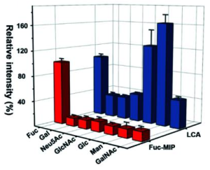
A new strategy called triple MIP-based PISA (triMIP-PISA) was developed for precise disease diagnosis by detecting changes of glycosylation of glycoprotein disease biomarkers. This method benefits greatly from the excellent protein-recognizing properties of MIPs that approach those of monoclonal antibodies, as well as the excellent monosaccharide-recognizing properties of MIPs, outperforming lectins.

This strategy utilized triple recognition of the glycoprotein biomarker by three different types of Molecular Imprinted Polymers(MIPs) integrated with plasmonic detection.
An N-terminal epitope-imprinted substrate was used to specifically extract the target glycoprotein from clinical samples, then the captured target molecules were labelled with C-terminal epitope-imprinted nanotags encapsulated with Raman reporter 1, while the fucosylated glycans of the glycoprotein were labelled with fucose (Fuc)-imprinted nanotags encapsulated with Raman reporter 2, which has characteristic Raman peaks distinct from those of reporter 1. Sandwich-like immunocomplexes formed on the substrate were subject to plasmonic detection.

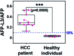
In this report, this method was applied to HCC diagnosis using AFP-L3 as the marker. Raman signal generated by reporter 1-containing nanotags reports the total AFP level, while that by the reporter 2-containing nanotags reports the level of fucosylated glycans of AFP (also referred to as L3). Thus, the relative expression level of fucosylated glycoforms over the total level of AFP in human serum (AFP-L3/AFP) can function as a reliable and specific indicator for HCC patients. The Kd value of the AFP N-terminal epitope imprinted substrate could reach 10−9 M level, which is much smaller than that of LCA lectin.

Sialylated Human milk oligosaccharides (HMOs) in mothers’ breastmilk are twice as risky in developing severe acute malnutrition

A group from Nutrition and Clinical Services Division, International Centre for Diarrhoeal Disease Research, Bangladesh, Mohakhali, Dhaka, Bangladesh, etc. has reported that a higher relative abundance of sialylated Human milk oligosaccharides (HMOs) in mothers’ breastmilk may have a negative impact on young infants’ nutritional status.
https://www.ncbi.nlm.nih.gov/pmc/articles/PMC9177541/

HMOs are not digestible by infants and arrive intact to the large intestine, where they exert prebiotic roles by supporting the development of selected beneficial groups of the gut microbiota, including Bifidobacterium sp and some Lactobacillus sp. by providing metabolic products for their existence, growth, and ultimate colonization in the gut of infant. In addition to helping in healthy gut microbiome, HMOs deliver a number of assistances to infants, including brain development, acting as decoys for harmful organism, and averting disease and infection.

Human milk oligosaccharides differ between secretor and non-secretor mothers, whereby fucosylation in the human milk oligosaccharides are a result of gene products that regulate Lewis and secretor blood group types.

In this study, a total of 45 breast milk samples, of which 26 were from the mothers of severe acute malnutrition (SAM) infants and 19 were from mothers of non-malnourished infants. Among the mothers of SAM infants, 14 were secretors and 12 were non-secretors.

It was found that sialylated HMOs were associated with higher odds of severe acute malnutrition status in age and sex adjusted model (AOR = 2.00, 90% CI 1.30, 3.06), in age, sex, secretor status adjusted model (AOR = 1.96, 90% CI 1.29, 2.98) and also among non-secretor mothers when age and sex adjusted model was used (AOR = 2.86, 90% CI 1.07, 7.62). All these different statistical models show statistically significant association with sialylated HMO and severe acute malnutrition among the young infants. Fucosylated HMOs were less likely associated with severe acute malnutrition but there was no significant association between these.


model 1: adjusted odds ratio (aOR) (90% CI) was adjusted for age and sex.
model 2: adjusted odds ratio (aOR) (90% CI) was adjusted for age and sex and secretor status.
model 3: it was for secretor mothers only and adjusted odds ratio (aOR) (90% CI) was adjusted for age and sex.
model 4: it was for non-secretor mothers only and adjusted odds ratio (aOR) (90% CI) was adjusted for age and sex.

Galectin-1 plays important role in the process of H-1 Parvovirus infection

A group from Laboratory of Oncolytic Virus Immuno-Therapeutics, German Cancer Research Centre, Im Neuenheimer Feld 242, 69120 Heidelberg, Germany, etc. has reported about the importance of Galectin-1 in the process of H-1 Parvovirus infection.
https://www.ncbi.nlm.nih.gov/pmc/articles/PMC9146882/

Oncolytic viruses selectively infect and destroy cancer cells while sparing normal tissues. They can also stimulate strong anti-tumour immune responses and destroy tumour vasculature. No fewer than 40 oncolytic viruses are currently under evaluation in clinical trials as treatments against a variety of cancers. Among them is H-1 rat protoparvovirus (H-1PV), a member of the Parvoviridae family in the genus Protoparvovirus.

It was found that H-1PV enters cancer cells via clathrin-mediated endocytosis, a process that involves dynamin and requires a low pH in the endocytic compartments. It was also found that laminins, in particular those containing the laminin γ1 chain, act as attachment factors at the cell surface for a successful H-1PV infection. In particular, sialic acid moieties in the laminins provide a docking place for the virus to anchor to at the cell surface, and Gal-1 promotes the efficient internalisation of virus particles into a clathrin-coated pit. After engagement of these factors, H-1PV penetrates the cells preferentially via clathrin-mediated endocytosis.

Unstudied function and diversity of bacterial lectins found in the human intestinal commensal microbiome

A group from Department of Medicine, Icahn Genomics Institute, Icahn School of Medicine at Mount Sinai, New York, NY, USA, etc. has reported about unstudied function and diversity of bacterial lectins found in the human intestinal commensal microbiome.
https://www.nature.com/articles/s41467-022-29949-3

One commensal bacterial effector gene (Cbeg) family that was found in commensal intestinal bacteria of multiple patient stool samples is composed of uncharacterized genes predicted to encode for lectins (e.g., Cbeg4 and Cbeg5).

A predicted domain analysis of Cbeg4 and Cbeg5 revealed both genes encode proteins containing a secretion signal peptide, a fibronectin type 3 domain (Fn; IPR003961) and a carbohydrate-binding module domain (CBM, CBM6-CBM35-CBM36_like_2, IPR033803). Among 865 proteins found in the Uniprot database that are predicted to contain a CBM6-CBM35-CBM36_like_2 carbohydrate-binding domain, 108 different domain architectures and only 2 functionally characterized proteins were identified. The two functionally characterized proteins with a CBM6-CBM35-CBM36_like_2 domain are enzymes and not lectins; one is a xanthan lyase isolated from Bacillus sp. GL1 and the other is a golgi trafficking enzyme (golvesin) isolated from Dyctostelium discoideum. Analysis of the Cbeg4 and Cbeg5 protein sequence by SWISS-MODEL suggests Cbeg4 and Cbeg5 exist as monomeric proteins. The domain architecture of Cbeg4 and Cbeg5 is specific to commensal Bacteroides species and is distinct from any functionally characterized lectins.

Glycan structures that bound to either CBM4 or CBM5 share a common Galβ1–3GlcNacβ1–2Manα1–3Man motif. In human glycomics data from the Glyconnect database, this N-linked glycan sub-structure is most frequently seen in datasets generated from peripheral blood mononuclear cells (PBMC). Based on their domain content and glycan-binding properties Cbeg4 and Cbeg5 appear to be lectins that bind leukocyte-associated N-linked glycan motifs.

Actually, for CD14+ monocytes, CD16+ monocytes, and cDC2 dendritic cells Cbeg5 increased IL-1β, IL-6, IL-8, IL-10 and TNFα in a dose-dependent manner. Cbeg5 also affected cytokine production in CD1c−CD14−CD16−CD11c+ myeloid cells (mCD11c). The induction of cytokines in these four cell populations (CD14+ monocytes, CD16+ monocytes, cDC2 dendritic cells, and mCD11c cells) was striking with a >100-fold increase relative to the PBS control. To date the role of lectins in the human microbiome has focused on their function as adhesins or in the binding and transport of glycans for bacterial metabolism. The functional analysis of Cbeg5 exampled in interaction with PBMCs mentioned above suggests the diversity of lectin functions in the human microbiome may extend to regulation of the mucosal immune system.

Those lectins encoded by human microbiota represent a biologically relevant, functionally diverse, but yet largely unstudied. The systematic investigation of these lectins will improve our understanding of how the human microbiome contributes to health and disease.

Spatial and abundance mapping of plant N-glycosylation cellular heterogeneity inside Soybean Root Nodules

A group from Environmental Molecular Sciences Laboratory, Earth and Biological Sciences Directorate, Pacific Northwest National Laboratory, Richland, WA, USA, etc. has reported about spatial and abundance mapping of plant N-glycosylation cellular heterogeneity inside Soybean Root Nodules.
https://www.ncbi.nlm.nih.gov/pmc/articles/PMC9150855/

Although glycans are significant mediators of the nodulation process and plant-microbe interactions, there are no studies on whether protein N-glycosylation is affected by nitrogen fixation. Therefore, we compared N-glycome spatial and abundance profiles in soybean nodules infected with wild-type rhizobia and infected with nifH-mutant rhizobia incapable of efficiently fixing atmospheric nitrogen. The nifH-mutant still infects the root and forms the nodules, but cannot fix the nitrogen.

It was found that the majority of N-glycans showed an overall higher abundance in the nifH-mutant nodules, only a few N-glycans showed non-significant changes between WT and nifH-nodules, while no N-glycan was found significantly more abundant in WT. Notably, all glycans with LewisaN-がた-epitope showed significantly higher abundance in the nifH-mutant nodule tissue. In contrast, the level of truncated N-glycans was conserved between the two types of nodules.

Proteomic results identified nine glycoproteins as potentially major carriers of Lewisa glycans as possible candidates for further characterization.
The nine proteins identified were as follows; phytocyanin domain-containing protein (I1LWP0), amidohydro-rel domain-containing protein (I1L921), dirigent protein (I1JL51), peroxidase (I1MP39), germin-like protein (C7S8D5), an uncharacterized copper ion binding protein with oxidoreductase activity (I1MUX7), an uncharacterized enzyme with mannosyl-oligosaccharide glucosidase activity (I1K3K7), and two uncharacterized proteins with unknown molecular function (I1K380, and C5HU39).

Glycoproteins with those specific glycans may be involved in mediating efficient biological nitrogen fixation or downstream effects during bacteroid infection. Indeed, all these nine proteins have known or predicted functions related to biological nitrogen fixation or root development, and it is plausible that their Lewisa-type glycosylation may be involved in their function.

Serum O-glycosylated hepatitis B surface antigen (HBsAg) level could be used to evaluate serum HBV virion levels

A group from Department of Gastroenterology and Hepatology, Juntendo University Shizuoka Hospital, 1129 Nagaoka, Izunokuni-shi, Shizuoka, 410-2295, Japan, etc. has reported that the serum O-glycosylated hepatitis B surface antigen (HBsAg) level can be used to evaluate serum HBV virion levels through conventional immunoassay and may be a novel potential biomarker of viral kinetics, especially in patients receiving NA therapy.
https://pubmed.ncbi.nlm.nih.gov/35641912/

Currently, oral administration of nucleos(t)ide analogs (NAs) is the most popular treatment strategy for patients with CHB because of the excellent virologic efficacy and safety profile of NAs. Long-term administration of NAs suppresses HBV replication in most patients, resulting in biochemical remission and histological improvement, including the regression of fibrosis and cirrhosis. However, HBV infection cannot be completely eliminated because of the persistence of intrahepatic covalently closed circular DNA (cccDNA). Measuring the intrahepatic cccDNA concentration would be the most direct way to assess the replication-competent viral reservoir. However, there are limitations, including the need for liver biopsy and the lack of a standardized method to quantify cccDNA.

HBsAg has long served as a qualitative serological marker for the diagnosis of HBV infection. Quantitative HBsAg assay has demonstrated that serum HBsAg levels are correlated with serum HBV DNA levels and intrahepatic cccDNA levels, and show prognostic significance. However, currently available HBsAg assays cannot distinguish between HBV virions and non-infectious subviral particles (SVPs). Recently, O-glycosylation of the PreS2 domain of M-HBsAg was identified as a distinct characteristic of genotype C HBV virions via a glycan-based immunoassay, and a recombinant antibody that specifically recognizes O-glycosylated M-HBsAg (anti-Glyco-PreS2 antibody) was developed.

Authors have found that the serum O-glycosylated HBsAg level can be used to evaluate serum HBV virion levels through conventional immunoassay and may be a novel potential biomarker of viral kinetics, especially in patients receiving NA therapy.

Palmitoylethanolamide (PEA) would be a promising adjuvant in the therapy of COVID-19

A group from Retrovirus Center, Department of Translational Research and New Technologies in Medicine and Surgery, University of Pisa, 56100 Pisa, Italy, etc. has reported that palmitoylethanolamide (PEA) would be a promising adjuvant in the current therapy of COVID-19 or against emerging RNA viruses that share the same route of replication as coronaviruses.
https://pubmed.ncbi.nlm.nih.gov/35632821/

One of the candidate inhibiting molecules against SARS-CoV-2 was PEA, one of the members of the N-acyl-ethanolamine family. PEA has been shown to inhibit peripheral inflammation and mast cell degranulation as well as to exert neuroprotective and antinociceptive effects in rats and mice. Recently, it was discovered that the anti-inflammatory activity of PEA does not follow the same route as anandamide. PEA-induced analgesic and anti-inflammatory activities are mediated by the activation of peroxisome proliferator receptor alpha (PPAR-α). PPAR-α generates a signaling cascade that leads to the disruption of lipid droplets by the activation of β-oxidation within mitochondria and peroxisomes, and the concomitant stimulation of omega-oxidation in microsomes. The efficacy of PEA in the prevention or treatment of bacterial and viral infections has also been reported. This encouraging evidence in the literature has stimulated research as to whether PEA can be used to inhibit the pathogenesis of SARS-CoV-2.

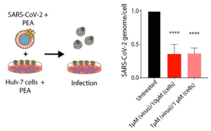
No PEA toxicity was detected, even when PEA was present as high as 100 μM in Huh-7 and 293T cells. When Huh7 cells were infected with SARS-CoV-2 virions incubated or not with PEA at 1 μM, the amount of SARS-CoV-2 genomes were reduced by ~64%, as detected by qRT-PCR 72 h after infection.

The effect of PEA was evaluated against SARS-CoV-2 two variants (Delta and Omicron). It was shown that PEA-pretreated Huh-7 cells at 10 and 1 μM reduced the numbers of viral genomes by 62.4% and 51.2% for SARS-CoV-2 Delta and by 43.4% and 77.3% for the Omicron variant, respectively. Interestingly, it was found that SARS-CoV-2 Delta and Omicron virions exposed to PEA showed a reduction in viral genomes/cell up to nearly 65% for both variants with 10 μM PEA. Also, when PEA was applied to both cells and virions simultaneously. PEA administered at 1 μM led to significant reductions in the number of viral genomes in cell lysate that were quantified as 75.3% and 72.5% for the Delta and Omicron VOCs, respectively.