Glycosylation of adeno-associated virus (AAV) used for gene therapy was investigated using Lectin microarrays

A group from Department of Biotechnology, Graduate School of Engineering, Osaka University, Osaka, Japan has reported about glycosylation of recombinant adeno-associated virus serotype 6 (rAAV6) which is used for Gene therapy.
https://www.ncbi.nlm.nih.gov/pmc/articles/PMC11107246/

Glycosylation of recombinant adeno-associated viruses (rAAVs) that are used for gene theraphy has been evaluated by Lectin microarrays.
It was found that rAAV6 was mainly O-glycosylated with the mucin-type glycans, O-GalNAc (Tn antigen), and mono- and di-sialylated Galβ1-3GalNAc (T antigen).

The purpose of this study was to see if glycosylation of rAAV6 can affect gene therapy safety in terms of immunogenecity and overall transduction efficacy of this method.
However, unfortunately, this study still could not evaluate the direct influence of mucin-type O-glycans on transduction efficiency.

Galectins bind to the N-glycan of FGFR1 and can directly activate the downstream signaling of FGFR1.

A group from Department of Protein Engineering, Faculty of Biotechnology, University of Wroclaw, Wroclaw, Poland has reported that galectin-1, -7, and -8 can activate FGFR1 signaling and control endocytosis.
https://biosignaling.biomedcentral.com/articles/10.1186/s12964-024-01661-3

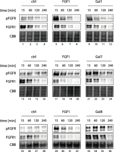
N-glycans of FGFR1 are recognized by extracellular galectins (Gal-1, Gal-7, and Gal-8), which are not authentific ligand of FGFR1 (i.e., FGF1), and the binding of those galectins to FGFR1 trigers activation of the receptor and initiation of downstream signaling cascades. Subsequent endocytosis of activated FGFR1 serves as a major cellular mechanism for the downregulation of FGFR1 signaling.

Both FGF1 and Gal-1 directly activate FGFR1 and after short and intensive pulse of FGFR1 signaling, the receptor is shut down due to the induction of clathrin medited endocytosis, followed by lysosomal degradation of the receptor. Gal-7 and -8 also directly activate FGFR1 by the receptor clustering mechanism, but by inhibiting FGFR1 endocytosis and degradation, these galectins largely prolong FGFR1 signaling.


pFGFR means tyrosine-phosphorylated FGFR1

T-antigen could be a biomarker of progression-free survival in patients with glioblastoma ?

A group from Department of Neurosurgery, the First Affiliated Hospital of Anhui Medical University, Hefei, China has reported that T-antigen could be a biomarker of progression-free survival (PFS) in patients with glioblastoma (GMB).
https://onlinelibrary.wiley.com/doi/10.1002/acn3.52082

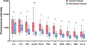
They concluded from studies using Lectin microarrays that serum Jacalin-probed T-antigen levels, which were positively correlated with those in GBM tissues, may be used as a non-invasive biomarker of PFS, predicting GBM recurrences.

However, blog author thinks that their conclusion is problematic, because the glycan binding specificity of Jacalin is quite broad covering GlcNAcβ1-3GalNAc (Core3), Siaα2-3Galβ1-3GalNAc (sialyl T), Galβ1-3GalNAc (T-antigen), α-GalNAc (Tn-antigen), and also PNA could not discriminate glioblastoma so well, although it has high binding specificity to Galβ1-3GalNAc (T-antigen).

Combining lectin microarrays and machine learning will be a trend?

A group from Laboratory for Functional Glycomics, College of Life Sciences, Northwest University, China has reported about employing a combination of lectin microarrays and machine learning to identify alterations in serum glycopatterns with a special emphasis on its early detection.
https://pubmed.ncbi.nlm.nih.gov/38698681/

In recent years, the number of Chinese papers has increased rapidly, and the probability of finding Japanese papers has decreased considerably when searching for papers.
This work from China proposes a method that uses the glycan profile of blood glycoproteins as a serum biomarker combining machine learning with lectin microarrays for the early detection of non-alcoholic fatty liver disease.

However, this type of method was already developed by us about six years ago, and although the target was different from the above paper, its excellent effectiveness has been demonstrated.
The following website explains that it is possible to characterize target cells quite accurately by using deep learning and lectin microarrays for glycoproteins secreted by cells into the culture media.
Combining Deep Learning and Lectin microarrays