A single vaccination is sufficient to boost immunity to high levels among previously infected individuals

A group from Northwestern University Feinberg School of Medicine, USA, etc. has reported on neutralizing antibody responses after one or two doses of COVID-19 mRNA vaccine in previously infected and uninfected individuals.
https://www.ncbi.nlm.nih.gov/pmc/articles/PMC8276631/

The following three groups were compared:
(1)COVID-19 group: previously recovered from a confirmed outpatient case of COVID-19,
(2)Seropositive group: seropositive for anti-SARS-CoV-2 receptor binding domain (RBD) IgG but no acute virus diagnostic test for COVID-19,
(3)Seronegative group: seronegative for prior SARS-CoV-2 infection prior to vaccination.

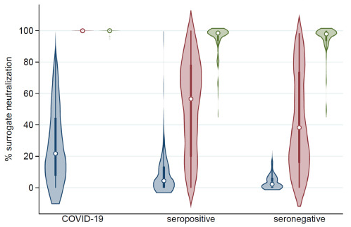
The COVID-19 group had median% neutralization (22.2 vs. 4.4, p < 0.001) in comparison with the seropositive group. The COVID-19 group had significantly higher median% neutralization (99.9 vs. 56.5, p < 0•001) in comparison with the seropositive group. Post-dose 1 responses were higher for the seropositive group in comparison with the seronegative group, but %neutralization (56.5 vs. 38.2, p = 0•12) were not significantly different. The COVID-19 group had higher post-dose 2 median% neutralization (99.9 vs. 98.5, p<0•001) in comparison with the seropositive group. The seropositive and seronegative groups did not differ significantly in post-dose 2 median% neutralization (98.5 vs. 97.9, p = 0.46). As a result, a single dose is sufficient to boost immunity to high levels among previously infected individuals.

Inhibition of SARS-CoV-2 infection with seaweed polysaccharides and those cytotoxicity

A group from Marine Biotechnology Research Center, Korea, etc. has reported on inhibition of SARS-CoV-2 infection with seaweed polysaccharides and those cytotoxicity.
https://www.ncbi.nlm.nih.gov/pmc/articles/PMC8071526/

Seaweeds are an excellent source of bioactive compounds such as polysaccharides, dietary fibers, amino acids, essential fatty acids, carotenoids, phlorotannins, vitamins, and minerals. These compounds have been reported to have a variety of pharmacological activities such as antitumor, antiviral, antioxidant, antimicrobial, anticoagulant, and immune-inflammatory effects

Authers investigated the inhibitory activities of the following crude polysaccharides (CPs):
Undaria pinnatifida sporophyll (CPUP),
Laminaria japonica (CPLJ),
Hizikia fusiforme (CPHF),
Sargassum horneri (CPSH),
Abalone viscera (CPAV),
Codium fragile (CPCF),
Porphyra tenera (CPPT), and
Fucoidan.

Cytotoxicity Assay with the viability of HEK293/ACE2 cells was assessed. When the HEK293/ACE2 cells were treated with each CP at the final concentration range from 1 ng/mL to 1 mg/mL (serial diluted 1/10) for 96 h, most of the CPs did not show severe cytotoxicity. However, CPAV, CPCF, and CPPT were slightly cytotoxic at a concentration of 1 mg/mL. CPAV showed notable cytotoxicity when applied at a concentration of 1 mg/mL, following a degree of cytotoxicity by CPPT, CPCF, CPHF, CPLJ, CPUP, CPSH, and fucoidan. Nevertheless, the CC50 of all CPs was over 500 μg/mL.

The testing of the inhibition of viral infection by CPs was performed with a SARS-CoV-2 pseudovirus. All CPs, except CPPT, inhibited SARS-CoV-2 pseudovirus infection of HEK293/ACE2 cells at various concentrations. Among the CPs tested, CPSH showed the strongest antiviral activity with an IC50 of 12 μg/mL, followed by CPAV (33 μg/mL), CPHF (47 μg/mL), CPCF (74 μg/mL), CPLJ (105 μg/mL), fucoidan (142 μg/mL), and CPUP (289 μg/mL), respectively.

As a conclusion, Sargassum horneri (CPSH) would be the best as an anti-SARS-CoV-2 agent.

Lentil lectin has potent anti-SARS-CoV-2 activity against mutant strains and epidemic variants. 

A group from National Institutes for Food and Drug Control (NIFDC), Beijing, China, etc. has reported that lentil lectin has potent anti-SARS-CoV-2 activity against mutant strains and epidemic variants.
https://www.tandfonline.com/doi/full/10.1080/22221751.2021.1957720

A pseudovirus based neutralization assay was performed on Huh7 cells by preincubating SARS-CoV-2 pseudoviruse with lectins. WGA, lentil lectin, PHA-L and PHA-E showed most potent antiviral activity against SARS-CoV-2 pseudovirus with IC50 range from 8.5 μg/mL to 22.0 μg/mL.

Hemagglutination and cytotoxicity activity of lectins are always problematic in practical applications. PHA-L and PHA-E showed hemagglutination activity at 3.91 μg/mL, and WGA showed it at 7.81 μg/mL. The lentil lectin showed weak hemagglutination activity at the highest concentrations tested (at 1 mg/mL). Cytotoxicity was evaluated with using Huh7 or 293T cells in 96-well plates and incubated at 37°C for 24 h. These lectins showed no cytotoxicity at 500 μg/mL.

Taking these things into consideration, lentil lectin would be more suitable to be a candidate as SARS-CoV-2 inhibitor.

It is of note that elimination of individual N- or O-linked glycosylation site on SARS-CoV-2 S protein had no influence on neutralization susceptibility to lentil lectin, suggesting that lentil lectin may bind to glycans at multiple sites on S trimer. The lentil lectin has strong binding to both oligomannose-type glycans (Man-5 to Man-9), and N-glycans containing GlcNAc at the non-reducing end terminus including both the complex- and hybrid-type glycans.

Glycosylation sites at N165, N234 and N343 were located around the RBD, and the majority of glycans at these three sites are lentil lectin binding glycans, especially the glycans at N234 are totally oligomannose-type which could be efficiently bound by lentil lectin. Interestingly, removal of any one of glycosylation sites at N165, N234 and N343 had no effect on neutralization susceptibility to lentil lectin, suggesting that the existence of two of these glycosylation sites could support neutralization by lentil lectin.

Although a number of mutations have been emerging, glycosylation sites at N165, N234 and N343 were 100% conserved so far. Therefore, the use of lentil lectin might to be a GOOD selectin because of its high tolerance for SARS-CoV-2 variants. 

Age-dependent T-cell telomere length shortening affects COVID-19 severity 

A group from University of Washington, Seattle, USA, etc. has reported that age-dependent T-cell telomere length shortening affects COVID-19 severity.
https://www.ncbi.nlm.nih.gov/pmc/articles/PMC8282112/

Transient lymphopenia is a common feature of acute viral respiratory infections. The drastic and prolonged lymphopenia of COVID-19, however, is distinctive and largely stems from falling counts of T cells. Although the primary causes of this T-cell lymphopenia is still poorly understood, the decline in T-cell count in COVID-19 demands fast and massive T-cell clonal expansion, which is a telomere length (TL)-dependent issue. As hematopoietic cell TL (HCTL) shortens with age, T-cells from some older adults might lack the clonal expansion capacity required to offset the development of COVID-19 lymphopenia or recover from it.

In absence of an acute infection, the T cell turnover is slow because of the relatively long biological half-lives of naïve T cells and memory T cells in the circulation, i.e., ~ 5 years and ~ 5 months, respectively. In the face of SARS-CoV-2 infection, however, diminished TL-dependent T-cell proliferative capacity in older adults could result in a shortfall between T-cell depletion and repletion. Moreover, the clearance of SARS-CoV-2 requires clonal expansion and differentiation of naïve T cells into SARS-CoV-2 antigen-specific effector/memory (henceforth memory) T cells . Short naïve T-cell telomeres might thus limit adaptive immunity against the virus even without infection-mediated T-cell lymphopenia.

The following assumptions on T-cell replication and T-cell clone size (CS) are used in the following author’s model:
(i) T-cell TL-dependent cessation of replication is defined by a “telomeric brink” (TLB) that stops replication at 5 kilobases, (ii) TL of a naïve T cell at age 20 years (TL20) progressively shortens at a rate g of 0.03 kb/year until it reaches the TLB, (iii) In exponential growth, a single naïve T cell can generate a maximal CS (MCS) of ~ 106 (one million) memory T cells through ~ 20 replications (Nmax), (iv) the maximal TL shortening due to clonal expansion is defined as Δmax, (v) Due to age-dependent TL shortening, a naïve T- cell TL reaches the “telomeric onset” (TLO = 6.4 kb) at “age of onset” (XO), (vi) Most memory T cells are formed in response to new antigens during childhood and early adulthood.


TL20 = TL at age 20 years,
TLB = Telomeric brink, stopping cell replication,
TLO = TL at onset of clone size limitation,
XO = Age in years when TLO is reached,
MCS = Maximal clone size (~106 T cells),
Δmax = TL shortening required for achieving MCS (~1.4kb)

In conclusion, the TL effect on naïve T-cell clonal expansion probably influences the outcome of SARS-CoV-2 infection, because it becomes difficult to supply enough amount of T cell clone size with aging (with shortening TL).

The most potent neutralizing antibody developed with antibody phage display from COVID-19 convalescent patients 

A group from Technische Universität Braunschweig, Institut für Biochemie, Biotechnologie und Bioinformatik, Abteilung Biotechnologie, Germany, etc. has reported on the most potent neutralizing antibody developed with antibody phage display from COVID-19 convalescent patients.
https://www.ncbi.nlm.nih.gov/pmc/articles/PMC8260561/

Antibody phage display is now widely used as in vitro technology to select human antibody fragments for the development of therapeutic antibodies against target disease.
In this author’s work, immune phage display libraries from six COVID-19 convalescent patients were constructed and RBD-binding antibodies have been selected, to develop SARS-CoV-2-inhibiting and -neutralizing antibodies.

30 antibodies were screened in a cytopathic effect (CPE)-based neutralization screening assay to select antibodies for further characterization as IgG. This assay was performed with 250 plaque-forming units (pfu)/well SARS-CoV-2 and 1 μg/mL (∼10 nM) scFv-Fc. CPE is characterized by rounding and detachment clearly visible in phase contrast microscopy upon SARS-CoV-2 infection within 4 days, while uninfected cells maintained an undisturbed confluent monolayer. The best neutralizing 19 scFv-Fc were re-cloned and produced as human IgG in 50-mL culture scales. Half-maximal effective concentration (EC50) values of binding to RBD, S1, or S1-S2 were determined. Antibodies named STE90-C11, STE90-B2-D12, STE94-F6, and STE94-H2 showed EC50 values ranging between 0.2 and 0.5 nM on all of the antigens tested. As a result, STE90-C11 antibody was selected as the most potent one.

Excitingly, STE90-C11 was tolerant to most known RBD mutants as shown below, especially those of the mutants B.1.429/B.1.427, B.1.526, B1.258Δ, B.1.535, B.1.617, and B.1.1.33, which are currently emerging.

To get further insight into the neutralizing mechanism of STE90-C11, a complex of STE90-C11 Fab and SARS-CoV-2 RBD was prepared and subjected to crystallization screening. X-ray diffraction images collected from the resulting crystals yielded a dataset to an overall resolution limit of 2.0 Å. After solving the structure by molecular replacement, a model was built into the electron density. Roughly 60% of this binding area can be contributed to the VH segment, forming up to 10 hydrogen bonds at the same time. The remaining 40% are provided by the VL segment contributing 8 additional hydrogen bonds to stabilize the interaction. The superposition of the RBDs of the STE90-C11:SARS-CoV-2 RBD complex with a ACE2:SARS-CoV-2 RBD complex revealed that the neutralization mechanism of STE90-C11 is based on directly competing for the ACE2 binding side, as the interaction interfaces on the RBD of both molecules almost completely overlap. 

Adhesion mechanism of Staphylococcus epidermidis to corneocytes: L-type lectin domain in Asp binds to glycans expressed on corneocytes 

A group from University of Nebraska Medical Centergrid.266813.8, Omaha, Nebraska, USA, etc. has reported on adhesion mechanism of Staphylococcus epidermidis to corneocytes.
https://journals.asm.org/doi/10.1128/mBio.02908-20

Staphylococcus aureus (S. aureus) is the most significant cause worldwide of skin and skin structure infections. S. aureus is infrequently isolated from the skin from healthy subjects. This suggests that Staphylococcus epidermidis (S. epidermidis) and other bacteria colonizing the skin are highly effective at inhibiting colonization of this pathogen.

It was found that S. epidermidis expresses a full-length accumulation-associated protein (Aap) on its cell membrane, and the Aap adheres to glycans expressed on corneocytes. Aap is composed of two distinct A and B domains. The A domain is comprised of a repeat region and a conserved L-type lectin domain. A domain-mediated binding is dependent upon the lectin subdomain but requires the A-repeat domain and the B domain for maximum ligand-receptor interaction.

The glycan binding specificity of Aap was investigated by changing glycan structures with glycosidases. As a result, it was found that Neu5Ac, fucose, and galactose contribute binding to Aap, either individually or together, but no affinity to GlcNAc and mannose.

Since S. aureus contains an Aap orthologue, the common mechanism for corneocyte adherence will be also applied in this strain. Therefore, it will be likely that S. epidermidis has the ability to outcompete S. aureus, potentially via adherence to carbohydrate molecules decorating corneocytes.


Today is the 1st Anniversary of Mx homapage

Today is the 1st Anniversary of Mx homepage. The homepage was uploaded on July 19th, 2020. Thank you so much for your continuous support, and we look forward to your continued patronage moving forward.

Currently, Mx is transmitting information through this homepage focusing on mechanisms of SARS-CoV-2 infection and causes developing serious conditions, especially focusing on topics around glycan and lectin related matters.

The following picture is a scene trying to extract lectins from Capitulum mitella.

By the way, blog Admin has finished two times vaccinations recently.

A gene therapy-based strategy to prevent COVID-19 using an ACE2-decoy protein encoded in an AAV vector delivered by nasal spray. 

A group from University of Pennsylvania, Philadelphia, US, has proposed a gene therapy-based strategy to prevent COVID-19 using an ACE2-decoy protein encoded in an AAV vector delivered by nasal spray.
https://journals.plos.org/plospathogens/article?id=10.1371/journal.ppat.1009544

An affinity-matured ACE2 decoy achieving 1000-fold binding improvements across a wide range of SARS-CoV-2 variants has developed and fused with IgG4 Fc. The ACE-decoy as named CDY14-Fc as the most improved ACE2 decoy variant. The affinity maturation was done with using a diverse (>108 transformants) ACE2 variant libraries in a yeast-display format.

It was demonstrated that the CDY14-Fc could broadly neutralizes SARS-CoV-2 variants as follows. it seems that unlike antibodies, decoy inhibitors could achieve broader neutralization and escape mutant resistance; 501Y from B.1.1.7/alpha variant, 417N/484K/501Y from B1.351/beta variant, 452R/484Q from B.1.617.1/kappa variant, and so forth.

The goal in this strategy is to get decoy proteins expressed internasal delivering the decoy genes with AAV vectors by nasal spray. This idea was confirmed by in vivo experiments with nonhuman primates.

A bivalent protein targeting oligo-mannose in SARS-CoV-2 Spike and HR1 domain in S2 subunit to inhibit SARS-CoV-2 infection 

A group from Shanghai Institute of Infectious Disease and Biosecurity, Fudan University, Shanghai, China, has developed a bivalent protein targeting oligo-mannose in SARS-CoV-2 Spike and HR1 domain in S2 subunit to inhibit SARS-CoV-2 infection.
https://www.ncbi.nlm.nih.gov/pmc/articles/PMC8264481/

To enter target cells, SARS-CoV-2 binds to its receptor ACE2 on the host cell through the receptor-binding domain (RBD) in S1 subunit of spike (S) protein. Such binding triggers conformation changes in the S2 subunit of S protein, resulting in the formation of a six-helix bundle (6-HB) between the heptad repeat 1 and 2 (HR1 and HR2) domains, thus bringing viral and target cell membranes together for fusion. Therefore, both S1 and S2 subunits can serve as important targets for the development of SARS-CoV-2 fusion and entry inhibitors. The sequence of S2 subunit is more conserved than that of S1 subunit, making it a better target for developing broad-spectrum CoV entry inhibitors.

Authors aimed to design and construct a bivalent protein consisting of antiviral lectin GRFT and pan-CoV fusion inhibitory peptide EK1 and evaluate its inhibitory activity and mechanism of action against infection by SARS-CoV-2 and its mutants, as well as other human coronaviruses (HCoVs). Three types of recombinant plasmids encoding bivalent proteins (GRFT and EK1) with different linkers in the length, GRFT-L15-EK1 (GL15E), GRFT-L25-EK1 (GL25E), and GRFT-L35-EK1 (GL35E), containing linkers L15 (GGGGS)3, L25 (GGGGS)5, and L35 (GGGGS)7 between the GRFT and EK1 components.
It was found that GL25E was the best to inhibit SARS-CoV-2 infection as shown below.

Niclosamide inhibits SARS-CoV-2 infection: Inhibition of SARS-CoV-2 infection mediated by endocytosis pathway 

A group from National Centre for Biological Sciences (TIFR), Bengaluru, India, etc. has reported on SARS-CoV-2 infection mechanisms based on endocytosis.
https://journals.plos.org/plospathogens/article?id=10.1371/journal.ppat.1009706

A key step in successful virus infection is the release of viral genomic content into the host cell cytoplasm. To achieve this, viruses bind to specific cell surface receptors and subsequently undergo membrane fusion either directly at the plasma membrane or following endocytic uptake. Both alternatives of entry are feasible for SARS-CoV-2 infections depending on the availability of receptors and proteases at the host cell surface. Although angiotensin converting enzyme 2 (ACE2) is a well-studied receptor for SARS-CoV-2, other receptors and co-receptors have been discovered from a number of groups. Additionally, SARS-CoV-2 requires proteolytic processing of the viral envelope Spike protein by host cell proteases to gain entry. Therefore, these viruses can directly fuse at the cell surface if the Spike protein is cleaved by a cell surface serine protease like TMPRSS2, or utilize an endo-lysosomal route for fusion, where the Spike protein is primed by cysteine protease cathepsins. So, viral entry and infection in different host cells is dependent on the expression of these key host factors (receptors such as ACE2 and protease like furin, TMPRSS2, and cathepsin.

The CLIC/GEEC (CG) pathway is a clathrin-independent endocytic pathway mediated by uncoated tubulovesicular primary carriers called clathrin-independent carriers (CLICs) which arise directly from the plasma membrane and later mature into tubular early endocytic compartments called Glycosylphosphotidylinositol- anchored protein (GPI-AP) enriched compartments (GEECs).

Authors studied the endocytosis of receptor binding domain (RBD) of SARS-CoV-2 Spike protein in gastric epithelial cells (AGS) in the presence and absence of ACE2. AGS can be considered as a cell line with undetectable levels of endogenous ACE2. It was shown that RBD is endocytosed via the CG endocytic pathway (rather than clathrin-mediated endocytosis (CME) pathway) and its uptake is sensitive to pharmacological perturbations of this pathway in AGS cells.

To determine the effect of ACE2 on uptake of RBD in AGS cells, a stable AGS cell line ectopically expressing ACE2 (AGS-ACE2) was generated; the expression of ACE2 was confirmed using qPCR and western blot analysis. RBD uptake in AGS-ACE2 was about 3-fold higher than AGS cells. On characterizing the RBD endocytic itinerary in AGS-ACE2 cells, an increase in the co-occurrence of RBD with transferrin was observed, and slightly reduced co-occurrence of RBD with dextran compared to AGS cells was also observed. This indicates that in addition to trafficking via the CG pathway, RBD is now trafficked via the CME in AGS-ACE2 cells.

Finally, it was shown that niclosamide neutralizes endosomal pH and inhibits SARS-CoV-2 infection as follows.