Decay of Fc-dependent antibody functions in COVID-19 mild convalescent patients

A group from University of Melbourne, Australia, etc. has reported on decay of Fc-dependent antibody functions in COVID-19 mild convalescent patients.
https://www.ncbi.nlm.nih.gov/pmc/articles/PMC8106889/

The FcγR-binding, antibody-dependent cellular cytotoxicity (ADCC), and antibody-dependent phagocytosis (ADP) activities of SARS-CoV-2 Spike-specific antibodies decay during convalescence from COVID-19. The first sample was collected at a median of 41 days post-symptom onset, and the last sample was collected at a median of 123 days post-symptom onset. The decline of plasma ADCC and ADP activity correlated with the decay of SARS-CoV-2 Spike-specific IgG and FcγR-binding antibodies (see figure below).

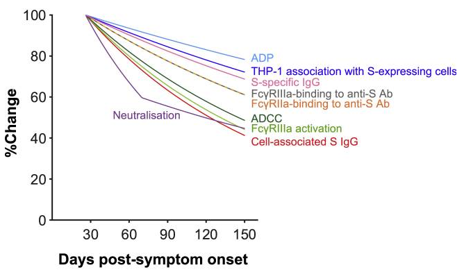
Importantly, Fc effector functions were readily detectable above uninfected controls in 94% of subjects for all assays at the last time point sampled, in contrast with neutralization activity, which remained detectable above background for only 70% of subjects. Overall, it seems that mild to moderate COVID-19 generates robust FcγR-binding, ADCC, and ADP antibody functions that decay at a slower rate than plasma neutralization activity.  

CD8+ T-cell activities get lower and anaphylatoxin expression increases significantly in severe COVID-19 

A group from Medical University of Innsbruck, Innsbruck, Austria, etc. has reported on T cell immunity and related humoral immune responses of COVID-19.
https://www.ncbi.nlm.nih.gov/pmc/articles/PMC8237940/

It was clearly demonstrated that SARS-CoV-2 specific CD8+ T lymphocytes and higher IFNγ production was significantly induced in patients with mild compared to patients with severe or critical COVID-19. In all patients, SARS-CoV-2-specific antibodies with similar neutralizing activity were detected, In other words, no significant differences in viral neutralization could be observed between patients with mild, severe or critical COVID-19. And further, elevated anaphylatoxin C3a and C5a levels were identified in severe and critical COVID-19 patients probably caused by aberrant immune complex formation due to elevated antibody titers in these patients. Normally, complements should be protective during viral infections, but with respect to COVID-19, local complement activation might be related to tissue damages leading to severe.

This study indicates that early and polyfunctional CD8+ T cell immunity along with low anaphylatoxin expression is associated with mild.

In addition, authors proposed an idea of blocking the C5a–C5aR1 axis as therapeutic strategy to limit excessive lung inflammation and tissue damage caused by infiltrating myeloid cells.
 

H522 human lung adenocarcinoma cells were infected with SARS-CoV-2, despite no evidence of detectable ACE2 and TMPRSS2 expression: What is the infection mechanism? 

A group from Washington University in St. Louis, School of Medicine, USA, etc. has reported a new mechanism of SARS-CoV-2 infection.
https://www.ncbi.nlm.nih.gov/pmc/articles/PMC8220945/

Human lung and head/neck cancer cell lines express varying levels of ACE2 and TMPRSS2. Quite interestingly, H522 human lung adenocarcinoma cells were infected with SARS-CoV-2, despite no evidence of detectable ACE2 and TMPRSS2 expression. Using orthogonal assays, it was confirmed that H522 cells are infected independently from the existence of ACE2, which suggests the utilization of an alternative receptor in a cell line of lung origin. Although recent findings establish Neuropin 1 (NRP1), AXL, and heparan sulfate as mediators of ACE2-dependent SARS-CoV-2 entry, it was found that SARS-CoV-2 infection of H522 cells is independent of Neuropin 1 (NRP1) and AXL but dependent on heparan sulfate.

To decipher the mechanisms of SARS-CoV-2 entry into H522 cells, infection inhibition experiments were performed in the presence of compounds that potentially interfere with SARS-CoV-2 entry, including camostat mesylate (TMPRSS2 inhibitor), E64D (broad spectrum inhibitor of proteases, including endosomal cathepsins: proteases (enzymes that degrade proteins) found in all animals as well as other organisms), bafilomycin A (inhibitor of vATPase), and apilimod (inhibitor of PIKfyve), SGC-AAK1-1 (specific inhibitor of AAK1 kinase 1, which promotes clathrin-mediated endocytosis (CME) through phosphorylation of the AP2M1 subunit of the AP2 complex).
E64D, bafilomycin A, SGC-AAK1-1, and apilimod reduced cell-associated viral RNAs in a dose-dependent manner, whereas camostat mesylate increased viral RNA levels. So, these data support an infection mechanism through CME and endosomal cathepsins in H522 cells.

Characteristics of antibodies (targeting RBD, NTD, and S2) induced by two times Pfizer mRNA vaccination: Effectiveness against current SARS-CoV-2 variants 

A group from Icahn School of Medicine at Mount Sinai, New York, USA, etc. has reported on characteristics of Pfizer mRNA vaccinated SARS-CoV-2 antibodies.
https://www.ncbi.nlm.nih.gov/pmc/articles/PMC8185186/

The antibody response peaked 1 week after the second vaccine dose, followed by a decline in titers over the following weeks as expected from an antibody response to vaccination. Interestingly, titers against the spike proteins of β-coronaviruses OC43 and HKU1 increased substantially after vaccination.

RBD and NTD were co-dominant epitopes of the mRNA vaccine-induced antibodies, and the majority of remaining epitopes was mapped to S2. All antibodies were tested for neutralizing activity against the USA-WA1/2020 strain of SARS-CoV-2. Only a minority of the binding antibodies showed neutralizing activity (see figure below, obtained from three individuals: V1, V5, V6).

For sera from the six vaccinated individuals, binding between vaccinee sera and variant RBDs were evaluated. The highest reduction was only 2-fold for E406Q, N440K, E484K, and F490K (see figure below, six vaccinated individuals: V1-V6).
This would be a kind of good news showing that Pfizer mRNA vaccine would be effective against current SARS-CoV-2 variants.

It is confusing which is which, because of  name changes of SARS-CoV-2 variants

You may be familiar with SARS-CoV-2 variants named UK variant, South African variant, Brazilian variant, Indian variant, and so forth. Although these naming are easy to understand the variants closely rather than using virus strain names, it tends to put a stigma on those countries. Recently, it seems to be decided to use Greek letters to identify virus variants.

I have cited a figure showing mutations occurring in those variants from the following paper, and added relationships among several virus variants names as follows.

Institute of Microbiology and Immunology, Faculty of Medicine, University of Belgrade
https://www.mdpi.com/1999-4915/13/7/1192/htm

Alpha(α)= B1.1.7 strain, UK variant
Beta(β)= B1.351 strain, South African variant
Gamma(γ)= P.1 strain, Brazilian variant
Delta(δ)= B.1.617 strain, Indian variant
other is for instance,
Epsilon(ε)=B.1.427/29 strain, US variant

losatran, ACE2 inhibitor, did not show any therapeutic effects on against SARS-CoV-2 in Phase II clinical trial 

A group from Department of Emergency Medicine, University of Minnesota, etc. has indicated that losatran, ACE2 inhibitor, did not show any therapeutic effects on against SARS-CoV-2.
https://www.ncbi.nlm.nih.gov/pmc/articles/PMC8225661/

This was a multicenter prospective double blind randomized placebo-controlled trial for the treatment of symptomatic outpatients with confirmed COVID-19 conducted across 3 hospital systems in Minnesota, United States between April and November 2020.

The intervention was losartan 25 mg versus equally appearing placebo. Participants self-administered study drug orally twice daily for 10 days for participants with eGFR >60 mL/min/1.73 m2, and once daily for those with eGFR 30–60 mL/min/1.73 m2. The threshold for angiotensin receptor blockade is 20 mg daily, with twice daily dosing more effective than once daily due to a 6–9 h half-life of the active metabolite. This dose was chosen in lieu of a 50 mg twice daily (maximum) dose after the FDA raised safety concerns regarding the higher dose regimen. This dose is expected to provide 37% inhibition of the angiotensin receptor.

The primary outcome did not differ significantly, and also viral loads were not statistically different between treatment groups at any time point. This clinical trial was terminated.

Why Δ mutation (Indian mutant strain named B.1.617) in SARS-CoV-2 has higher infectivity ?

A group from Kumamoto University, etc. has reported about why Δ mutation (Indian mutant strain named B.1.617) in SARS-CoV-2 has higher infectivity.
https://www.sciencedirect.com/science/article/pii/S1931312821002845?via%3Dihub

L452R is a key mutation observed in SARS-CoV-2 Δ mutation. It was found that the L452R mutant significantly increased the binding affinity to human ACE2 (Kd = 1.20 ± 0.06 nM). The L452 residue is not directly located at the binding interface (see figure below). Structural analysis and in silico simulation suggested that the L452R substitution promotes electrostatic complementarity.

Because residue 452 is located in close proximity to a negatively charged patch of ACE2 residues (E35, E37, and D38), the electrostatic interaction with ACE2 becomes larger in positively charged R452 than electrically neutral L452 (see figure below).

It was also found that the L452R mutation significantly increased fusion efficacy compared to the parental S, using a SARS-CoV-2 S-based fusion assay, suggesting that the L452R mutation promotes viral replication by increasing viral fusogenicity.

Transient receptor potential vanilloid 2 (TRPV2) accelerates SARS-CoV-2 infection in the presence of fever: A novel mechanism of SARS-CoV-2 Infection

A group from Key Laboratory of the Ministry of Education for Conservation and Utilization of Special Biological Resources in the Western, China, etc. has reported a novel mechanism of SARS-CoV-2 infection in the presence of fever.
https://www.ncbi.nlm.nih.gov/pmc/articles/PMC8210595/

Transient receptor potential vanilloid 2 (TRPV2) was originally isolated as a molecule sensitive to temperatures. TRPV2 plays an important role in innate immunity and is mainly distributed in the endoplasmic reticulum in the absence of stimulation. Under certain stimulatory conditions, such as stimulation by chemokines, growth factors, or stress conditions, TRPV2 can be transferred from the endoplasmic reticulum to the cell membrane to promote Ca2+ entry into the cell and accumulation in pseudopodia, which can promote the migration of macrophages towards an inflamed area and promote the phagocytic function of macrophages.

It was examined if TRPV2 was involved in SARS-CoV-2 infection. The interaction between TRPV2 and SARS-CoV-2 Spike was examined at 37 °C and 39.5 °C with co-IP assays using PBAMs, human THP-1 cells, and mouse macrophages (RAW264.7) as host cells. As shown below, the co-IP analysis confirmed the interaction between TRPV2 and SARS-CoV-2 Spike occurs at 39.5 °C but not at 37 °C.

The febrile temperature (39.5 °C) significantly enhanced the expression of macrophage-secreted cytokines, including IFN-α, IFN-γ, IL-13, IL-α, TNF-α, IL-10, IL-18, IL-2, IL-4, IL-17A, MCP-1, and IL-15, and further it was confirmed that the increased cytokine secretion was independent from existence of ACE2 and Neuropilin-1 at 39.5C.

Then, it was tested whether an inhibitor of TRPV2 (SKF-96365) could suppress such cytokine release or not, and it was confirmed that SKF-96365 effectively suppresses the cytokine storm caused by SARS-CoV-2 in the presence of fever.

Thus, authors discovered a novel mechanism of SARS-CoV-2 infection of macrophages and showed that SKF-96365 is potential candidate for treating SARS-CoV2 in febrile conditions. 

Relationship between Cholesterol and Severity of COVID-19:  the lower the cholesterol, the higher the severity

In the case of COVID-19, it is said that the lower the cholesterol, the higher the severity.

A group from Malmö University, Sweden, etc. has reported that the Spike protein removes lipids and cholesterol from model membranes, and upon co-incubation of both the SARS-CoV-2 spike protein and HDL on model membranes, the levels of lipid removal from the membrane was suppressed, from experiments using model membranes and through interactions with SARS-CoV-2 Spike with and without HDL.

https://www.ncbi.nlm.nih.gov/pmc/articles/PMC8195693/

This suggests that the higher the concentration of cholesterol existing on the membrane and in serum, the lower the adsorption of SARS-CoV-2 spike onto the membrane, in other words, the higher the cholesterol, the lower the infectivity.

Incidentally, the model membrane used here was deuterated 1,2-dimyristoyl-D54-3-sn-glycerophosphatidylcholine (dDMPC) and perdeuterated cholesterol (dcholesterol) at a molar ratio of 80:20 mol%.


SARS-CoV-2 Spike is a key inducing proinflammatory response of macrophages 

A group from Washington State University, USA, etc. has reported that SARS-CoV-2 Spike is a key inducing proinflammatory response of macrophages.
https://www.ncbi.nlm.nih.gov/pmc/articles/PMC8219098/

Virus replication and release of infectious progeny was determined by TCID50 assay in supernatants from SARS-CoV-2 infected Vero E6 cells and THP-1 human-derived macrophage-like cells as shown below. While THP-1 macrophages did not support productive SARS-CoV-2 replication, Vero E6 cells effectively replicated SARS-CoV-2.

To clarify the role of SARS-CoV-2 Spike in proinflammatory responses in macrophages, expression of the pro-inflammatory cytokines TNF-α, CXCL10 and IFN-γ and the antiviral cytokine IFN-β were evaluated with using THP-1. While Spike did not induce gene expression of antiviral IFN-β or IFN-γ in THP-1 macrophages, expression of proinflammatory TNF-α and CXCL10 was upregulated. TNF-α was upregulated by 30-fold at 4h post treatment with Spike (p < 0.05) and remained upregulated by two-fold at 16h post treatment. CXCL10 expression was consistently upregulated by 3 to 8-fold in THP-1 macrophages exposed to Spike up to 16h post treatment (p<0.05).