The severity of influenza correlates with Host Cell’s high Mannose modification

For influenza viruses, it is well known that the binding of α2-6Sia in human respiratory tract cells and HA (hemaggulutinin) with influenza virus causes infection. However, the relationship between the severity of influenza and the glycosylation is not well known.
The following groups use lectin microarrays to study the severity of influenza and the glycosylation of Host Cells using ferret model.
https://www.pnas.org/content/117/43/26926.long


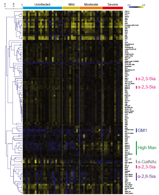
The result is that the glycosylation of Host cells changes when infected with the influenza virus, and the increase in the high mannose structure is correlated with the severity of the disease.
This is an interesting phenomenon. The high Mannose-binding C-type lectin is expressed on immune cells, so its association resulting in self-tissue damages might be suspected.

S-protein of SARS-CoV-2 has a staphylococcal enterotoxin-like sequence in the shadow of the severity of the new coronavirus (COVID-19)

The fact that the S-protein sequence of the new coronavirus (SARS-CoV-2) contains an unique sequence “PRRA” not found in other coronaviruses is inserted at the boundary between the S1 and S2 domains. It has also been frequently pointed out that the presence of this sequence has increased the infectivity of this virus to humans.

There is a paper discussing about the presence of a staphylococcal enterotoxin-like sequence around the inserted “PRRA” sequence (661-685) of SARS-CoV-2, and its potential effect on hyperinflammation of COVID-19.
https://www.pnas.org/content/117/41/25254.long

By binding the T cell receptors (TCR) to this bacterial toxin-like sequence, the resulting toxic shock syndrome might be behind of the severe hyperinflammation of COVID-19? It says. This inference was made from molecular structural analysis of proteins, and is expected to be tested in practice.


SEB represents staphylococcal enterotoxin and has a quite structure similar to the 661-685 sequence of SARS-CoV-2.

Relationship between the incidence rate of the new coronavirus (COVID-19), temperature, and BCG

Using data on the new coronavirus (COVID-19) in the United States, Canada, Italy, Spain, Germany, France, Australia, Japan, South Korea, Malaysia, and Thailand, the results of analyzing the effects of temperature and BCG vaccination on the incident rate of COVID-19 have been reported.
https://journals.plos.org/plosone/article?id=10.1371/journal.pone.0240710

Effects of Temperature (Average Temperature Model):
The temperature of -21 to 5.5 degrees Celsius is used as a reference.
In the region from 6 to 10.5 degrees C, the incident rate ratio is 1.18 (95% CI: 0.87 – 1.59)
In the region from 11 to 18.5 degrees C, the incident rate ratio is 0.97 (95% CI 0.65 – 1.45)
In the region from 19 to 36.5 degrees C, the incident rate ratio is 0.87 (95% CI: 0.47 – 1.62)

About BCG Vaccination:
If vaccinated with BCG vaccine, the incident rate ratio is 0.37 (95% CI: 0.17 -0.79)

In conclusion, although the average temperature of 6 to 10.5 degrees Celsius tends to increase the incident rate slightly, there is little change in the incident rate even if the average temperature rises. As for the inoculation of BCG vaccine, it is clearly effective in suppressing the incident rate.

Serum-IgG response in mild and severe COVID-19 patients infected with the new coronavirus (SARS-CoV-2)

In COVID-19 patients infected with the new coronavirus (SARS-CoV-2), IgG antibody tests of (Architect and iFlash) were used to measure serum-IgG in mild and severe cases. In some mild cases, IgG was not detected. However, when a method of testing for a neutralizing antibody was used, it was really detected from such mild cases in which IgG was not detected. So, this means that we must be careful in choosing IgG assay for COVID-19.

IgG rises up on the average 11th day (range of 7 to 20 days) after symptoms appear in severe patients, while on average the 22nd day (range of 14 to 79 days) for mild patients, and the total amount of IgG produced was higher in severe patients than in the mild.
https://journals.plos.org/plosone/article?id=10.1371/journal.pone.0241104

The universal tendency of age to be involved in the severity of the new coronavirus (COVID-19) and its reasons

The fact that the new coronavirus (COVID-19) is more likely to develop severe respiratory illness with the age is well known. This behavior is not specific to COVID-19 and shows a similar trend also in SARS and MARS that have occurred in the past.
Below is the result of the latest COVID-19 fatality rates by ten-year age-groups in seven countries.
https://journals.plos.org/plosone/article?id=10.1371/journal.pone.0241031


The figure above proves splendidly that the elderly persons are really dangerous. But why does the fatality rate increase as the age increases? Unfortunately, we still don’t know exactly what the mechanism is for this.

However, generally speaking, when the age increases, (1) dendritic cells and alveolar macrophages decrease, (2) the expression level of TLR7 and MDA5 is reduced, which are virus RNA receptors to activate innate immunity, and (3) although neutrophils and alveolar macrophages are on the increase, it can be said that the immune function of these cells such as phagocytosis etc. tends to decrease with the age. In other words, it seems to be a rough picture that the impaired innate and adaptive immune responses develops severe disease.
https://www.jci.org/articles/view/144115

Relationship between the new coronavirus (COVID-19) and ABO/Rh± blood types

A group of University of Bologna has evaluated the effects of ABO blood types on the risk of infection with the new coronavirus (COVID-19) using 7,503 cases and 296,216 controls.
https://www.ncbi.nlm.nih.gov/pmc/articles/PMC7500631/

The reported results are as follows,

Type A: OR: 1.23 (95% CI: 1.09-1.4)
Type B: OR: 1,05 (95% CI: 0.96-1.15)
AB Type: OR: 1.09 (95% CI: 0.94-1.26)
Type O: OR: 0.77 (95% CI: 0.67-0.88)

Further, a group of Sakarya University has reported that the Rh+ blood group was found in a significantly high number of patients who were admitted to ICU.
https://pubmed.ncbi.nlm.nih.gov/32965363/

Using CRISPR/Cas12a to detect new coronavirus (COVID-19:SARS-CoV-2): Sensitization effect adding Mn2+

CRISPR/Cas9 won the 2020 Nobel Prize in Chemistry about a week ago.
We have found several papers that used this technique as a high sensitive detection method for the new coronavirus. The following paper reports that detection using CRISPR/Cas12a could be further 13 times more sensitive by adding Mn2+.
https://www.ncbi.nlm.nih.gov/pmc/articles/PMC7536916/#advs2020-bib-0005

A brief description of the protocol is as follows:

  • Extract an RNA sample from a sample.
  • Make cDNA using reverse transcription, and amplify by PCR (dsDNA is made).
  • Pre-design crisper RNA (crRNA) using RNA sequences specific to the E protein of the new coronavirus.
  • Add Cas12a enzyme and crRNA to the dsDNA sample, the new coronavirus-specific sequence is cut out when it is in the sample dsDNA.
  • Thus, since the inhibitory interaction between the fluorescent probe and the quencher that have been incorporated in the PCR process is lost, the fluorescence comes out by applying the excitation light.

Of course, this fluorescence is read by a detector, but it can be confirmed by the eyes.
The addition of Mn2+ to this system increased the sensitivity by 13 times.

Why is the humoral response (antibody-centric adaptive immune response) in the new coronavirus (COVID-19) non-persistent?

A group of Harvard medical school reported about why the humoral response (antibody-centric adaptive immune response) is non-persistent in the new coronavirus (COVID-19)?
https://pubmed.ncbi.nlm.nih.gov/32877699/

In addition to innate immunity, adaptive immunity centered on antibodies plays an important role in the elimination of foreign substances such as viruses that invade the living body. In the early stages of infection, low affinity IgG antibodies are produced, but IgG antibodies produced over time mature and increase affinity for antigens. This is because B cells that produce high affinity antibodies differentiate in germinal center (a small structure formed in the immune system, such as the spleen and lymph nodes, during the immune response). In the early stages of infection, antigen-specific B cells quickly differentiate into plasma cells to produce low-affinity antibodies, but some B cells express transcription factor Bcl6 (Bcl-6 Tfh cells) to form the germinal center.

In fact, in COVID-19, the fact that the formation of this germinal center is suppressed seems to be the cause of the title. Why is the formation of the cup center suppressed? The detailed mechanism is not known, however,  it is said that excessive production of cytokines such as TNF-α suppresses the differentiation of Bcl6-Tfh cells and results in loss of germinal center.

This behavior seems to occur not only in COPID-19, but also in Ebora, Marburg disease, and H5N1 influenza.

How glycan targeted drug discovery for cancer should be

Glycans, as is said to be the face of cells, greatly change the structure depending on the state of the cell. It is known that specific changes in the glycan structure occur in canceration, and various diagnostic and therapeutic drugs are under development using glycans as the drug discovery targets. Changes in glycan structure caused by canceration depend on the type of cancer, but common characteristics include followings:

(1) Increase in multi-branching of N-type sugar chains
(2) Shortening of O-type glycan chains
(3) Increase in terminal-sialic acid modification
https://jitc.bmj.com/content/8/2/e001222.long

However, in addition to these, there seems to be a feature that the amount of glycan modification increases when it becomes cancerous, and the high mannose structure increases, too.
https://pubmed.ncbi.nlm.nih.gov/32143591/

Based on the cited papers above, I will summarize the strategy of glycan targeted drug discovery as follows.

  • Antobody-drug conjugates (ADC): target on cancerous cell surface expressing targeted glycans using glycan binding agents (i.e., lectins etc.). In this case, it might work better to adopt bispecific antibodies to glycans on target cancerous cell surface more precisely  (e.g., bispecific to GD2 ganglioside and MUC1)
  • Siglec inhibitors: Siglec is expressed on most of the immune cells, the immune response is suppressed when the signal is triggered by binding to sialoglycan.
  • Galectin inhibitors: tumor cells highly express galectins, and bind to immune checkpoint CTLA-4 to suppress the immune response. Galectin-1 induces apoptosis of T- cells.
  • Immune checkpoint inhibitors: PD-1/PD-L1 are also strongly glycosylated, and molecular-targeted drugs with those excellent inhibitory effects are promising.
  • C-type lectins: DC-SIGN, Dectin-1 and others promote anti-tumor activity, while NK62DG and Mincle work immuno-suppressively

Characteristics of IgG Responses in The New Coronavirus (COVID-19)

A group of Chulalongkorn Univ. reported the IgG response of patients with the new coronavirus (COVID-19) in Thailand as follows:
The study is based on data from 384 patients analyzed between March 10 and May 31, 2020.
https://journals.plos.org/plosone/article?id=10.1371/journal.pone.0240502

IgG in serum comes up two weeks after infection.
In patients with mild infection, IgG was not detected in 20% of patients 2 weeks after infection.
The expression level of IgG increases as mild, moderate, severe and symptoms become heavier.
Men have higher expression of IgG than women.


The reported tendency on IgG correlates with other reports published.