HepaMN Cells (Hepatocytes)
Hepatocytes are an important tool for in vitro toxicology testing.
Hepatocytes have been freshly isolated from livers, generated from hepatomas, induced from pluripotent stem cells such as embryonic stem cells (ESCs) and induced pluripotent stem cells (iPSCs), and converted from other somatic cells such as fibroblasts. However, a steady supply of hepatocytes dissociated from livers for use as in vitro models cannot be guaranteed because of limited supply and lot-to-lot variations due to genetic and environmental backgrounds. Even when hepatocytes are generated from a bank of undifferentiated iPSCs or ESCs, the direct reprogramming technology requires complex protocols and a relatively long period for complete differentiation, and yields a limited number of mature hepatocytes among a heterogeneous population.
Only a limited number of immortalized hepatocytes has been used so far. Immortalized hepatocytes derived from normal hepatocytes would be ideal to ensure of a steady supply. From this viewpoint, HepG2 and HepaRG cells have been used for evaluating the toxicity of chemicals and drugs.
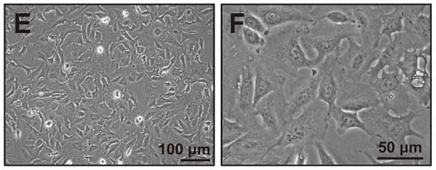
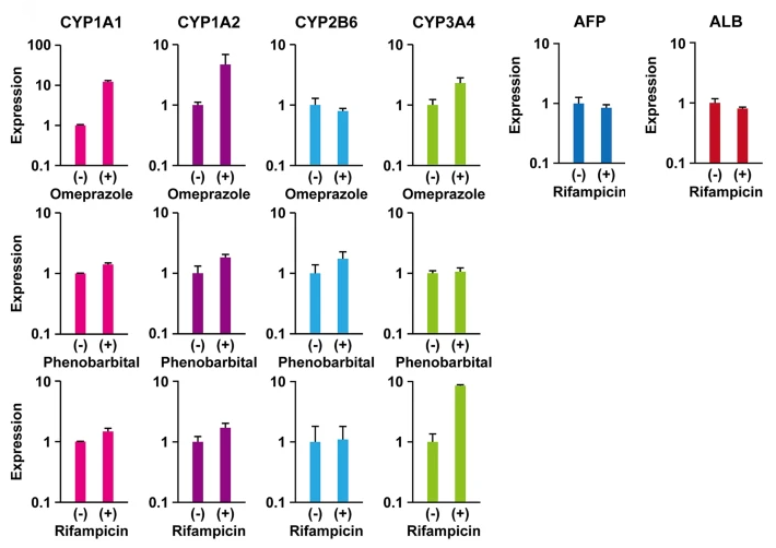
HepaMN cells, which were established from a patient with biliary atresia, are the first immortalized hepatocyte line with normal function and diploid chromosomes (see Fig.1). It is most noteworthy that HepaMN cells exhibited comparable expression levels of the albumin gene with HepaRG, and exhibited liver-like morphology in vivo. Existing cell lines such as HepG2, Huh7, THLE-2, PLC-PRF-5, and AML-12 have been used for drug interaction studies. Likewise, HepaMN cells could serve another hepatocyte model cell because of its stable CYP3A4 induction and constitutive albumin gene expression (see Fig.2).
Most importantly, HepaMN cells resembled normal hepatocytes with regards to two important hepatic functional features: (1) maintenance of efficient proliferation accompanied by constitutive expression of albumin, and (2) the ability to display metabolic functions.
Information about how to get HepaMN cells and its exclusive culture media is written below.
How to get HepaMN cells
https://cellbank.nibiohn.go.jp/ Registered cell name = JCRB1697 (HepaMN)
Special Culture Media for HepaMN cells
Culture Media = EMUKK-15
Reference: https://www.nature.com/articles/s41598-020-73992-3
Contact info: Inquiries
ProliHH Cells (Hepatocytes)
ProliHH cells, which originated from a DILI patient’s liver, were established as proliferating human hepatocytes by utilizing a liver-specific drug-metabolizing function (i.e., puromycin treatment).
Expression of hepatocyte- and BEC markers of the puromycin-selected ProliHH cells were examined after long-term culture . The cells continued to express genes for hepatocyte and bile duct markers. The expression of cytochrome P450 enzymes, CYP1A2, CYP2B6, and CYP3A4, in puromycin-treated cells at P4 was higher compared to non-treated cells. Hepatocyte markers such as ALB and AAT were also upregulated in puromycin-treated cells at P11 and P17. In contrast, the expression levels of mesenchymal cell markers were significantly decreased in puromycin-treated cells, probably due to the elimination of mesenchymal cells and the selection of puromycin-resistant cells.
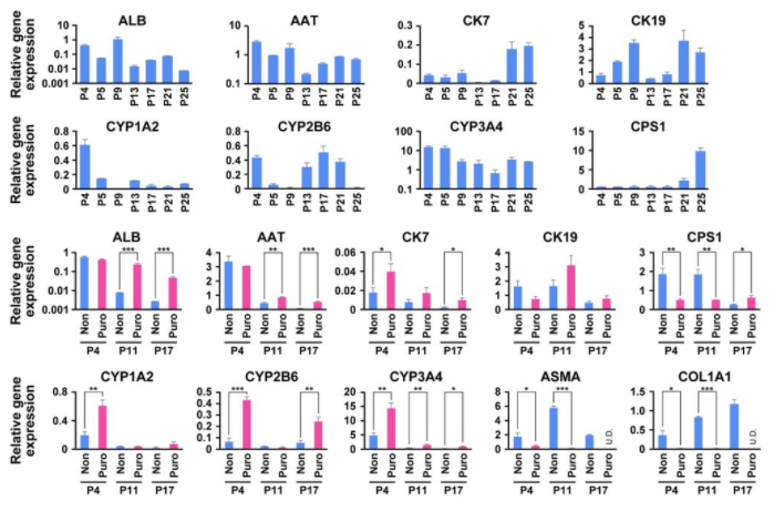
In the next, it was examined whether puromycin-treated and non-treated ProliHH cells could acquire reversible mature hepatocyte properties. Puromycin-treated and non-treated ProliHH cells at early (P5), middle (P11), and late (P21 or P25) passages were cultured in low-attachment plates for 10 days for three-dimensional (3D) culture. Under 3D-culture conditions, puromycin-treated and non-treated ProliHH cells formed spheroids irrespective of the number of passages. Glycogen storage, which could not be detected in two-dimensional culture of puromycin-treated and non-treated ProliHH cells, was confirmed in 3D culture. Hepatocyte markers (ALB, CYP3A4, and MRP2) were expressed in 3D-cultured puromycin-treated and non-treated ProliHH cells. Expression levels of hepatocyte markers, especially ALB, AAT, CYP1A2, CYP2B6, and CYP3A4, were significantly increased with maturation in 3D culture, whereas the expression of CK7, BEC and hepatocyte progenitor marker, was substantially suppressed. It is noteworthy that maturation of puromycin-treated ProliHH cells was observed in both early and late passages of ProliHH cells.
These results indicate that puromycin-treated ProliHH cells is highly capable of regaining characteristics similar to mature hepatocytes, suggesting that the addition of puromycin is effective in maintaining the characteristics of mature hepatocytes.
How to get ProliHH cells
to be prepared
Special Culture Media for ProliHH cells
Culture Media = EMUKK-05
Reference: https://www.biorxiv.org/content/10.1101/2021.04.18.440311v2.full