Methods to promote colonization and activation of Bacillu. in the rhizosphere: SynCom and Prebiotics

A group from College of Resources and Environmental Sciences, Nanjing Agricultural University, Nanjing, China etc. has reviewed about plant biocontrol mechanisms of Bacillus.
https://ami-journals.onlinelibrary.wiley.com/doi/10.1111/1751-7915.14348

As is well known, species of the genus Bacillus have been widely used for the biocontrol of plant diseases in the demand for sustainable agricultural development.

The “cry for help” mechanism in plant means that plants fight pathogen attack by assembling health-promoting beneficial microbes by releaseing specific signals. This mechanism is very similar to human’s immunity that immune cells secrete cytokines/chemokines, and thereby recruit immune cells further and activate immunity.
The root exudates are extremely crucial for recruiting biocontrol agents (i.e., beneficial microbes like Bacillus) in response to plant diseases, and it has been known that L-malic acid, citric acid, fumaric acid, and tryptophan, threonic acid, lysine, pectin, xylan, and arabinogalactan are key exudates.

The use of Bacillus strains for the biocontrol of plant disease has achieved certain benefits worldwide. However, practical utilization of Bacillus is usually confronted with unstable disease suppression efficacy under field conditions. That is because complicated and dynamic factors, such as soil characteristics, plant genotypes, and indigenous microbiota, can all influence the colonization and functional efficacy of inoculated Bacillus agents.

To overcome this issue, two types of methods have been proposed.
One is to use a method called “SynCom” which is to use a bacterial consortium constructed by using some keystone strains from the genus of Bacillus, Burkholderia, Enterobacter, Lysobacter, Stenotrophomonas, Pseudoxanthomonas, Pseudomonas, and Acinetobacter.
The other is to use “Prebiotics”. As mentioed above, specific signals released from root exudates recruit Bacillus strains and induce their activities. Therefore, relevant compounds can be developed as prebiotics for enhancing root colonization and biocontrol performance, similar to those widely applied for stimulating beneficial bacteria in the human gut. So, exogenous addition of sucrose, L-glutamic acid, riboflavin could be used as prebiotics to promote rhizosphere colonization by beneficial Bacullus strains.

Comparative glycan profiling between drug-sensitive Pseudomonas aeruginosa strains and drug-resistant strains

A group from Laboratory of Functional Glycomics, College of Life Sciences, Northwest University, Xi’an, Shaanxi, China, etc. has reported about comparative glycan profiling between drug-sensitive Pseudomonas aeruginosa (DSPA) strains and carbapenem-resistant Pseudomonas aeruginosa (CRPA) strains.
https://pubmed.ncbi.nlm.nih.gov/37861315/

lectin microarrays were used to analyze the differences in glycan alterations between 53 drug-sensitive DSPA strains and 57 carbapenem-resistant CRPA strains obtained from clinical isolates, with the goal of identifying important glycopatterns associated with carbapenem resistance.

In this experiment, whole bacterial cell lysates, which were fluorescently labeled with Cy3, were applied onto Lectin microarrays to take glycan profiles of bacteria.

As a result, it was found that LCA could be an strong biomarker detecting differential expression of glycan structures on the bacterial surface between DSPA with CRPA.

Averrant glycosylation (reduced fucosylation) of Mesoderm-specific transcript (MEST) is related to pregnancy failure.

A group from Liaoning Provincial Core Lab of Glycobiology and Glycoengineering, College of Basic Medical Science, Dalian Medical University, Dalian, China, etc. has reported that averrant glycosylation (reduced fucosylation) of Mesoderm-specific transcript (MEST) is related to pregnancy failure.
https://www.nature.com/articles/s41419-023-06166-4

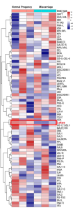
By using Lectin microarray, it was discovered that the α1,3-fucosylation, especially difucosylated Lewis Y (LeY: Fuc α1–2 Gal-β1–4[Fuc α1–3]GlcNAcβ1) glycan, in the villus tissues of miscarriage patients decreased comparing with normal pregnancy women. And a novel evidence was shown that decreasing LeY on MEST hampers the binding of MEST and eukaryotic initiation factor (eIF4E2) and inhibits implantation-related gene translation, which leads to pregnancy failure.

MEST, also named as PEG1, belongs to the alpha/beta hydrolase superfamily. MEST is a single chain glycoprotein composed of 335 amino acids. As a maternal imprint gene, MEST is widely expressed throughout the embryonic developmental period. MEST plays a crucial role in the development of embryo and placenta, as well as fetal growth. Inappropriate MEST expression is linked to the increased probability of early spontaneous miscarriage and severe fetal defects, such as growth abnormality, low birth weight, or metabolic disorders in human.

Recognition of N-glycans of the porcine whipworm by the immune system

A group from Institut für Biochemie, Department für Chemie, Universität für Bodenkultur, Wien, Austria, etc. has reported about recognition of N-glycans of the porcine whipworm by the immune system.
https://www.ncbi.nlm.nih.gov/pmc/articles/PMC10542551/

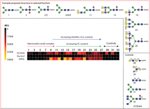
In this work, a natural glycan microarray using N-glycans (27 species) from porcine nematode parasite Trichuris suis was developed, and the interactions of these glycans with C-type lectins (DC-SIGN, Dectin-2, MGL), etc. were explored. Obtained N-glycans were fucosylated LacdiNAc motifs (bi/tri/tetra-antennary) with and without phosphorylcholine moieties and phosphorylcholine-modified oligomannose structures.

DC-SIGN recognises a rather wide range of oligomannosidic and fucosylated ligands, in the present study, its binding correlated generally with the occurrence of Man5-9GlcNAc2 in the relevant fractions.
Dectin-2 binding was lower on the array relativeto other innate immune system lectins.
MGL bound the majority of fractions, regardless of the presence of phosphorylcholine-modifications of the putative LacdiNAc-containing ligands.

Lectin microarrays and lectin-based methods continue to be important for developing biomarkers from Exosomes

A group from Department of Life Technologies, Division of Biotechnology, University of Turku, Finland, etc. has repoprted that lectins, lectin microarrays, and lectin-based methods continue to be important for screening, targeting, separation, and identification of improved biomarkers from the surface of Extracellular Vesicles (EVs).
https://pubmed.ncbi.nlm.nih.gov/37773167/

The EVs field has grown rapidly in the last decade. Although significant progress has been made in EV-based cancer biomarker discovery, translating these findings into clinical practice (including therapeutics) faces several challenges. These include the technical challenges of EV separation and detection and the need for more insights into molecular mechanisms governing EV release and EV uptake by target cells.
In each of these areas, it is thought that glycosylation holds a key to progress and that lectin-based approaches will unlock doors with their specificity and ease of implementation.

Some lectins have overlapping binding affinities and specificities towards multiple glycan structures which lead to difficulties in precisely identifying the glycan structures of interest. These ambiguities influence the assay results which lead to false-positive or false-negative results. Despite this kind od limitation, lectins, lectin microaerrays, and lectin-based methods definitely remain valuable tools in glycan research. To overcome some of these challenges, combination with other techniques such as MS and HPLC will be useful for a better understanding of detailed glycan structures and their applications. Moreover, glycan microarray technology will be complementaly to study glycan interactions and their functions in a systematic way.

To ensure accurate and reliable results from lectins and their glycan interaction, it is essential to verify this interaction through competitive inhibition, through modification of the glycan structures or by comparison with lectins which have been modified to show different binding specificities. This combination approach helps to avoid false positive or negative results and provides concrete evidence for glycan-mediated binding.