Archive 23/4/29

Antiviral activities of nanoparticle-like κ-Carrageenan polyelectrolyte complex with Chitosan

A group from G.B. Elyakov Pacific Institute of Bioorganic Chemistry, Far-Eastern Branch of the Russian Academy of Sciences, Vladivostok, Russia, etc. has reported about antiviral activities of κ-Carrageenan polyelectrolyte complex with chitosan
https://www.mdpi.com/1660-3397/21/4/238

Some of the well known polysaccharides of marine origin are the polysaccharides of red algae—carrageenans (CRGs). It is known that CRGs, mimicking heparan sulfate, are potential antivirals that can interfere with the early stages of viral replication, including virus entry, by masking the positive charge of the virus surface receptors to prevent them from binding to the heparan sulfate proteoglycans in the host cell surface. CRGs can be included in pharmaceutical compositions for the prevention or treatment of viral infections with no side effects.

Nanoparticles formation is one of the ways to modulate the physicochemical properties and enhance the activity of original polysaccharides. For this purpose, based on the polysaccharide of red algae, κ-carrageenan (κ-CRG), its polyelectrolyte complex (PEC), with chitosan (CH), were obtained. Obtained average diameter of nano@articles were about 150–200 nm.

The antiviral effect of these compounds was assessed by the inhibition rate of the cytopathogenic effect of the herpes simplex virus type 1 (HSV-1) in Vero cells. As a result, a two-fold increase in the antiherpetic activity (selective index) of PEC compared to κ-CRG and a 13 times increase compared to CH were shown, which was considered to be due to a change in the physicochemical characteristics of κ-CRG in PEC.

Comparative functional genome analysis of plant beneficial bacteria in NCBI Databases

A group from State Key Laboratory of Pharmaceutical Biotechnology, School of Life Sciences, Nanjing University, Nanjing, China, etc. has reported about comparative functional genome analysis of plant growth-promoting bacteria (PGPB) in NCBI Databases.
https://journals.asm.org/doi/10.1128/spectrum.05007-22?url_ver=Z39.88-2003&rfr_id=ori:rid:crossref.org&rfr_dat=cr_pub%20%200pubmed

PGPB are a group of beneficial microorganisms that include 60 bacterial genera, such as Bacillus, Pseudomonas, and Burkholderia, which widely colonize plant leaves and soil, promote plant growth, and/or inhibit pathogen infection. PGPB colonize either leaves (leaf associated [LA]; 195 strains) or rhizospheric soil (soil associated [SA]; 283 strains).

The results of this study demonstrate that PGPB generally contain a large quantity of carbohydrate enzymes (CAZymes), which enhance their ability to colonize plants. Among the LA PGPB strains, Pseudomonas strains exhibited a higher abundance of CAZymes than other strains, indicating that this genus could be developed into an ideal interfoliar biocontrol agent. The CAZymes can break down the cell walls of plant pathogens, leading to pathogen death. Conversely, among the SA PGPB strains, Burkholderia strains possessed a higher concentration of genes encoding carbohydrate metabolism enzymes, suggesting that they have diverse mechanisms for carbohydrate utilization. The genera Bacillus and Paenibacillus, found in the LA and SA habitats, produced a higher number of secondary metabolite clusters, making them suitable for both leaf and soil environment. The greater the number of secondary metabolic gene clusters in bacteria, the stronger their ability to perform biological defense. The majority of Bacillus strains of PGPB possess more abundant secondary metabolic clusters than other taxonomic groups in the LA and SA habitats. Actually, Bacillus strains are dominant biocontrol agents in the market.

Proteoglycan link protein-1 (HAPLN1) would be a driver for peritoneal metastasis in Pancreatic ductal adenocarcinoma (PDAC) and could be a GOOD prognostic marker

A group from Division Vascular Signaling and Cancer, German Cancer Research Center (DKFZ), Heidelberg, Germany, etc. has reported that Proteoglycan link protein-1 (HAPLN1) would be a driver for peritoneal metastasis in Pancreatic ductal adenocarcinoma (PDAC) and could be a GOOD prognostic marker.
https://www.nature.com/articles/s41467-023-38064-w

Cellular plasticity is an important feature of tumore cells. Cellular plasticity is characterized by the ability of cells to convert between different (intermediate) cellular states by inducing epithelial-to-mesenchymal transition (EMT), as well as mesenchymal-to-epithelial transition (MET) and features of cancer cell stemness5. Such plasticity not only makes cancer cells more prone for invasion and adaption to the microenvironment, but also protects them from apoptosis, immune attack and chemotherapy.

A key regulator of metastasis is the microenvironment that tumor cells are facing during their journey to the metastatic site. The tumor microenvironment (TME) consists of several cellular and non-cellular components, including cancer-associated fibroblasts (CAFs), endothelial cells, immune cells and extracellular matrix (ECM). In PDAC, the TME is strongly desmoplastic, with substantial accumulation of ECM components. Hyaluronic Acid (HA), which is one major component of the ECM, facilitates tumor progression and metastasis through promoting partial EMT, invasion, immunomodulation and therapy resistance. CAFs are the main producers of ECM.

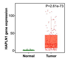
HAPLN1 is a HA and chondroitin sulfate proteoglycan (CSPG) crosslinker in the ECM, but its role has been so far poorly understood in cancer. In this study it was identified that HAPLN1 was the most upregulated genes in PDAC compared to adjacent tissue. It was thought that HAPLN1 could be defined as a mediator of peritoneal dissemination in PDAC, by inducing a highly plastic phenotype in cancer cells, which leads to a pro-tumoral metastatic niche.

Rhizosphere bacteriome associated with tobacco black shank disease and its biocontrol with Bacillus

A group from Key Laboratory of Microbial Resources Collection and Preservation, Ministry of Agriculture and Rural Affairs, Institute of Agricultural Resources and Regional Planning, Chinese Academy of Agricultural Sciences, Beijing, China, etc. has reported about rhizosphere bacteriome associated with tobacco black shank disease and its biocontrol with Bacillus velezensis S719.
https://www.ncbi.nlm.nih.gov/pmc/articles/PMC10108594/

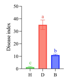
The tobacco rhizosphere soil samples obtained from the diseased field was regarded as the disease group (D group),
the samples obtained from the healthy control field were regarded as the healthy group (H group), which were 600 m away from the diseased field,
and the biocontrol group (B group) was defined as the diseased field treated with biocontrol agent consisting of Bacillus velezensis S719 after tobacco seedlings were transplanted.

The desease index was the owest in H groups and the highest in D groups as shown below. The effect of Bacillus innocuration was clearly obserbed in B groups comparing with D groups.

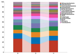
The relative abundance of the bacterial taxa was compared at the class levels.
In B groups, Alphaproteobacteria accounted for 27.2% of the ASVs, at least two folds more than other terms in the same group;
In D groups, Actinobacteria was abundant, accounting for 13% of the ASVs, while the proportions in group B and H were 10.6% and 9.5%, respectively;
In H groups, Sphingobacteria and Cytophagia were abundant, accounting for 6.7% and 3.5% of the ASVs, respectively.

Protective antibodies to schistosome infections include IgG responses to the core Xyl/Fuc epitopes in surface-expressed N-glycans

A group from Department of Biochemistry, Emory University School of Medicine, Atlanta, GA, USA, etc. has reported that protective antibodies to schistosome infections in brown rats and rhesus monkeys include IgG responses to the core Xyl/Fuc epitopes in surface-expressed N-glycans, and raise the potential of novel glyco-based vaccines that might be developed to combat this disease.
https://pubmed.ncbi.nlm.nih.gov/37081851/

Schistosomiasis, caused by infection with parasitic helminths of the genus Schistosoma, infects over two hundred million people worldwide and causes up to seventy million disability-adjusted-life years (DALYs)—more DALYs than those caused by malaria. Therefore, it is thought that novel vaccine targets are urgently needed. Many different mammals can host schistosomes. Humans become chronically infected, while Rodents can clear the worms soon after infection.

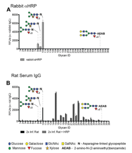
Human N-glycans typically contain a core modification of the GlcNAc residues in GlcNAc-Asn by α6-linked fucose. Core α2-Xylose (CX) and core α3-Fucose (CF) are not found in humans, but are common in N-glycans from insects (CF), plants (CX, CF), and worms (CX, CF).

In ths work, it was demonstrated that antibodies including rabbit anti-horseradish peroxidase (rabαHRP) to CX/CF promote complement-dependent and glycan-specific killing of schistosomula in vitro.

A new lectin (HiL) isolated from the tropical sponge Haliclona (Reniera) implexiformis shows antimicrobial activity against Staphylococcus spp.

A group from Universidade Federal do Ceará, Departamento de Engenharia de Pesca, Laboratório de Biotecnologia Marinha, Brazil, etc., has reported about a new lectin (HiL) isolated from the tropical sponge Haliclona (Reniera) implexiformis showing antimicrobial effect.
https://pubmed.ncbi.nlm.nih.gov/37075356/

Marine sponges are multicellular, sessile and filtering organisms, belonging to the phylum Porifera, the oldest of the Metazoans. More than 8,500 species found in marine and freshwater ecosystems have been described。A lectin from the marine sponge Haliclona (Reniera) implexiformis was isolated by affinity chromatography on Sepharose™ matrix, and HiL showed specificity for galactose and its derivatives.

It seems that the recognition of glycans in the bacterial membrane by lectins may cause an inhibitory effect in the biofilm formation. Some studies have shown that galactose-binding lectins may recognize glycans in bacterial surface and distinguish bacteria Gram-positive from Gram-negative bacteria。The mechanism of activity of lectins on biofilm formation is not yet fully determined, but as shown below, HiL showed antimicrobial activity potential to prevent infections mainly caused by Staphylococcus spp (no antimicrobial activity for E. coli: Gram-negative bacteria).

 The vertical axis shows the total mass of biofilmms quantified by measuring the intensity of crystal violet stain

Core fucose-binding lectin PhoSL inhibits HBV infection

A group from Department of Molecular Biochemistry and Clinical Investigation, Osaka University Graduate School of Medicine, Osaka, Japan, etc. has reported that core fucose-binding lectin Pholiota squarrosa lectin (PhoSL) inhibits HBV infection.
https://www.ncbi.nlm.nih.gov/pmc/articles/PMC10105536/

Current anti-HBV therapies include nucleoside and nucleotide analogs, which can competitively inhibit HBV replication2,3, and pegylated interferons, which can modulate the host immune response to HBV infection and induce the degradation of covalently closed circular DNA (cccDNA) in hepatocytes.

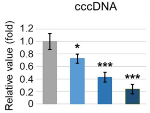
In this study, it was investigated the effects of PhoSL on HBV infection of the human cell line HepG2-hNTCP-C4, and also analyzed the molecular mechanisms underlying PhoSL-mediated inhibition of HBV infection. It was shown that the treatment with PhoSL dramatically decreased the levels of human HBV e antigen (HBeAg) , cccDNA (as shown belwo), HBV DNA, and HBV RNA), markers of HBV infection, in a dose-dependent manner without cytotoxicity.

There are two possibilities: (1) PhoSL affects the protein dynamics of the host cells, or (2) PhoSL binds to HBV.
As aresult, it was found that PhoSL inhibits EGFR activation by blocking the binding of EGF to EGFR, and also PhoSL binds to HBV particles directly. HBV particles bound by PhoSL are internalized into host cells, and PhoSL seems to inhibit HBV infection after internalization.

PhoSL treatment could contribute to the development of novel anti-HBV therapies.

Binding of galectins to the N-glycans of fibroblast growth factor receptors (FGFRs) affects cellular activity

A group from Department of Protein Engineering, Faculty of Biotechnology, University of Wroclaw, Poland, etc. has reported that the clustering of FGFR triggered by galectins (Gal-1, Gal-s, Gal-7 and Gal-8) binding to those N-glycans can activate the receptor and initiate downstream signaling cascades.
https://www.ncbi.nlm.nih.gov/pmc/articles/PMC10070233/

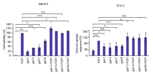
It was demonstrated that galectins modulate FGF/FGFR cellular processes through direct action on the receptor and the cellular consequences of galectin-induced FGFR signaling largely differ from those achieved by the canonical ligand (FGF).

Galectin-1, -3, -7, and -8 were the most effective binders of FGFRs (FGFR1–FGFR4) among human galectins, and FGFR1 clustering promoted by multivalency of galectins was essential for FGFR1 activation and initiation of downstream signaling cascades. Interestingly, the combination of galectin-1, -3, and -8 with FGF1 enhanced the number of viable cells more effectively than each single protein. And further, glucose uptake was enhanced by mixtures of FGF1 with galectin-1 and -3 compared to treatments with each single protein.

Specific MUC1 glycan marker (sialylated core-2 O-glycan) in mucoepidermoid carcinoma (a type of salivary gland tumors)

A group from Department of Oral Oncology, Oral and Maxillofacial Surgery, Ichikawa General Hospital, Tokyo Dental College, Ichikawa-Shi, Chiba, Japan, etc. has reported that MUC1 modified with α2,3-linked sialic acid-containing core-2 O-glycans could be a new potential diagnostic marker of Mucoepidermoid carcinoma (MEC).
https://www.ncbi.nlm.nih.gov/pmc/articles/PMC10082819/

Salivary gland tumours account for approximately 1% of all tumours and 3–6% of head and neck tumours. MEC is the most frequent of the rare salivary gland malignancies. MEC has a distinct feature of producing mucus and abnormally expressing Mucin 1 (MUC1), a type of mucin that is a heavily glycosylated, high-molecular-weight glycoprotein. It was found that MEC produced MUC1 distinctively modified by sialylated core-2 O-glycans (GlcNAcβ1-6(Galβ1-3)GalNAcαSer/Thr), and further, the MUC1 modified with core-2 O-glycans containing α2,3-linked sialic acid is expressed in mucous cells and non-mucous cells of MEC.

Relationship between peanut root exudates and changes in gene expression of the plant growth-promoting effects of burkholderia pyrrocinia strain P10

A group from College of Life Sciences, Guizhou University, 550025 Guiyang, Guizhou, China has reported about relationship between peanut root exudates and changes in gene expression of the plant growth-promoting effects of burkholderia pyrrocinia strain P10.
https://www.ncbi.nlm.nih.gov/pmc/articles/PMC10061817/

The peanut root exudates were mainly composed of organic acids and amino acids, but it also contained sugars, alcohols, fatty acids, sugar alcohols, sugar acids, and other components. The detected compounds included low-molecular-weight organic acids, such as malic acid, lactic acid, succinic acid, pyruvic acid, oxalic acid, and citric acid, which were present at relatively high concentrations. Various amino acids were also detected, including alanine, glycine, proline, valine, phenylalanine, isoleucine, tyrosine, methionine, threonine, glutamic acid, serine, lysine, asparagine, glutamine, and aspartic acid. Xylose, allose, lyxose, and ribose were the most prominent sugars in the peanut RE, which also contained fatty acids (e.g., palmitic acid, stearic acid, myristic acid, oleic acid, and palmitoleic acid), alcohols (e.g., 4-hydroxyphenylethanol, myo-inositol, and phytol), sugar alcohols (e.g., threitol, xylitol, sorbitol, and arabitol), sugar acids (e.g., galactonic acid, gluconic acid, and threonic acid), and some other components (e.g., indole-3-acetamide and urea).

And, the up-regulated genes of burkholderia pyrrocinia strain P10 tended to be associated with ATP binding cassette (ABC) transporters, steroid degradation, quorum sensing (QS), biosynthesis of siderophore group nonribosomal peptides, and galactose metabolism. More specifically, 47 genes with expression levels that were up-regulated by 1.01- to 5.83-times were associated with the transport of minerals and organic ions, oligosaccharides, monosaccharides, amino acids, peptides, iron-siderophores, and ATP binding cassette subfamily C (ABCC) subfamily members. In some cases, the transcription of an entire gene cluster was observed, including the ssuA-C-B genes responsible for alkanesulfonate transport, afuA-B-C genes (Fe3+ transport), proX-W-V genes (glycine betaine/proline transport), and araF-H-G genes (L-arabinose transport). Additionally, the expression of 19 genes involved in QS and four genes contributing to steroid degradation were up-regulated. The expression levels of three genes involved in the biosynthesis of siderophore group nonribosomal peptides and five genes related to galactose metabolism were also up-regulated. The expression of genes associated with strain P10 adhesion and biofilm formation were also up-regulated. In the pathways mediating the metabolism of glucose and mannose, amino sugars, and ribose, the expression of the mannose-1-phosphoguanyltransferase gene algA, which encodes the enzyme that converts mannose-1-phosphate to GDP-mannose, was up-regulated. The GDP-mannose is an important exopolysaccharide (EPS) constituent and the main component of biofilms.

Powered by WordPress |Copyright © 2020 Emukk. All rights reserved