Archive 21/3/2

How long the neutralizing antibody titer could be maintained in the case of SARS-CoV-2

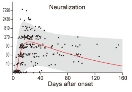
A group from Medical School University of Tokyo, etc. has reported on changes in neutralizing antibody titers from sequential blood samples collected from Jan. 2020 to Apr. 2020 in Japan.
https://www.thelancet.com/journals/eclinm/article/PIIS2589-5370(21)00014-6/fulltext

After the infection onset, it seems that the neutralization titer is maintained for a several months. In the figure below, 95% CI is shown in gray color, and the red line shows the median value. As for the pathogenicity of SARS-CoV-2 via antibody-dependent enhancement (ADE) activity, numerous studies are ongoing in the world. But, no studies describing an increase in pathogenicity due to ADE have been reported yet, so that authors are thinking that the possibility of ADE would be limited.

Lactoferrin is to be a quite GOOD nutrient to inhibit SARS-CoV-2 infection

A blog article about Lactoferrin was already uploaded on Feb. 12th, 2021, focusing on the antiviral function of Lactoferrin. It is know that Heparan sulfate is a co-receptor for SARS-CoV-2 positioning ACE2 as the primary receptor. The antiviral function of lactoferrin is considered to inhibit SARS-CoV-2 binding to heparan sulfate.

A group from Kagawa Nutrition University is recommending to take 2 capsules of enteric lactoferrin (1 capsule = 30mg) purchasable without doctor’s prescription everyday. You could purchase it easily even from Amazon. Actually, Mongolia shows the world lowest fatality rate, and the group pointed out that Mongolians are taking larger amount of lactoferrin than any other nations in the world.
https://www.eiyo.ac.jp/ions/?p=4407  

Asymptomatic HIV infected Children have distinct IgG Fc Glycosylation patterns

A group from University of Oxford, etc. has reported quite interesting immune response of HIV infected children.
https://www.ncbi.nlm.nih.gov/pmc/articles/PMC7763548/

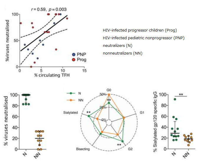
There are some asymptomatic children, HIV-infected pediatric nonprogressors (PNPs), who maintain normal CD4 counts HIV despite ongoing viral replication at high rates in the absence of antiretroviral therapy (ATR). It is also very interesting that some children develop broadly neutralizing antibodies (bnAb). It is also characteristic that the effector functions (ADCC and ADCP) mediated by IgG Fc binding to effector cells such as NK cells and macrophages are strong in the PNPs.

As for IgG glycosylation, PNPs have similar glycosylation to health controls, or rather, slightly higher sialylation and lower core fucosylation, although HIV infected adults usually develop agalacto type N-glycans.

It was also found that bnAb collelated with circulating Tfh cells and sialylation of gp120-specific IgG.
It is well know that the effector functions via IgG Fc binding get higher with increasing core fucosylation.

Taking these things into consideration, the immune activation in PNPs is rather low, but it might be compensated by the existence of bnAb and stronger effector functions. 

Effectiveness of Pfizer and Moderna vaccines for various SARS-CoV-2 variants occuring in the world: B.1.351 and P.1 variants are critical

A group from Massachusetts General Hospital, etc. has reported on effectiveness of two typical vaccines, BNT162b2 (Pfizer) and mRNA-1273 (Moderna), for various SARS-CoV-2 variants occuring in the world.
https://www.ncbi.nlm.nih.gov/pmc/articles/PMC7899476/

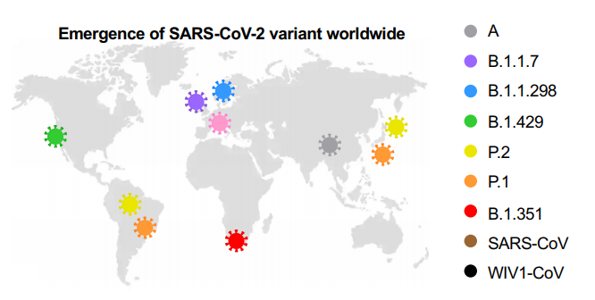
Distribution of typical SARS-CoV-2 variants in the world.

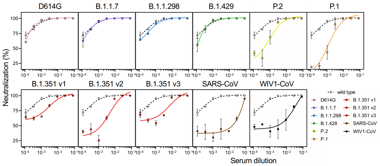
Effectiveness of Pfizer and Moderna vaccines is shown by the neutralization response. There seems to be almost no change for B.1.1.7 variant, but the neutralization activity of these vaccines are significantly reduced in B.1.351 v2 and P.1 variants. Two common mutations, K417N and E484K, exist in B.1.351 and P.1 variants, and these mutations do not exist in B.1.1.7. 

Anti-complement Therapy would be effective for Membranous Nephropathy

A group from Ohio State University Wexner Medical Center, etc. has reported that excess compliments are deposited in Membranous Nephropathy (MN).
https://www.ncbi.nlm.nih.gov/pmc/articles/PMC7879111/pdf/main.pdf

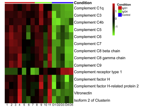
Glomeruli tissue samples were captured by laser microdissection, and IgGs and complements in the samples were analyzed by mass spectrometry comprehensively. As a result, elevated glomerular levels of all complements (except for CR1) and IgG1~IgG4 were observed in MN patients comparing with health controls. On the other hand, mannose-binding lectin (MBL), Ficolin, and Collectin were not detected. These results suggest that Glomeruli tissue could be damaged by excessively activated complements not through the lectin pathway. Loss of CR1 at the site of the damaged filtration barrier might accelerate excess complement activation resulting in significant damages. In addition, it is thought that M-type phospholipase A2 receptor (PLA2R) could be an antigen in membranous nephropathy.

Cross-reactivity of  hCoV-specific IgGs against SARS-CoV-2 uninfected individuals

A group from NMI Natural and Medical Sciences Institute at the University of Tübingen, etc. has reported on the cross-reactivity of hCoV-specific IgGs against SARS-CoV-2 infected and uninfected individuals using 1,173 serum/plasma samples.
https://www.nature.com/articles/s41467-021-20973-3.pdf

Up to now, it has been known that there are 4 kinds of epidemic human coronaviruses (hCoVs: NL63, 229E, OC43, HKU1) which cause seasonal colds.
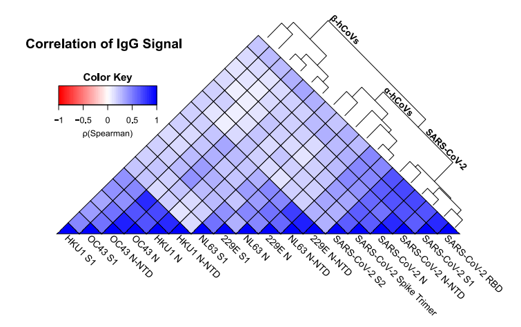
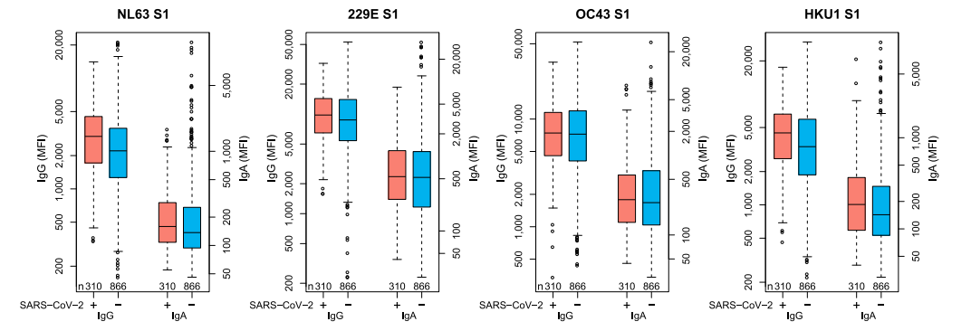
As shown below, there is certainly correlation between SARS-CoV-2 and hCoVs specific IgGs to a certain degree. 10% of SARS-CoV-2 infected individuals do not develop detectable SARS-CoV-2-specific IgG, but it has not been identified if this result could come from cross-reactivity of hCoVs-specific IgGs or just from innate immunity against SARS-CoV-2.


Once again: C-type Lectins, CD209/DC-SIGN, CD209L/L-SIGN, could be infection receptors of SARS-CoV-2 as well as ACE2

There have been some papers reporting that C-type Lectins could be SARS-CoV-2 infection receptors as well as well-know ACE2.
Harvard Medical School, July 30, 2020
https://www.biorxiv.org/content/10.1101/2020.07.29.227462v1
Boston University, Dec. 9, 2020
https://www.biorxiv.org/content/10.1101/2020.06.22.165803v2
There was a review paper from Boston University, (Dec. 22, 2020) in addition to the above paper, let me introduce two kinds of interesting data for your reference.
https://www.mdpi.com/2079-7737/10/1/1

Although ACE2 is a really important virus receptor, the expression level of ACE in lung is very low, however, CD209 and CD209L are more broadly expressed in human tissues rather than the ACE2.
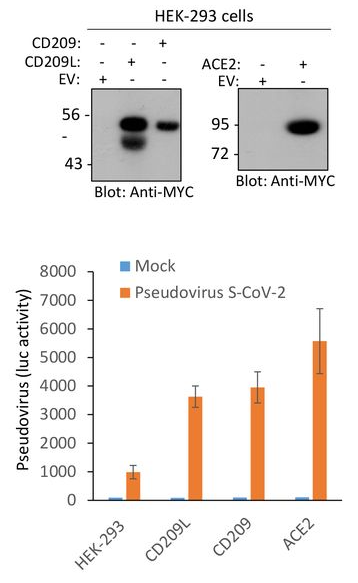
A result of infection experiments using SAR-CoV-2 pseudotyped lentivirus and HEK293 cells overly expressing CD209L, CD209, and ACE2 is also shown below.
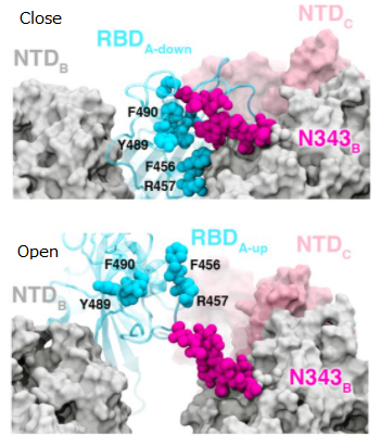
Taking these things into consideration, it would be clear that CD209 and CD209L could mediate SARS-CoV-2 entry as infection receptors even for tissues lacking ACE2 expression.
The CRDs of C-type Lectins (DC-SIGN, L-SIGN) recognize strongly high mannose type glycans.

In order not to develop COVID-19 even if infected with the new coronavirus (SARS-CoV-2): A way to coexist with SARS-CoV-2, survive virus wars

What I am always thinking about COVID-19 was written in the following paper published from a group of Central University of Tamil Nadu etc.
https://academic.oup.com/femspd/article/79/1/ftaa076/6027506

In the case of COVID-19, there are so many asymptomatic individuals (some studies have estimated that up to 80%).
Generally speaking, the incubation period for COVID-19 is reported to be between 5 to 6 days, although this can be up to around 14 days, but, asymptomatic individuals show an extended median duration time of 19 days.

SARS-CoV-2 activates the STING pathway and NLRP3 inflammasome assembling, and induces cytokine storm as a result of excess production of IL-1β, and IL-18, TNF-α, IFN-γ, IL-6, etc. However, bats do not develop any disease, although they are reservoir of various viruses. In the case of bats, they are coexisting with virus preparing the following features evolutionally.
(1)limiting the assembly of the NLR family pyrin domain containing three (NLRP3) in monocytes、
(2)loss of the ability to effectively sense exogenic cytoplasmic DNA via loss of PYHIN genes
(3)excessively high levels of the anti-inflammatory cytokine IL-10,
(4)lack of functional killer cell Ig-like (KIR) and killer cell lectin-like (KLR) receptors.

It is know that the levels of inflammatory cytokines are relatively low, and the SARS-CoV-2-specific IgG and neutralizing antibody levels are significantly lower in asymptomatic individuals. This might mean that asymptomatic is a result of weak immune response. And on the contrary, it might be a result of existing antibody cross-reactivity of classical seasonal cold-causing coronaviruses 229E, HKU1, OC43 and NL63.

In the context of coexistence with virus, mechanisms uncovered in bats likely hint the development of therapeutic strategies against COVID-19 in humans. That could be a wisdom, to survive virus wars.
 

Agalacto (i.e. GlcNAc) binding lectins: BLL, PVL, GSL-II, and BGL 

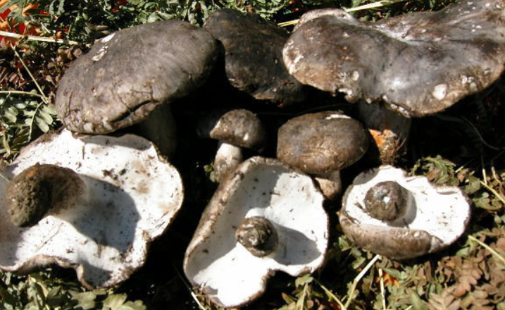
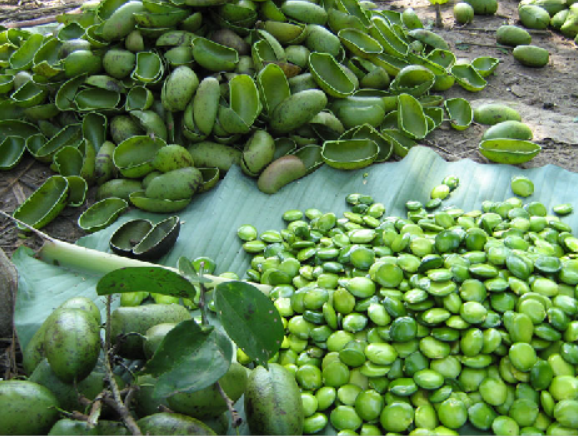
Agalacto type complex N-glycans without terminal Gal are often seen in autoimmune disease. Well known Lectins with glycan binding specificity to agalacto are BLL, PVL, GSL-II as shown below. BLL and PVL are mushroom derived, and GSL-II is bean derived.

Boletopsis leucomelaena: BLL lectin


Psathyrella velutina: PVL lectin


Griffonia simplicifolia: GSL-II lectin


A group from New England Biolabs, Inc. has identified gene sequence of Boletopsis grisea, a close North American relative of Boletopsis leucomelaena, and investigated glycan binding specificities of recombinant lectin (named rBGL) with using glycan arrays. Curiously, rBGL has bi-specific glycan binding characteristics, not only to agalacto type N-glycans but also to O-glycans with Thomsen–Friedenreich antigen (TF-antigen; Galβ1,3GalNAc-α-). Depending on applications, the binding specificity to O-glycan would be interesting.
https://www.nature.com/articles/s41598-020-80488-7

Powered by WordPress |Copyright © 2020 Emukk. All rights reserved