Effects of synthetic microbial consortia derived from rhizosphere soil of wheat against a pathogen causing wheat root rot disease

A group from North Central Agriculture Research Laboratory, USDA-ARS, Brookings, SD, USA, etc. has reported about the effects of synthetic microbial consortia derived from rhizosphere soil of wheat against rhizoctonia solani AG8, a pathogen causing root rot disease.
https://www.ncbi.nlm.nih.gov/pmc/articles/PMC9473337/

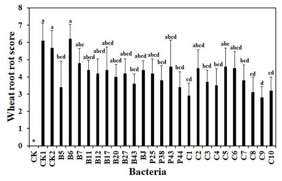
The effects of bacteria, including 14 single strains and 10 SynComs, on wheat root rot disease caused by R. solani AG8 were evaluated in soil in the greenhouse. After 3 weeks, compared with wheat growth in AG8 inoculated soil (CK1, CK2), wheat treated with some single bacterial strains and SynComs reduced root rot at different levels. Among them, wheat treated with single bacterial strains (Pseudomonas sp. B5, Rhodococcus erythropolis B43, Chryseobacterium soldanellicola P38, and Pedobacter sp. P44) and SynComs (C1, C3, C4, C7, C8, C9, and C10) significantly reduced wheat root rot score, compared with the controls (CK1 and CK2) in AG8 inoculated soil. Further, the fresh root weight of wheat treated with single bacterial strains (Pseudomonas spp. B5 and B11, and Chryseobacterium sp. B7) and SynComs (C1, C4, C7, C8, C9, and C10) were greater than those of the controls in AG8 inoculated soil.

SynCom 1(C1): Pseudomonas sp. B5, Pseudomonas sp. B11, Pseudomonas sp. B12, Pseudomonas sp. P25
SynCom 2(C2): Chryseobacterium sp. B7, Chryseobacterium soldanellicola P38, Chryseobacterium sp. P43
SynCom 3(C3): Sphingomonas sp. B17, Cupriavidus campinensis B20, Asticcacaulis sp. B27, Rhodococcus erythropolis B43
SynCom 4(C4): Cupriavidus campinensis B20, Asticcacaulis sp. B27, Rhodococcus erythropolis B43, Chryseobacterium soldanellicola P38
SynCom 5(C5): Cupriavidus campinensis B20, Rhodococcus erythropolis B43, Janthinobacterium lividum BJ, Chryseobacterium soldanellicola P38
SynCom 6(C6): Streptomyces sp. B6, Chryseobacterium sp. B7, Pseudomonas sp. B12, Sphingomonas sp. B17
SynCom 7(C7): Pseudomonas sp. B5, Streptomyces sp. B6, Chryseobacterium sp. B7, Pseudomonas sp. B11
SynCom 8(C8): Pseudomonas sp. B12, Sphingomonas sp. B17, Cupriavidus campinensis B20, Asticcacaulis sp. B27
SynCom 9(C9): Pseudomonas sp. B12, Rhodococcus erythropolis B43, Janthinobacterium lividum BJ, Pedobacter sp. P44
SynCom 10(C10): All 14 bacterial strains
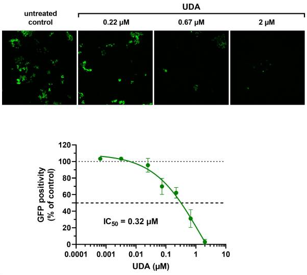
Antiviral activity of UDA llectin against SARS-CoV-2

A group from Laboratory of Virology and Chemotherapy, Rega Institute, Department of Microbiology, Immunology and Transplantation, KU Leuven, Belgium, etc. has reported about antiviral activity of UDA against SARS-CoV-2.
https://www.ncbi.nlm.nih.gov/pmc/articles/PMC9468479/

UDA is one of the smallest plant lectins reported, and has a binding preference for GlcNAc and high-mannose sugars on target glycoproteins.

It was shown that UDA prevents viral replication of the early Wuhan-Hu-1 strain in Vero E6 cells (IC50 = 225 nM), but also the replication of SARS-CoV-2 variants of concern, including Alpha, Beta and Gamma (IC50 ranging from 115 to 171 nM). In addition, UDA exerts antiviral activity against the latest circulating Delta and Omicron variant in U87.ACE2+ cells (IC50 values are 1.6 and 0.9 µM, respectively).

It was found that UDA is not acting as a direct receptor-attachment competitor, and its strongest interaction site is not located in the RBD from SPR analyses.

Site-specific N-Linked glycan characterization of PSMA from metastatic prostate cancer cells

A group from Leroy T. Canoles Jr. Cancer Research Center, Eastern Virginia Medical School, Norfolk, Virginia 23507, USA, etc. has reported about site-specific N-Linked glycopeptide characterization of Prostate-Specific Membrane Antigen from metastatic prostate cancer cells.
https://www.ncbi.nlm.nih.gov/pmc/articles/PMC9435049/

The prostate-specific membrane antigen (PSMA), also known as folate hydrolase 1 (FOLH1) or glutamate carboxypeptidase 2, was previously studied as a PCa biomarker in tissues and body fluids using conventional biochemical methods with mixed results. PSMA is not exclusively expressed in prostatic tumors; the protein is expressed in low abundance in prostate epithelium, in the neo-vasculature of exclusive solid tumor types, and in some healthy tissues including proximal renal tubes, duodenum, and ganglia of the nervous system. In PCa, PSMA abundance increases with disease severity, and up to 100-fold higher abundance has been observed in advanced aggressive forms of the disease compared to normal tissue.

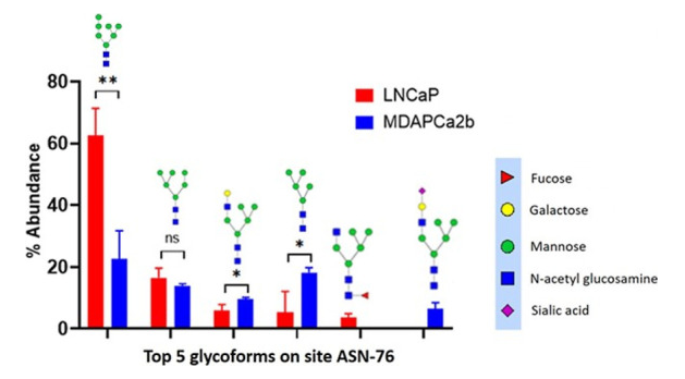
In this study, two PCa cell lines, LNCaP cells (CRL-1740) and MDAPCa2b (ATCC CRL-2422) were used to compare site-specific N-linked glycopeptide characterization of PSMA.
It was demonstrated that there are significant differences in the expression of several glycans in two cell lines LNCaP and MDAPCa2b (see below), which have different phenotypes, and further there are significantly more glycans identified in MDAPCa2b compared to LNCaP cells.
These studies will form the basis of developing site-specific PSMA glycoform-based prognostic markers for PCa disease stratification in the future.

How extract the features of the complex systems of rhizobacterial microbiomes, and efficiently control the microbiomes?

For self-organizing complex systems, feature extraction methods such as principal component analysis and clustering analysis are often used. Conversely, it means that there is no other good analysis methodology close at hand. In self-organizing complex systems, the properties of the whole can often be neither understood nor predicted by knowledge of its constituents alone, and the interactions between the constituents of the system give rise to new structures and functions, and those will appear on a large scale.
In the case of the rhizobacterial microbiomes, it is possible that when good bacteria is exceeded a certain threshold, opportunistic bacteria become allies and a synergistic effect appears remarkably. The reverse is also true, and when the number of bad bacteria exceeds a certain threshold, the plant dies at once.

The problem is how to perform feature extraction (in other words, pattern recognition) of such a complex system of rhizobacterial microbiome in the field (farm). At the research level, 16S rRNA read analysis of the rhizobacterial microbiomes and computer analysis on the full use of the obtained big data might be able to visualize the hidden patterns which could not be identified with  conventional methods. But, can such a method be used in the field? No, this kind of work will remain in the world of papers. If the technology is not easy for anyone to use, it will not succeed in reality as a business. To be used on site, it has to be cheap, and it has to give immediate results.

Bacillus, Pseudomonas, Streptomyces, Arthrobacter are said to be representative good bacteria as rhizobacteria, and Enterobacter and Barkholderia are also to be good bacteria.
The bacterial classification of such representative good bacteria and bad bacteria is written below.
First of all, good bacteria,
Actinobacteria phylum→ Actinobacteria class → Streptomyces order → Streptomycetae → Streptomyces genus (good guys)
Proteobacteria phylum → γ-Proteobacterial class → Pseudomonad order → Pseudomonadidae → Pseudomonas genus (typical good guys)
Proteobacteria phylum → γ-Proteobacterial class → Enterobacter order → Enterobacteraceae → Enterobacter genus (good guys)
Proteobacteria phylum → β-Proteobacterial class → Barkholderia order → Barkholderidae → Balkholderia genus (good)
Firmicutes phylum→ Bacillus class → Bacillus order → Bacillus family → Bacillus genus (typical good guy)
Actinobacteria phylum→ Actinobacteria class → Actinobacteria family→ Micrococcaceae → Arthrobacter genus (good guys)
and bad bacteria,
Ascomycota phylum → Hydrangea class → Pistanthus → Asteraceae → Fusarium genus (typical bad guys)

If we can sense the phylum Actinobacteria, Proteobacteria, and Firmicutes, it will be possible that we can grasp major patterns in the rhizosphere, I think.
For your information, Proteobacteria are Gram-negative bacteria, while Actinobacteria and Firmicutes are Gram-positive bacteria.

Humic acid could be a good organic fertilizer in greenhouse-planted cherry tomato

A group from College of Environment, Zhejiang University of Technology, Hangzhou 310032, China, etc. has reported that Humic acid could be a good organic fertilizer in greenhouse-planted cherry tomato.
https://www.ncbi.nlm.nih.gov/pmc/articles/PMC9434616/

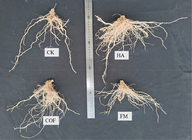
Due to the adverse effects of chemical fertilization on greenhouse soils, nowadays, special attention has been paid to organic and biological fertilizers or those technical managements that avoid soil fertility reduction in greenhouses. Humic acid is a component of humus, and it is also a natural component and contributes toward the reduction of the diseases and stresses and increase of the crop yield. Actually, several studies have shown that Humic acid application in vegetable production can improve the primary and secondary metabolism of plants, increase crop yields and quality, and decrease pests and diseases. Humic acid can accelerate soil organic material decomposition by increasing the microbial activity in soil. Furthermore, Humic acid can also alter the root exudation profile by affecting the plant metabolism and thus influence the structure of the rhizosphere microbial community. Principal coordinate analysis of the bacterial communities revealed that control, Humic acid application, and organic fertilizer formed different clusters.


where, humic acid (HA), farmyard manure (FM), commercial organic fertilizer (COF), and control check (CK)

Changes in high-mannosylated gylcan structures of complement C3 could be a good biomarker for children’s type 1 diabetes

A group from Faculty of Pharmacy and Biochemistry, University of Zagreb, Zagreb, Croatia, etc. has reported that plasma high-mannose glycan change of complement C3 showed notable discriminative power between children with early onset type 1 diabetes and their healthy siblings with AUC of 0.879.
https://www.mcponline.org/article/S1535-9476(22)00215-8/fulltext

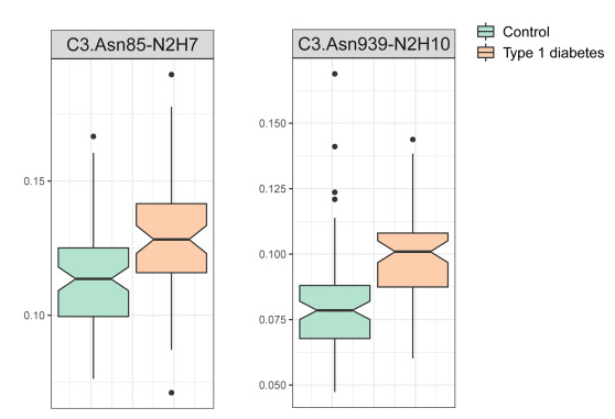
Children’s type 1 diabetes was associated with an increase in the proportion of high-mannose structures of complement C3 with more mannose units.
In this experiment, high-mannosylated C3 was enriched by ConA lectin, and the glycan structures were analyzed by LC-MS/MS. C3 has two glycosylation sites at Asn85 and Asn939, and the high-manosylated structure gets longer with developing type 1 diabates as shown below.

A novel monoclonal IgG1 antibody specific for α-Gal epitope

A group from Technical University of Munich, School of Medicine, Neuherberg, Germany, etc. has reported about a novel monoclonal IgG1 antibody specific for α-Gal epitope.
https://www.ncbi.nlm.nih.gov/pmc/articles/PMC9391071/

The alpha-Gal (α-Gal) epitope is a carbohydrate immunogen in humans that has relevance in allergy and xenotransplantation. Interestingly, antibodies of different isotypes against the α-Gal epitope are quite abundant in humans with IgG levels estimated to range between 1% to 0.1% of total plasma IgG with high variability between subjects and lowest abundance in individuals carrying the blood type B antigen. This observation is likely due to the structural similarity between the α-Gal epitope and blood type B antigen, which contains an additional fucose molecule on the second last galactose molecule. These human anti-α-Gal antibodies pose a challenge for xenotransplantation, in particular for pig organ transplantation, which was overcome to some extend with developing GGTA1 knockout (KO) pigs.

In this paper, the development of a novel IgG1 antibody called 27H8 was reported, which is highly specific for both synthetic and naturally occurring α-Gal epitopes. In order to generate a monoclonal antibody specific for the α-Gal epitope determining structure Gal-α1,3-Gal that is equally able to bind to the naturally occurring α-Gal epitope Gal-α1,3-Gal-β1,4-GlcNAc, α-galactosyltransferase knockout mice (Ggta1 KO) were immunized with Gal-α1,3-Gal-β1,4-GlcNAc coupled to ovalbumin as carrier protein (α-Gal-OVA).

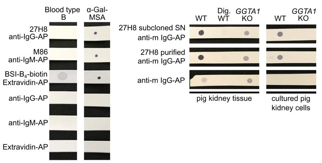
To verify the specificity, the 27H8 monoclonal antibody was compared to Bandereia simplifolica isolectin B4 (BSI-B4: GSL-I B4) and to the monoclonal IgM antibody M86, which are both widely used to detect the α-Gal epitope. BSI-B4 is specific for terminal α-galactose oligosaccharides and therefore recognizes also the blood group B antigen, which differs from the α-Gal epitope only in the addition of one fucose residue and is thus structurally very similar. To assess whether 27H8 also binds to the blood group B antigen we blotted lysates of whole blood from a type B donor on a membrane and applied the antibodies 27H8 and M86 or biotinylated BSI-B4 for detection. While BSI-B4 bound to the blood type B specimen as expected, neither 27H8 or M86 did. Next, it was investigated whether 27H8 also binds to natural α-Gal epitopes. As pig kidney is naturally rich in α-Gal and reactions in α-Gal allergic patients are severe after ingestion, it was also tested if 27H8 recognizes α-Gal in pig kidney lysates in a dot blot assay. 27H8 binding to wildtype (WT) pig kidney lysate was observed with strong staining intensity. However, there was no signal on a sample digested with α-Galactosidase (Dig. WT).

 refer to the original paper for detailed explanation

Comparison of SPR detection of SARS-CoV-2 spike protein with using gold nanorods (AuNRs) and gold nanoparticles (AuNPs)

A group from Department of Chemistry, University of Warwick, Gibbet Hill Road, Coventry, U.K., etc. has reported about comparison of SPR detection of SARS-CoV-2 spike protein with using gold nanorods (AuNRs) and gold nanoparticles (AuNPs).
https://pubs.acs.org/doi/10.1021/acsmacrolett.1c00716

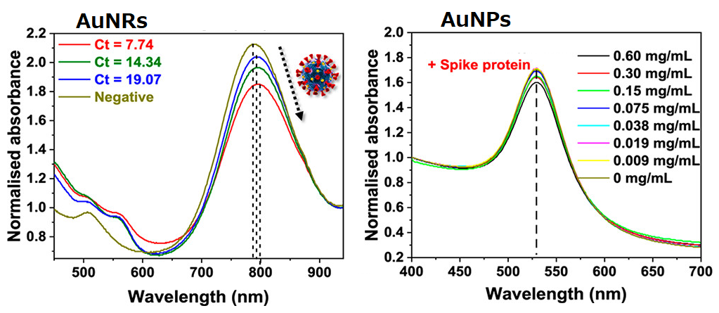
2,3-sialyllactose was immobilized onto AuNPs and AuNRs to detect SARS-CoV-2 with using a SPR detection method.

To highlight the importance of anisotropic particles (e.g., nanorods), spherical AuNRs were compared with AuNPs.
(1)AuNPs (40nm) showed the maximum aborbance at ∼520 nm, and AuNRs (10 × 38 nm) showed the maximum absorbance at ~780 nm.
(2)Although AuNPs (which generates a signal due to aggregation) did not show significant spectral changes with this spike protein, AuNRs showed spectral change with increasing concentration of spike protein.
(3)The signal output from AuNRs using the primary clinical samples was correlated with the Ct (cycle threshold) values from RT-PCR.

Thus, it has clearly shown that AuNRs are better than AuNPs.

Ceruloplasmin with bisecting GlcNAc could be a good biomarker for pancreatic cancer

A group from College of Basic Medical Sciences, Dalian Medical University, Dalian, China, etc. has reported that ceruloplasmin with bisecting GlcNAc could be a good biomarker for pancreatic cancer.
https://www.mdpi.com/2073-4409/11/15/2453/htm

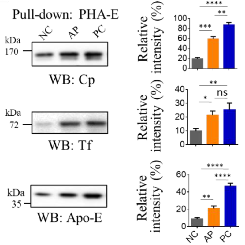
Compared with the normal controls (NC) group, the relative abundances of bisecting glycans, mannosylation, and fucosylation were significantly increased in the pancreatic cancer (PC) group. Compared with the acute pancreatitis (AP) group, the relative abundances of bisecting glycans and fucosylation were significantly enhanced in the PC group; however, the relative abundances of fucosylation seemed similar between the NC and AP groups.

Since glycans are difficult to be developed as biomarkers alone, the serum glycoproteins containing bisecting glycans were pulled down with biotinylated PHA-E lectin and streptavidin agarose beads, and analyzed by nano LC-MS/MS. As a result, three proteins, Ceruloplasmin (Cp), apolipoprotein E (Apo-E), and transferrin (Tf) were listed as biomarker glycoproteins.

Among these candidates, Cp showed the highest performance: the AUC for Cp between NC and AP, NC and PC, and PC and AP, were 0.917, 0.972, and 0.757, respectively.

The crystal structure of Cry78Aa from Bacillus thuringiensis: consists of a lectin domain and a pore-forming domain

A group from Institute of Plant Protection, Chinese Academy of Agricultural Sciences, Beijing, China, etc. has reported about the crystal structure of Cry78Aa from Bacillus thuringiensis.
https://www.nature.com/articles/s42003-022-03754-6

Biological control methods using Bacillus thuringensis (Bt) as well as genetically modified plants expressing insecticidal proteins from Bt have been proven effective and economic against some insect pests. Cry78Aa is a novel protein identified from the Bt C9F1 strain that effectively kills rice planthoppers, with median lethal concentration (LC50) values against Laodelphax striatellus and Nilaparvata lugens of 6.89 and 15.78 μg ml−1, respectively. The activity of Cry78Aa does not require in vitro activation or any additional components, making it convenient for application in field trials.

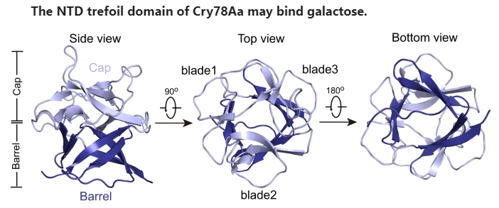
In this paper, the crystal structure of Cry78Aa was analyzed in detail. This structure consists of two independent domains: a trefoil domain at the N-terminus, which shares the highest identity with S-type lectin, and a pore-forming domain belonging to the aerolysin family. Bioassays showed that the NTD or CTD of Cry78Aa alone has no toxicity against planthopper nymphs, indicating that its insecticidal activity is dependent on the cooperation of both domains. The NTD of Cry78Aa plays a vital role for its insecticidal activity, probably by recognize galactose derivatives linked to proteins or lipids on the surface of the cell membrane.

Powered by WordPress |Copyright © 2020 Emukk. All rights reserved