Archive 22/9/7

Changes in high-mannosylated gylcan structures of complement C3 could be a good biomarker for children’s type 1 diabetes

A group from Faculty of Pharmacy and Biochemistry, University of Zagreb, Zagreb, Croatia, etc. has reported that plasma high-mannose glycan change of complement C3 showed notable discriminative power between children with early onset type 1 diabetes and their healthy siblings with AUC of 0.879.
https://www.mcponline.org/article/S1535-9476(22)00215-8/fulltext

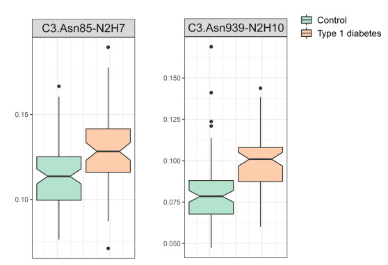
Children’s type 1 diabetes was associated with an increase in the proportion of high-mannose structures of complement C3 with more mannose units.
In this experiment, high-mannosylated C3 was enriched by ConA lectin, and the glycan structures were analyzed by LC-MS/MS. C3 has two glycosylation sites at Asn85 and Asn939, and the high-manosylated structure gets longer with developing type 1 diabates as shown below.

Tamoxifen (TAM)-resistant breast cancer cells bind to WFA lectin which recognizes LacdiNAc glycan

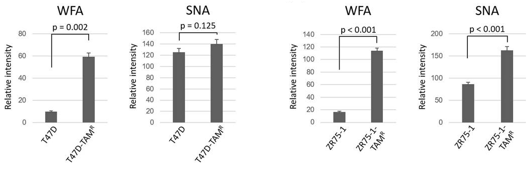
A group from Department of Breast Oncology, Juntendo University Faculty of Medicine, Tokyo, Japan, etc. has reported that Tamoxifen (TAM)-resistant breast cancer cells bind to WFA lectin which recognizes LacdiNAc glycan.
https://www.ncbi.nlm.nih.gov/pmc/articles/PMC9409572/

Some breast cancers require estrogen (female hormone) for the growth of cancer cells, and account for 60-70% of all breast cancers. Anti-estrogen drugs (tamoxifen), which suppress the action of estrogen, are expected to be effective against such types of breast cancer that proliferate with estrogen. However, some breast cancer cells result in developing resistance to tamoxifen.

Glycosylation is a major post-translational modification of proteins through the sequential action of glycosyltransferases, and alterations in glycosylation are well known to be associated with carcinogenesis, malignant progression and metastasis.
Therefore, in this study, it was investigated the glycan profiles of TAM-resistant human breast cancer cells were evaluated by using lectin microarrays, to investigate whether changes in glycosylation can be used as predictive biomarkers for endocrine therapy.

As a result, it was shown that TAM-resistant breast cancer cells have LacdiNAc glycosylation and strongly react with WFA lectin.

High bacterial diversity and siderophore-producing bacteria are important in suppressing Fusarium oxysporumin

A group from Key Laboratory of Plant-Soil Interactions, China Agricultural University, Beijing, China, etc. has reported that high bacterial diversity and siderophore-producing bacteria collectively suppress Fusarium oxysporumin maize/faba bean intercropping.
https://www.ncbi.nlm.nih.gov/pmc/articles/PMC9389221/

It is generally known that global food production is subject to various threats, in which soil-borne pathogens account for approximately 20% of yield losses. Intensive large-scale monocultures are prone to disease outbreaks, which cause yield losses. Fungal disease can be effectively controlled by sustainable diversified cropping systems such as rotations and/or intercropping.

In this study, three planting patterns were compared:
(1) monocultured maize,
(2) monocultured faba bean, and
(3) maize/faba bean intercropping.

As a result, intercropping increased the yields of maize and faba bean over monocropping by 21.3 and 14.4%, respectively, intercropping significantly decreased the gene copies of Fusarium oxysporum over monoculture by 2.91, 7.33 and 9.56% in bulk soil, rhizosphere soil and root endosphere, respectively, and the suppression of F. oxysporum was much stronger in faba bean than in maize.

From the analysis of bacterial diversity and community composition, the followings were highlighted:
(1) rhizosphere bacterial diversity and network connections are enhanced in the intercropping, which correlates the decrease of Fusarium oxysporum,
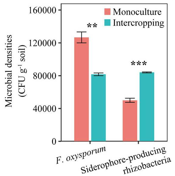
(2) Number of siderophore-producing rhizobacteria in rhizosphere of maize and faba bean was inversely correlated with the the abundance of Fusarium oxysporum.

A novel monoclonal IgG1 antibody specific for α-Gal epitope

A group from Technical University of Munich, School of Medicine, Neuherberg, Germany, etc. has reported about a novel monoclonal IgG1 antibody specific for α-Gal epitope.
https://www.ncbi.nlm.nih.gov/pmc/articles/PMC9391071/

The alpha-Gal (α-Gal) epitope is a carbohydrate immunogen in humans that has relevance in allergy and xenotransplantation. Interestingly, antibodies of different isotypes against the α-Gal epitope are quite abundant in humans with IgG levels estimated to range between 1% to 0.1% of total plasma IgG with high variability between subjects and lowest abundance in individuals carrying the blood type B antigen. This observation is likely due to the structural similarity between the α-Gal epitope and blood type B antigen, which contains an additional fucose molecule on the second last galactose molecule. These human anti-α-Gal antibodies pose a challenge for xenotransplantation, in particular for pig organ transplantation, which was overcome to some extend with developing GGTA1 knockout (KO) pigs.

In this paper, the development of a novel IgG1 antibody called 27H8 was reported, which is highly specific for both synthetic and naturally occurring α-Gal epitopes. In order to generate a monoclonal antibody specific for the α-Gal epitope determining structure Gal-α1,3-Gal that is equally able to bind to the naturally occurring α-Gal epitope Gal-α1,3-Gal-β1,4-GlcNAc, α-galactosyltransferase knockout mice (Ggta1 KO) were immunized with Gal-α1,3-Gal-β1,4-GlcNAc coupled to ovalbumin as carrier protein (α-Gal-OVA).

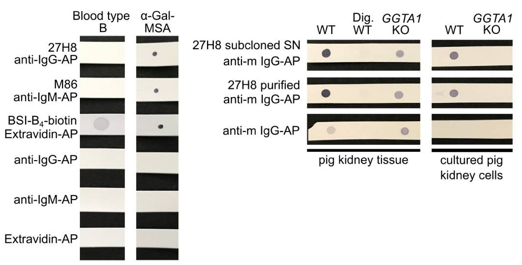
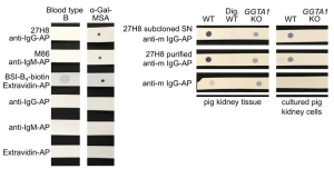
To verify the specificity, the 27H8 monoclonal antibody was compared to Bandereia simplifolica isolectin B4 (BSI-B4: GSL-I B4) and to the monoclonal IgM antibody M86, which are both widely used to detect the α-Gal epitope. BSI-B4 is specific for terminal α-galactose oligosaccharides and therefore recognizes also the blood group B antigen, which differs from the α-Gal epitope only in the addition of one fucose residue and is thus structurally very similar. To assess whether 27H8 also binds to the blood group B antigen we blotted lysates of whole blood from a type B donor on a membrane and applied the antibodies 27H8 and M86 or biotinylated BSI-B4 for detection. While BSI-B4 bound to the blood type B specimen as expected, neither 27H8 or M86 did. Next, it was investigated whether 27H8 also binds to natural α-Gal epitopes. As pig kidney is naturally rich in α-Gal and reactions in α-Gal allergic patients are severe after ingestion, it was also tested if 27H8 recognizes α-Gal in pig kidney lysates in a dot blot assay. 27H8 binding to wildtype (WT) pig kidney lysate was observed with strong staining intensity. However, there was no signal on a sample digested with α-Galactosidase (Dig. WT).
 refer to the original paper for detailed explanation
Altered sialidase expression in human myeloid cells undergoing apoptosis and differentiation

A group from Baltimore Veterans Affairs Medical Center, Research Service, Baltimore, MD, USA, etc. has reported about altered sialidase expression in human myeloid cells undergoing apoptosis and differentiation.
https://www.nature.com/articles/s41598-022-18448-6

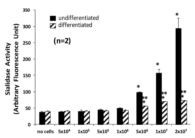
The sialidase/neuraminidase (NEU) activity and expression in mature human neutrophil (PMN)s and the HL60 promyelocytic leukemic cell line were studied, and changes in sialic acid modification that would occur in PMNs undergoing apoptosis and HL60 cells during their differentiation into PMN-like cells were observed.

PMNs are part of the first line of host defenses against invasive prokaryotic pathogens4. Myeloid progenitors undergo maturation within the bone marrow into mature PMNs. PMNs undergo profound shape changes permitting these cells to squeeze through the small caliber microvasculature and interendothelial cell junctions to enter extravascular tissues, where they adhere to and engulf bacteria for successful phagocytosis and intracellular killing. The HL60 promyelocytic leukemia cell line has been used as a model for myelopoiesis.

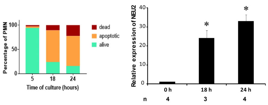
In proapoptotic PMNs, NEU2 protein expression increased >30.0-fold as shown below.

The total NEU activity in differentiated HL60 (dHL60) cells was dramatically reduced compared to that of nondifferentiated cells as shown below.

Thus, changes in PMN surface sialylation could regulate accessibility of both Siglecs and Galectins.

Comparison of SPR detection of SARS-CoV-2 spike protein with using gold nanorods (AuNRs) and gold nanoparticles (AuNPs)

A group from Department of Chemistry, University of Warwick, Gibbet Hill Road, Coventry, U.K., etc. has reported about comparison of SPR detection of SARS-CoV-2 spike protein with using gold nanorods (AuNRs) and gold nanoparticles (AuNPs).
https://pubs.acs.org/doi/10.1021/acsmacrolett.1c00716

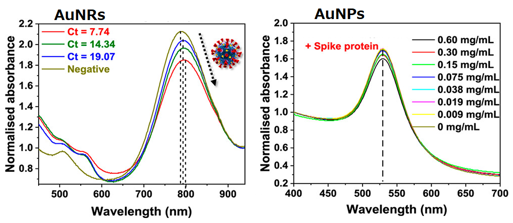
2,3-sialyllactose was immobilized onto AuNPs and AuNRs to detect SARS-CoV-2 with using a SPR detection method.

To highlight the importance of anisotropic particles (e.g., nanorods), spherical AuNRs were compared with AuNPs.
(1)AuNPs (40nm) showed the maximum aborbance at ∼520 nm, and AuNRs (10 × 38 nm) showed the maximum absorbance at ~780 nm.
(2)Although AuNPs (which generates a signal due to aggregation) did not show significant spectral changes with this spike protein, AuNRs showed spectral change with increasing concentration of spike protein.
(3)The signal output from AuNRs using the primary clinical samples was correlated with the Ct (cycle threshold) values from RT-PCR.

Thus, it has clearly shown that AuNRs are better than AuNPs.

Human surfactant protein D facilitates SARS-CoV-2 binding and entry in DC-SIGN expressing cells

A group from College of Health, Medicine and Life Sciences, Brunel University London, Uxbridge, UK, etc. has reported that human surfactant protein D (human SP-D) facilitates SARS-CoV-2 binding and entry in DC-SIGN expressing cells
https://www.frontiersin.org/articles/10.3389/fimmu.2022.960733/full

The ability of a recombinant fragment of human SP-D (rfhSP-D) to mediate the binding of SARS-CoV-2 to DC-SIGN expressing cells was evaluated. HEK 293T cells were transfected with a construct containing a DNA sequence of full-length human DC-SIGN to induce DC-SIGN cell surface expression (DC-HEK cells). To assess the effect of rfhSP-D on pseudotypes binding to DC HEK cells, the DC-HEK cells were challenged with rfhSP-D (20µg/ml) treated SARS-CoV-2 Spike protein-expressing pseudotype. Increased binding (~50%) in the treated samples (DC-HEK + SARS-CoV-2 spike Pseudotypes + rfhSP-D) compared to their untreated counterparts (DC-HEK + SARS-CoV-2 spike Pseudotypes) was observed. Similar experiments were done using THP-1 cells treated with PMA and IL-4 to induce the expression of native DC-SIGN. rfhSP-D treatment was found to increase the binding efficiency of the pseudotypes to the THP-1 cells expressing DC-SIGN by ~25%, compared to the untreated controls.

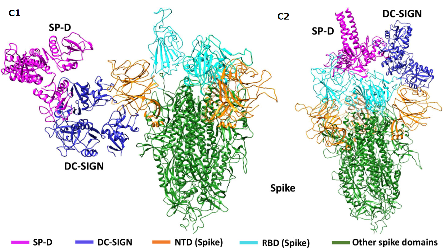
A blind docking approach was attempted to generate SARS-CoV-2 and DC-SIGN complexes. Analysis of the top ranked docked poses revealed that NTD (N-terminal domain) of spike protein interacted with the CRD domain of DC-SIGN. Tripartite complexes were generated by docking C-SIGN and SP-D with Spike protein. The top two docked poses (C1 and C2) were analysed for intermolecular interactions. In both C1 and C2 complexes, DC-SIGN (CRD) interacted with NTD domain of Spike protein. In C1, there were no molecular interactions between Spike protein and rfhSP-D. In C2, Spike protein interacted with rfhSP-D through RBD.

As a conclusion, it was shown that SP-D interacts with RBD and DC-SIGN interacts with NTD of SARS-CoV-2 spike protein, and also SP-D stabilises DC-SIGN and SARS-CoV-2 spike protein interaction.

Ceruloplasmin with bisecting GlcNAc could be a good biomarker for pancreatic cancer

A group from College of Basic Medical Sciences, Dalian Medical University, Dalian, China, etc. has reported that ceruloplasmin with bisecting GlcNAc could be a good biomarker for pancreatic cancer.
https://www.mdpi.com/2073-4409/11/15/2453/htm

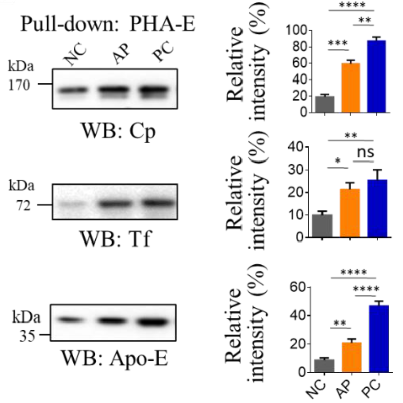
Compared with the normal controls (NC) group, the relative abundances of bisecting glycans, mannosylation, and fucosylation were significantly increased in the pancreatic cancer (PC) group. Compared with the acute pancreatitis (AP) group, the relative abundances of bisecting glycans and fucosylation were significantly enhanced in the PC group; however, the relative abundances of fucosylation seemed similar between the NC and AP groups.

Since glycans are difficult to be developed as biomarkers alone, the serum glycoproteins containing bisecting glycans were pulled down with biotinylated PHA-E lectin and streptavidin agarose beads, and analyzed by nano LC-MS/MS. As a result, three proteins, Ceruloplasmin (Cp), apolipoprotein E (Apo-E), and transferrin (Tf) were listed as biomarker glycoproteins.

Among these candidates, Cp showed the highest performance: the AUC for Cp between NC and AP, NC and PC, and PC and AP, were 0.917, 0.972, and 0.757, respectively.

Bacilli’s biofilm formation capability is critical to fulfill biocontrol activity against phytopathogens

A group from College of Food Science and Light Industry, Nanjing Tech University, Nanjing, China has reported that the biofilm formation enhanced Bacillus root colonization and its biocontrol activity against phytopathogens.
https://www.frontiersin.org/articles/10.3389/fmicb.2022.972393/full

Bacilli are used as biocontrol agents (BCAs) against phytopathogens and most of them can produce poly-γ-glutamic acid (γ-PGA) as one of the major extracellular polymeric substances (EPSs).

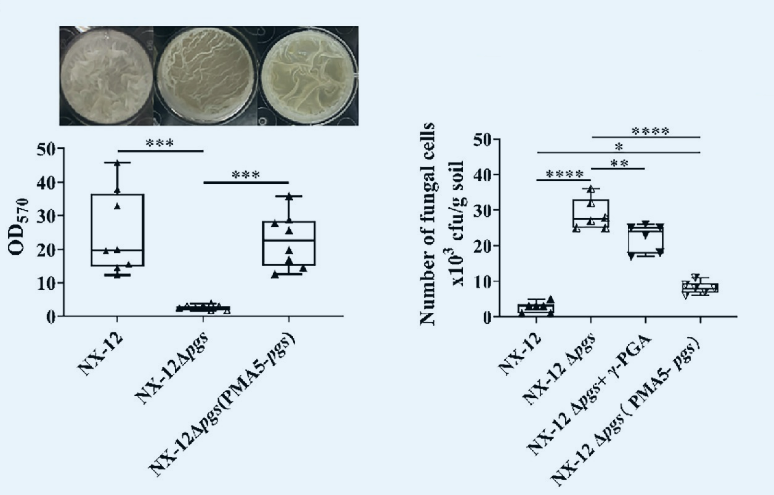
In this study, Bacillus atrophaeus NX-12 (γ-PGA yield: 16.8 g/l) was compared with its γ-PGA synthesis gene knocked out strain NX-12Δpgs from a view point of antifungal ability. The antifungal ability of γ-PGA synthetase-deficient strain NX-12Δpgs (γ-PGA yield: 1.65 g/l) was improved in vitro, while the biocontrol ability of NX-12Δpgs was greatly diminished in situ.

It was proved that γ-PGA produced by NX-12 contributes to the biofilm formation and rhizosphere colonization, which effectively improved biocontrol capability suggesting that the effective colonization of Bacilli as biocontrol agents in the rhizosphere is very important for its function.


NX-12Δpgs (pMA5-pgs) means that the mutant NX-12Δpgs was complemented with a primer pMA5-pgsBCA, where pgsBCA is a γ-PGA synthase gene of NX-12.

The crystal structure of Cry78Aa from Bacillus thuringiensis: consists of a lectin domain and a pore-forming domain

A group from Institute of Plant Protection, Chinese Academy of Agricultural Sciences, Beijing, China, etc. has reported about the crystal structure of Cry78Aa from Bacillus thuringiensis.
https://www.nature.com/articles/s42003-022-03754-6

Biological control methods using Bacillus thuringensis (Bt) as well as genetically modified plants expressing insecticidal proteins from Bt have been proven effective and economic against some insect pests. Cry78Aa is a novel protein identified from the Bt C9F1 strain that effectively kills rice planthoppers, with median lethal concentration (LC50) values against Laodelphax striatellus and Nilaparvata lugens of 6.89 and 15.78 μg ml−1, respectively. The activity of Cry78Aa does not require in vitro activation or any additional components, making it convenient for application in field trials.

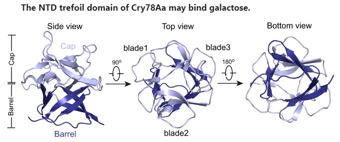
In this paper, the crystal structure of Cry78Aa was analyzed in detail. This structure consists of two independent domains: a trefoil domain at the N-terminus, which shares the highest identity with S-type lectin, and a pore-forming domain belonging to the aerolysin family. Bioassays showed that the NTD or CTD of Cry78Aa alone has no toxicity against planthopper nymphs, indicating that its insecticidal activity is dependent on the cooperation of both domains. The NTD of Cry78Aa plays a vital role for its insecticidal activity, probably by recognize galactose derivatives linked to proteins or lipids on the surface of the cell membrane.

Powered by WordPress |Copyright © 2020 Emukk. All rights reserved