Archive 22/3/22

Changes in gastric mucosal glycosylation before and after Helicobacter pylori Eradication: Studies using Lectin Microarray

A group from Department of Gastroenterology, Faculty of Medicine, Oita University, Oita, Japan, has reported about changes in gastric mucosal glycosylation before and after Helicobacter pylori Eradication.
https://pubmed.ncbi.nlm.nih.gov/35238778/

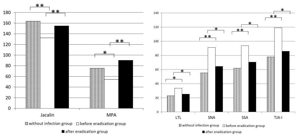
It was found that the jacalin and MPA signals in the gastric antrum were significantly lower in the H. pylori infection group than in the gastric mucosa of the group without H. pylori infection, and that, 1 year after eradication, the signals returned to the levels seen without infection.
The LTL, SNA, SSA, and TJA-I signals in the gastric body were significantly increased in the H. pylori infection group and returned to the levels seen without infection 1 year after eradication.

MPA and jacalin are 85% homologous, and these lectins have particularly high specificity for the TF-antigen that is expressed in more than 85% of human carcinomas.
LTL binds to Lewisx, Lewisy and H-antigen type II, and has been reported to be a marker for cancer progression in bladder cancer cell lines.
SNA has been reported to be a marker for the diagnosis, metastasis, and prognosis of colorectal and pancreatic cancers and hepatocellular carcinoma. SNA has also been reported to be a diagnostic marker for pneumonia and a predictive marker for diabetic nephropathy progression. SNA, SSA, and TJA-I have the same glycan binding specificity, α2-6Sia.

The results of this study showed that the signal levels for lectin binding to glycans resulting from H. pylori infection returned to the levels seen without infection as a result of H. pylori eradication. Thus, the data are intriguing in that they indicate that glycosylation is reversible.

How Bacillus subtilis manage to efficiently migrate in the semisolid or solid soil environment?

A group from Institute of Plant Protection, Tianjin Academy of Agricultural Sciences, Tianjin, China, etc. has reported on a novel sucrose induced signaling cascade promoting Bacillus subtilis rhizosphere colonization.
https://www.ncbi.nlm.nih.gov/pmc/articles/PMC8397739/

It has been known that Bacillus subtilis is an excellent biological control agent, capable of suppressing a number of soil-borne phytopathogens. But, how do such beneficial bacteria recognize the signals from the plant host, colonize the roots, and ultimately establish an intimate relationship with the plant? One of the specific focuses is on plant root-released nutrients and their impact on the rhizobacteria. Among those root-released nutrients, sucrose is found most abundantly released into the rhizosphere. Sucrose is uniquely important to the plants since photosynthetic plants primarily use sucrose as a fixed carbon transport and storage mechanism among different tissues.

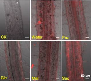
It is clearly shown in the figure below that sucrose induces more robust root colonization on tomato roots by Bacillus subtilis in the presence of sucrose than several other root-secreted sugars.

A red fluorescence-labeled Batillus subtilis strain was used with different sugars (Fru: fructose, Glc: glucose, Mal: maltose, and Suc: sucrose)

Further, in the rhizosphere soil with sucrose addition, the relative abundance of Bacillus reached ~10.1%, while this ratio was only ~0.1% without sucrose addition, indicating a 100-fold increase in the abundance of the native Bacillus species in the rhizosphere stimulated by sucrose. In addition to the strong positive influence of sucrose on the prevalence of Bacillus, the relative low abundance of Pseudomonas (0.05%) was also elevated to 2.7% when inoculated with the wild type B. subtilis cells and further increased to 12.5% with simultaneous B. subtilis inoculation and sucrose supplementation. It was also shown that supplementation of sucrose formula improved the suppression efficiency against the soil-borne disease caused by Fusarium oxysporum sp. Lycopersici (Fusarium wilt) .

Big question is how B. subtilis manage to efficiently migrate in the semisolid or solid soil environment?

Regarding this question, authors has shown that sucrose activates a signaling cascade to trigger solid surface motility leading to effective root colonization by B. subtilis. That is, sucrose initiates biosynthesis of levan, and levan is further hydrolyzed into levanoligosaccharides, and finally induces strong production of surfactin from B. subtilis resulting in acceleration of solid surface motility.

Activation of the lectin pathway plays a vital role in the development of Idiopathic Membranous Nephropathy

Ther are two types of Membranous nephropathy (MN), idiopathic MN (iMN) and atypical MN (aMN). A group from Department of Nephrology, Peking University People’s Hospital, Beijing, China has reported that activation of the mannose-binding lectin (MBL) pathway plays a vital role in the development of iMN.
https://www.ncbi.nlm.nih.gov/pmc/articles/PMC8920381/

There are three major complement pathways include the classical pathway, mannose-binding lectin (MBL) pathway, and alternative pathway. C4d is generated in both the classical and mannose-binding lectin complement pathways. C1q is the major precursor of classical complement activation.
The presence of both C4d and C1q indicates the activation of the classical pathway, but
identification of C4d without C1q is more consistent with MBL pathyway, and
C4d and C1q are both absent in alternative complement activation.

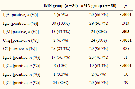
The renal pathology of iMN is characterized by the deposition of predominant IgG4 with low amounts of IgG1 and IgG3. IgG4 does not activate the classical complement pathway. C4d is detectable in essentially 100% of patients with iMN which was absent in alternative complement activation. These observations suggest that the MBL-initiated complement pathway may be the predominant complement activation in iMN. On the other hand, aMN characterized by cells proliferation, multi-site immune complex deposition, ‘full house’ in immunofluorescence, including IgA, IgG, IgM, C3, C1q positive, implies that the pathophysiological process involves complex complement system activation.

Lactobacillus crispatus Strain KT-11 S-Layer Protein can inhibit Rotavirus Infection: Sialic acid would be deeply involved

A group from Shinshu University, Nagano, Japan, etc. has reported that Lactobacillus crispatus Strain KT-11 S-Layer Protein can inhibit Rotavirus Infection and sialic acid could be involved in initiation of virus infection.
https://www.ncbi.nlm.nih.gov/pmc/articles/PMC8902352/

Lactic acid bacteria, including the genus Lactobacillus play a crucial role in the production of fermented dairy products such as cheese, yoghurt, and fermented milk. In recent years, there has been considerable focus on the action of the S-layer protein (SLP) of lactic acid bacteria as an antiviral component.

The effect of Lactobacillus crispatus KT-11 SLP on the infection of Rotavirus DS-1 strain in Caco-2 cells is shown below. DS-1 infection was significantly suppressed by pre-infection treatment with KT-11 SLP in a concentration-dependent manner. Conversely, KT-11 SLP did not suppress the infection of the Rotavirus Wa strain even after pre-infection treatment at 100 μg/mL.

The entry of rotavirus into cells is a complex multistep process, in which different domains of rotavirus surface proteins interact with cell-surface molecules that function as receptors for adhesion and entry. Among them, several carbohydrates, such as terminal sialic acids and histo-blood group antigens, have been reported to be involved in rotavirus attachment to target cells. As shown above, KT-11 SLP significantly inhibited the infection of the DS-1 strain in Caco-2 cells in a dose-dependent manner. The initial interactions of human rotavirus strains with host cells is dependent on the VP4 genotype. According to the classification based on the molecular properties of VP4 (P-types), the DS-1 strain is classified as the P[4] genotype. Increasing evidence indicates that the P[4] genotype rotaviruses, including the DS-1 strain, use H-type 1 and Lewis-b antigens for infection. Actually, H-type 1 and Lewis-b antigens have been reported in Caco-2 cells. This result suggests that the possibility of that DS-1 infection is inhibited by competitive binding of KT-11 SLP to these antigens. However, contrary to the expectations, infection of the Wa strain, another dominant P[8] subtype reported using the same H-type antigen and Lewis-b for infection, was not inhibited in the presence of KT-11 SLP.

Recently, it was reported that DS-1 strain infection was significantly inhibited by 3′-sialyl lactose and 6′-sialyl lactose, whereas the Wa strain was inhibited by 2′-fucosyl lactose. Therefore, compounds containing sialic acid could be deeply involved in the infection of DS-1 strain.

Changes in belowground Fungal flora in wheat field under long-term fertilization

A group from State Key Laboratory of Soil and Sustainable Agriculture, Institute of Soil Science, Chinese Academy of Sciences, Nanjing, China, etc. has reported about changes in belowground fungal flora in wheat field under long-term fertilization.
<a href=”https://pubmed.ncbi.nlm.nih.gov/35266812/”>https://pubmed.ncbi.nlm.nih.gov/35266812/</a>

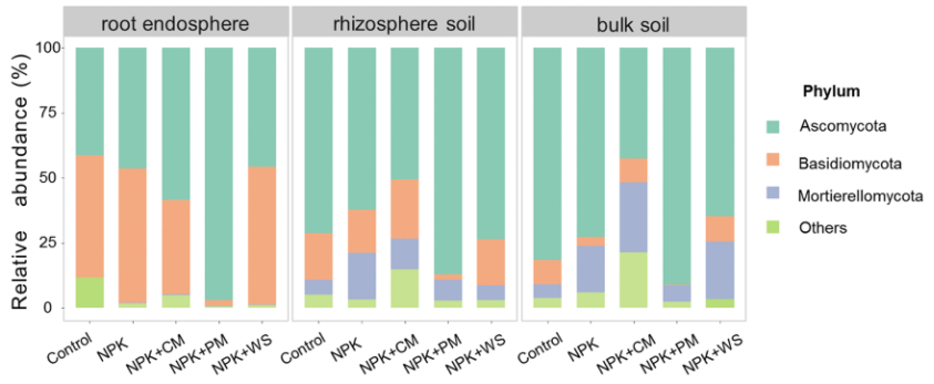
In this paper, new findings are reported from a long-running fertilization experiment in wheat field (more than 35 years) covering the following conditions: NPK fertilizer, NPK and cow manure (NPK+CM), NPK and pig manure (NPK+PM), NPK and wheat straw (NPK+WS), and no fertilizer (Control).

At the phylum level, Ascomycota, Basidiomycota, and Mortierellomycota dominated the fungal community, irrespective of the treatments and habitats, together accounting for over 80% of the total sequences obtained. Compared with the other treatments, NPK+PM treatment had the highest relative abundance of Ascomycota (96.9%, 87.1%, and 91.1% in root endosphere, rhizosphere soil, and bulk soil, respectively)

Then, the correlation between alpha diversity and physiochemical variables (such as Total P, Total C, Total N, Total K, P, Ca, Mg, Na, Fe, Mn etc.) was analyzed. The random forest analysis revealed that phosphorus and Zn always were the best predictors of diversity changes in root endosphere, rhizosphere soil, and bulk soil. There were significant correlations between phosphorus and alpha diversity in root endosphere, and between Zn and alpha diversity in rhizosphere soil and bulk soil. The higher the P and Zn concentration, the lower the alpha diversity in rhizosphere fungi.

This can be considered as follows. Pig manure had higher phosphorus content than cow pig manure and after adding pig manure, the phosphorus content of soils and wheat roots was significantly higher than that of cow manure addition. Hence, long-term fertilization, especially pig manure application, can supply adequate and readily accessible phosphorus and phosphorus-like nutrients across soil to roots, which may lessen the dependence of crops on microbiota, thereby lessening total fungal diversity probably. Similar to phosphorus, it is reasonable to link the competition for resources and survival among fungal communities to Zn-induced changes in fungal diversity.

Antagonist fungi isolated from soils of the rhizosphere of tomato crops against Sclerotium rolfsii: the causative agent of white rot

A group from Laboratoire Biologie et Santé, UFR Biosciences, Université Félix Houphouët-Boigny d’Abidjan (UFHB), Côte d’Ivoire, etc. has reported about antagonist fungi isolated from soils of the rhizosphere of tomato crops against Sclerotium rolfsii.
https://www.ncbi.nlm.nih.gov/pmc/articles/PMC8857420/

S. rolfsii is the causative agent of white rot is one of the destructive pathogens of nightshade crops. In Côte d’Ivoire, this fungal pathogen constitutes a major constraint for the cultivation of tomato (Solanum lycopersicum) with 41.01% crop losses in humid forest areas.

Antifungal activity of soil fungi against S. rolfsii
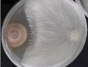
The screening carried out from the 153 soil-based fungal isolates by means of a direct confrontation test on the PDA medium made it possible to select 10 fungi which inhibit the growth of S. rolfsii. The percentages of inhibition ranged from 27.06 to 60.59%. The mechanisms of action observed during this inhibition were competition and antibiosis. The former is caused by the rapid growth of the antagonist will slow down or inhibit the growth of the pathogen, and the latter is caused by inhibiting biological activity of the pathogen. Molecular identification (ITS) of these antagonist fungi revealed that the isolates belonged to the genera Talaromyces sp. (n = 4), Trichoderma sp. (n = 3), Penicillium sp. (n = 2) and Clonostachys sp. (n = 1).


Among 10 antagonost fung, Talaromyces purpureogenus and Talaromyces assiutensis exerted the antibiosis mechanism.
T. purpureogenus secreted Mitorubrin and mitorubinol, and
T. assiutensis secreted Spiculisporic acid as molecules responsible for inhibiting S. rolfsii.

An ACE2-Fc fusion protein could be a good therapeutic drug overcoming viral escape of SARS-CoV-2 variants

A group from SystImmune Inc., 15318 NE 95th St., Redmond, WA, 98052, USA, etc. has reported that ACE2-Fc fusion protein could be a good therapeutic drug overcoming viral escape of SARS-CoV-2 variants.
https://www.ncbi.nlm.nih.gov/pmc/articles/PMC8882475/

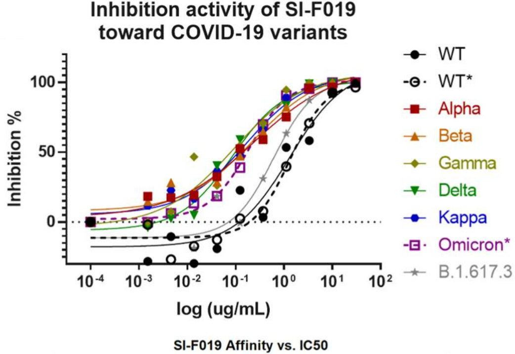
Interestingly, the pseudovirus neutralization data using the ACE2-Fc fusion protein named SI-F09 (shown below) shows that the higher the binding strength between SARS-CoV-2 RBD and ACE2, the lower the IC50 values. In other words, the IC50 value for WT is the highest among SARS-CoV-2 variants imcruding Omicron.

In the meantime, a phase I clinical trial evaluating safety of SI–F019 in healthy patients has been conducted (ClinicalTrials.gov Identifier: NCT04851444) and is being analyzed in preparation for future clinical studies.

Expression status of C-Type lectin receptor 5A (CLEC5A) on Monocyte-derived Macrophages and its functions

A group from Cancer Immunology & Immune Modulation, Boehringer Ingelheim Pharma GmbH & Co. KG, Germany, etc. has reported about expression status of C-Type lectin receptor 5A (CLEC5A) on Monocyte-derived Macrophages (MdM) and its functions.
https://www.ncbi.nlm.nih.gov/pmc/articles/PMC8896916/

CLEC5A also known as myeloid DAP12-associating lectin-1 (MDL-1) is a myeloid Syk-coupled pattern recognition receptor which preferentially binds to glycans highly expressed on the surface of pathogens. CLEC5A is mainly expressed on myeloid cells (monocytes, macrophages, neutrophils, and dendritic cells) and can be further upregulated by interferon-γ. The ligand for CLEC5A was identified as terminal fucose and mannose moieties of viral glycans, expressed by dengue virus, Japanese encephalitis virus or type A influenza virus. In addition, CLEC5A binds to disaccharides (N-acetylglucosamine and N-acetylmuramic acid) of bacterial cell walls (e.g., Listeria monocytogenes and Staphylococcus aureus). Functionally, the CLEC5A receptor activation triggered by dengue virus or by other pathogens induces the production of proinflammatory cytokines (TNF-α, IL-1, IL-6, IL-8, and IL-17A) and chemokines: macrophage inflammatory protein-1 alpha (MIP-1α/CCL3), interferon-gamma induced protein (IP-10/CXCL10), and macrophage-derived chemokine (CCL22/MDC) . It was also found that CLEC5A may recognize not only pathogen-associated antigens but also some endogenous danger signals and in consequence could contribute to the pathogenesis of the aseptic inflammation.

In this report, expression status of CLEAC5A on monocyte-derived macrophages and functional consequences of the selective activation of CLEC5A by α-CLEC5A Ab were discussed in aseptic conditions as well as their impact on the activation of autologous T cells.

Expression of CLEC5A on myeloid cells
Expression of CLEAC5A was compared among several MdM: proinflammatory M1, “neutral” M0, and protumorigenic M2c, and recently described in vitro tumor-associated macrophages (TAM). The CLEC5A expression was significantly elevated in proinflammatory M1 MdM as compared to monocytes and to other MdM subsets (M0 and M2c), while monocyte differentiation towards TAM resulted in a reduction of CLEC5A expression.

Functional effects of CLEC5A activation
In order to understand the functional effects of CLEC5A agonist under noninfectious conditions, the cytokine response in M0 MdM was evaluated. M0 MdM exposed to the α-CLEC5A Ab significantly upregulated the secretion of cytokines and chemokines such as TNF-α, IL-6, IL-10, IL-1b, CCL22/MDC, CCL17/TARC, and Matrix metalloproteinase (MMP1). Interestingly, the CLEC5A activation induced up-regulation of some myeloid cell-specific surface receptors, such as CD80, PD-L1, and CD206(MRC-1), and CD209(DC-SIGN).

Finally, the selective CLEC5A-mediated reprogramming of myeloid cells in aseptic conditions appeared insufficient to promote an autologous T cell activation.

Lectin Cytotoxicity

A group from Institute of Physiology, University of Zurich, Zurich, Switzerland has reported about lectin cytotoxicity.
https://www.ncbi.nlm.nih.gov/pmc/articles/PMC8866831/

While targeting surface glycans by lectins is a promising approach for cancer therapies, lectin-based approaches (lectin alone or in a combination with other therapeutic agents) are still at an early stage of development. The development of efficient therapies based on glycan targeting requires a deep understanding of the mechanisms of cell death induced by lectins on target tumor cells. It has been known that specific lectins induce distinct modes of cell death in different type of tumor cells. Wheat germ agglutinin (WGA), for example, induces apoptosis in melanoma and leukemic cell, whereas it kills cervical carcinoma cells through paraptosis-like cell death.

In this report, using murine adenocarcinoma cells (MC-38) and young adult mouse colon cells (YAMC), the multiple cell death pathways (apoptosis, necroptosis, proptosis, paraptosis, and autophagy-dependent cell death) activated in response to treatment of cells with lectins (MAL I, MAL II, SNA, AAL, WGA, and ECL) were studied with targeting different glycan structures. In order to characterize the signaling pathways mediating cell death induced by the cytotoxic lectins, a panel of MC-38 cells with knockouts in genes involved in cell death responses was used.

Knockouted genes are as follows;
pro-apoptotic proteins BCL2 antagonist/killer 1 (BAK1) and BCL2 associated X (BAX), which mediate intrinsic apoptosis, the intrinsic apoptosis signaling cascade is activated in response to various internal cell stress factors, such as DNA damage.

Fas-associated via death domain protein (FADD), which mediates extrinsic apoptosis, the extrinsic apoptosis pathway is induced in response to activation of cell death receptors, followed by formation of the death-inducing signaling complex that includes FADD and pro-caspase-8, which cleaves the executioner caspase-3.

tumor necrosis factor receptor type 1-associated death domain protein (TRADD), receptor-interacting serine/threonine protein kinases 3 (RIPK3), mixed lineage kinase domain-like protein (MLKL) and caspase-8 (CASP8), which mediate caspase-independent necroptosis.

caspase-1 (CASP1) and gasdermin D (GSDMD), which mediate pyroptosis, CASP1 is activated by the inflammasome in response to various microbial infections and non-infectious stimuli. The pathway leads to GSDMD cleavage, which embeds in the plasma membrane and forms pores that disrupt ionic gradients and facilitate water influx, hence leading to cell swelling and osmotic lysis.

Results:
The inactivation of the BAX/BAK1 complex decreased the cytotoxic response induced by WGA, MAL I and AAL treatment. The decrease in cytotoxicity by more than 50% was similar to the effect achieved in cells treated with cisplatin, which is a classical trigger of apoptosis. By contrast, the inactivation of FADD did not impacted the cell death mediated by WGA, MAL I and AAL.

The inactivation of TRADD, another adaptor molecule required for activation of apoptosis and necroptosis downstream of tumor necrosis factor receptor 1 (TNFR1), decreased cell death in cells treated with MAL I, but not when WGA and AAL was added. The loss of MLKL decreased the cytotoxic effect of WGA, MAL I and AAL, indicating the contribution of the necroptosis pathway in cell death induced by these lectins.

Inactivation of either CASP1 or GSDMD showed only a minor decrease in MAL I-mediated cytotoxicity, supporting a partial involvement of pyroptosis in response to MAL I treatment.

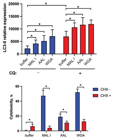
In addition to the induction of apoptosis, many lectins also up-regulate autophagy sometimes resulting in autophagy-dependent cell death. LC3-II is associated with autophagosome membranes. Treatment with MAL I, AAL and WGA up-regulated LC3-II in lysates from cells treated for 6 h with lectins. The increase in LC3-II levels indicated that cell death induced by MAL I, AAL and WGA was probably initiated through activation of the autophagic/lysosomal response rather than the classical apoptotic mitochondrial pathway. Actually by adding cycloheximide (CHX), is known to block starvation-induced autophagy, the cytotoxic response of the three lectins was significantly reduced.
Changes in rhizosphere microbiome with wheat evolution: from ancestral species to domesticated species

A group from Department of Plant Breeding and Biotechnology, Faculty of Agriculture, University of Tabriz, Tabriz, Iran, etc. has reported on changes in rhizosphere microbiome with wheat evolution (from ancestral wheat species of T. uratu and Ae speltoides (ancestral species, 1.5 to 2million years ago) to domesticated pecies of T. turgidum and T. aestivum).
https://www.ncbi.nlm.nih.gov/pmc/articles/PMC8881823/

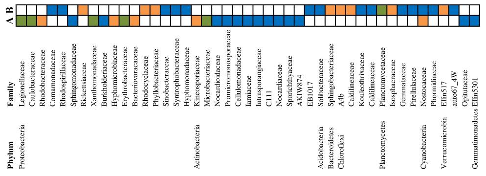
It was found that several families belonging to Actinobacteria and Proteobacteria revealed significant increases in domesticated crops (T. turgidum and T. aestivum) when compared to ancestral species (T. urartu, and Ae. speltoides) as shown below.


Differential family-level abundances in (A) domesticated and (B) ancestral wheat species during different developmental stages with color. Orange = vegetative, blue = reproductive, and green = comon in both developmental stages, respectively.

Powered by WordPress |Copyright © 2020 Emukk. All rights reserved