A drought-tolerant soil for wheat by adding silica particles with soil bacteria producing exopolysaccharides effectively

A group from Swedish University of Agricultural Sciences, Uppsala, Sweden, etc. has developed a drought-tolerant soil for wheat by adding silica particles with soil bacteria producing exopolysaccharides effectively.
https://www.ncbi.nlm.nih.gov/pmc/articles/PMC8229586/

Agriculture faces several challenges at the global level and, in coming decades, drought is expected to expand globally owing to increased evaporation and reduced rainfall, as well as or changes in the spatial and temporal distribution of rainfall. The scientific community across the world is earnestly looking for novel solutions to enhance crop plant stress tolerance under limited resource availability, and several environmentally friendly solutions have shown huge potential but need to be optimized for wide-scale field application. One such solution includes strengthening plants’ natural defence systems with plant growth promoting rhizobacteria. Predicting and controlling the rhizosphere has the potential to harness plant microbe interactions and restore plant ecosystem productivity, improve plant responses to environmental stress, and mitigate the effects of climate change.

First of all, soil bacteria named A26Sfp was created from its wildtype A26 by genetically silencing 4-phosphopantetheinyl transferase to improve drought tolerance ability. A26Sfp, compared to its wildtype A26, is enhanced in biofilm exopolysaccharides production consisting D-glucuronate. Polysaccharides are basically hydrophilic, and therefore are suitable to increase water retention.

Strains A26, A26Sfp were grown in 1/2 Tryptic soy broth (TSB) with 50 µg/mL silica particles (SN) at 30 ± 2 °C for 24 h. While the silica particles did not have a significant impact on the bacterial number, silica particles improved A26 and A26Sfp exopolysaccharides production by 46% and 29%, respectively. A26Sfp EPS production was 30–40% higher than its wildtype and A26Sfp SN treatment caused a further 20% increase in exopolysaccharides production. Why silica particles enhanced exopolysaccharides production was not clearly explained. However, morphological changes observed, bacterial elongation and cell aggregate formation, might reflect some changes in exopolysaccharides production capability.

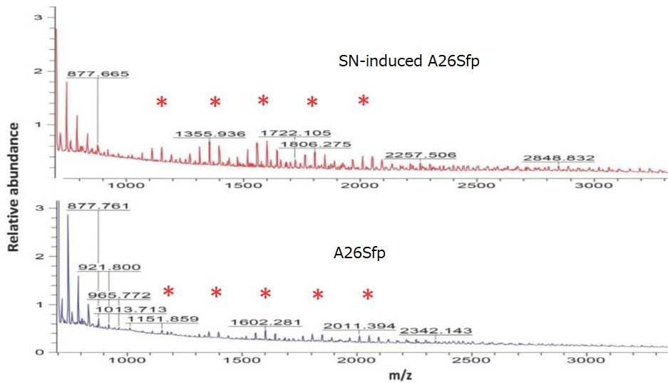
Quantification of exopolysaccharides using mass spectroscopy showed that the oligosaccharides produced by A26Sfp with SNs had longer chains than that of A26 wild type, suggesting that the improvement of the drought-tolerant soil will be due to increased production of longer chain polysaccharides.

Two groups with different COVID-19 immune profiles, that correlate with COVID-19 disease severity

A group from Sorbonne Universite, Inserm, Universite de Paris, France, etc. has reported on two groups with different COVID-19 immune profiles, that correlate with COVID-19 disease severity.
https://www.ncbi.nlm.nih.gov/pmc/articles/PMC8312547/

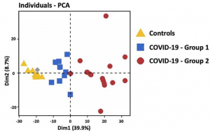
Strikingly, hospitalized COVID-19 patients segregated into two groups 1 and 2. Principal component analysis of transcriptomic features of whole blood at the onset of hospitalization revealed that group 1 (blue) was closer to healthy controls than group 2 (red).

Genes up-regulated in group 2 belong to signatures of myeloid cells, neutrophil activity, inflammatory response, TLR and type I IFN signaling pathways, and inhibition of T cell proliferation (i.e. arginase 1, PDL1, PDL2 and CD276/B7-H3). At the opposite, genes involved in CD8 and NK cell function, T cell activation, T helper (Th) differentiation, co-stimulatory receptors (TNFRSF4 (OX40), ICOSLG (ICOS ligand), TNFRSF18 (GITR) and TNFRSF11A (RANK)), and antigen presentation, were down-regulated in group 2. These results are compatible with the coexistence of patients with a distinct immune profile: (i) adaptive immune response triggering (i.e. group 1), (ii) exacerbated myeloid and innate responses, with dysfunctional adaptive immune response (i.e. group 2).

Patients of group 2 did not differ from group 1 for age and diabetes, but have a higher body mass index compared to group 1. The severity of respiratory distress in each group was estimated by the WHO score. Mild patients had a 4–5 WHO score, whereas patients requiring oxygen by NIV or high flow, mechanical ventilation or extracorporeal membrane oxygenation had >6 WHO score. Strikingly, almost all patients 94% from group 2 had severe respiratory distress (>6 WHO score) as compared to 25% (2/8) in group 1 (P = 0.0013). Differential biological variables between the two groups, reflecting other vital organs and tissues (i.e. liver, kidney, heart and blood vessels) were investigated. Among them Hepatic Steatosis Index (HSI) and prothrombin ratio (% PR) reflecting liver damage, urinary Na+ and Na+/K+ reflecting kidney injury, troponin for cardiac damage, and E-selectin and placental growth factor (PlGF) reflecting the vessel status were significantly different in group 2 patients as compared to group 1. Overall, multi-organ failure, potentially exacerbated by endothelium damage and thrombosis was more pronounced in group 2 patients. Finally, 88% of death belongs to group 2, underlying distinct clinical outcome associated with differential immune patterns of patients’ groups.

As for the immune populations between both groups, the group 2 was characterized by increased total leucocytes counts, with a profound lymphopenia, a decreased proportion of CD8+ T cells and increased proportion of CD4+ T cells, a decrease in NK cells and an increase of neutrophils. In addition, group 2 patients presented with higher plasma level of proinflammatory cytokines (IL-6, IL-8, TNF-α, soluble TNF receptor 2 (sTNFR2)). These data confirmed a higher inflammatory response, neutrophilia and reduced NK and T cell responses in group 2, whereas group 1 had a profile favoring an adaptive immune response. Of note, group 2 patients displayed a higher expression of TLR3 on PBMC.

Pulling force between RBD of various SARS-CoV-2 variants and ACE2 taking into consideration of glycans

A group from Lehigh University, Bethlehem, USA has reported on pulling force between RBD of various SARS-CoV-2 variants and ACE2 taking into consideration of glycans.
https://www.ncbi.nlm.nih.gov/pmc/articles/PMC8328061/

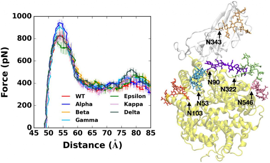
Pulling force analysis using molecular dynamics simulations was performed on RBD-ACE2 complexes as a function of distance between the centers of mass of RBD and ACE2 proteins. Fully-glycosylated S RBD-ACE2 complex model was employed for the pulling simulation, and the following variants were covered.

Alpha variant (first identified in United Kingdom, B.1.1.7: N501Y),
Beta variant (first identified in South Africa, B.1.351: K417N, E484K, N501Y),
Gamma variant (first identified in Japan/Brazil, P.1: K417T, E484K, N501Y),
Delta variant (first identified in India, B.1.617.2: L452R, T478K)),
Epsilon variant (first identified in US-California, B.1.427: L452R) ,
Kappa variant (first identified in India, B.1.617.1: L452R, E484Q)),

Alpha variant requires the highest force for initial separation from ACE2, followed by Beta and Gamma variants or Delta variant. K417N/T mutations of Beta and Gamma appear to make the RBD-ACE2 interactions less strong compared to Alpha variant. In addition, Epsilon variant is likely to be relatively easily dissociated from ACE2 than others due to its destabilized RBD structure upon the L452R mutation. In addition, Delta variant specifically shows stronger interactions with ACE2 than other variants at a relatively far distance between RBD and ACE2.

The Delta variant, interestingly, shows distinct features that are not found in other variants. Upon the T478K mutation, it requires the highest force for the RBD-ACE2 complex to be completely dissociated at D = 78 Å. In order to see what makes the difference, the number of contacts between RBD residue 478 and heavy atoms of selected key interacting residues of ACE2 was calculated. RBD Delta exclusively makes more contacts with ACE2 than other variants. Delta K478 retains contacts with ACE2 P84 and M82 at D = 78 Å, but Epsilon T478 already lost such interactions. It is possible that residue 478 located in the flexible loop could first have a chance to contact with ACE2, and the stronger interactions of Delta K478 with ACE2 could explain the reason why the proportion of Delta variant is recently dramatically being increased with high infectivity.

About importance of lectin complement pathway on severe COVID-19 pathology

A group from University of Cambridge, Cambridge, United Kingdom, etc. has reported on importance of lectin complement pathway on severe COVID-19 pathology.
https://www.frontiersin.org/articles/10.3389/fimmu.2021.714511/full

The complement system is an integral part of the innate and the adaptive immune systems, and three pathways are known: Classical pathway, Alternative pathway, and Lectin pathway.
The mannan-binding lectin-associated serine protease-2 (MASP-2) is the key enzyme of the Lectin pathway. The activated MASP-2 can cleave C4 efficiently to form C3 and C5 convertase complexes C4bC2a and C4bC2aC3b, leading to the formation of membrane attack complex (MAC). The pathogen-associated molecular patterns (PAMPs) recognition subcomponents (LP recognition molecules) are the key to activate MAPS-2. Those in humans are mannose-binding lectin (MBL), collectin-11 (CL-11), and heterocomplexes of CL-11 and CL-10 and ficolin. The Lectin pathway is initiated by the complexes of LP recognition subcomponent and MAPS-2 bind to PAMPs.

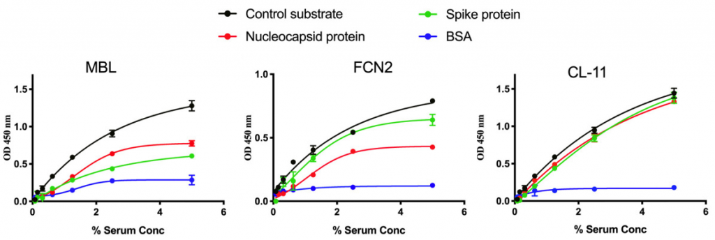
Binding of LP recognition molecules to SARS-CoV-2 proteins was studied. A MTP ELISA plate was coated with either SARS-CoV-2 Spike, SARS-CoV-2 N protein or control ligands (mannan for MBL, N-acetyl BSA for ficolin 2(FCN2), zymosan for CL-11), and serial dilutions of serum were added to the wells. It is clearly shown that LP recognition molecules such as MBL, ficolin2, and CL-11 bind to SARS-CoV-2 Proteins.

Although it was not mentioned if the LP was associated with COVID-19 severity in this report, this might be one of causes leading to acute respiratory distress syndrome (ARDS).

What differences in children and adults immunity drive COVID-19 pathogenesis?

A group from The University of Hong Kong, Hong Kong, SAR, China, etc. has reported on what differences in children and adults immunity drive COVID-19 pathogenesis.
https://www.nature.com/articles/s41467-021-24938-4

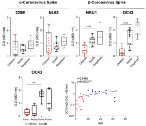
SARS-CoV-2 infection of children is associated with milder clinical outcomes than adults, and the immune mechanisms are unknown. Several hypotheses have been proposed to explain these differences such as innate cell recruitment and impairment by autoantibodies, mobilisation of antibody responses, differing levels of pre-existing cross-reactive immunity by common cold coronavirus exposure, or baseline total IgM levels.

Overall, it was found (1)total IFNγ CD4+ and CD8+ T cell responses are significantly lower in SARS-CoV-2 infected children than adults against the viral structural proteins, and in CD8+ T cells against ORF1ab proteins, and (2)prior β-coronavirus immunity is significantly lower in children than adults, indicating differing baseline immunity.

Therefore, reduced prior β-coronavirus immunity and reduced T cell activation in children might drive milder COVID-19 pathogenesis?

Importance of aberrant activation of alternative pathway of complement (APC) on COVID-19 severity

A group from Johns Hopkins School of Medicine, Baltimore, USA has reported the importance of aberrant activation of alternative pathway of complement (APC) on COVID-19 severity.
https://pubmed.ncbi.nlm.nih.gov/34289657/

Known complement pathways are three kinds: classical complement pathway, alternative pathway of complement (APC), and lectin pathway. The classical complement pathway needs activation with antibodies, however, in the case of APC, C3 directly adheres on the cell surface, and is activated by factors B and D directly.

It was demonstrated that serum from 58 COVID-19 patients (32 patients with minimal oxygen requirement, 7 on high flow oxygen, 17 requiring mechanical ventilation and 2 deaths) can induce complement-mediated cell death in a functional assay and increase membrane attack complex (C5b-9) deposition on the cell surface. 41.2% COVID-19 patients requiring intubation (n=7/17) were positive in the assay (>20% cell killing) and only 6.3% of COVID-19 patients requiring minimal oxygen support (n=2/32) were positive in the same evaluation. And further, C5 and factor D inhibition effectively mitigated the complement amplification induced by COVID-19 patient serum, and increased serum factor Bb level was associated with disease severity in COVID-19 patients, suggesting that APC dysregulation plays an important role.

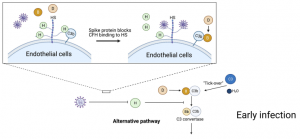
It was found that SARS-CoV-2 spike proteins directly block complement factor H from binding to heparin, which may lead to complement dysregulation on the cell surface. That is, early in infection, the SARS-CoV-2 spike protein binds heparan sulfate on the endothelial cell surface and interferes with the inhibitory function of complement factor H (CFH), leading to APC dysregulation. Suppression of CFH binding results in increased cleavage of factor B by factor D and generation of Bb. Factor Bb binds to C3b to form the alternative pathway C3 convertase (C3bBb), leading to the cleavage of C3 and generation of the C5 convertase (C4b2a3b or C3bBb3b). The C5 convertase cleaves C5 to generate C5a and C5b, which complexes with C6-9 to form the membrane attack complex (C5b-9) and kills the cell, leading to excess inflammation. As a conclusion, APC dysregulation due to SARS-CoV-2 contributes to the pathogenesis of COVID-19 and may be a marker of disease severity.

Rhizoshpere of Wheat: Phosphate solubilizing rhizobacteria stimulate wheat germination rate and seedlings growth

A group from Mohammed VI Polytechnic University (UM6P), Benguerir, Morocco, etc. has reported on Phosphate solubilizing rhizobacteria stimulating wheat germination rate and seedlings growth.
https://peerj.com/articles/11583/

Recently, rhizosphere is attracting so much attention. Rhizosphere is very much like the relationship between intestine and intestinal bacteria. Intestinal endothelium corresponds to root epithelium, and intestinal bacteria correspond to soil bacteria. However, there have been few researches on rhizosphere glycome, and blog admin is watching the progress in this field with great interests as a new frontier.

Phosphorus (P) is considered one of the most important elements in plant nutrition after nitrogen. It is an essential macronutrient to all major metabolic processes in plants growth e.g., photosynthesis, energy transfer, respiration, and signal transduction. Phosphate solubilizing microorganisms including bacteria play an important role in enhancing soil fertility and plant growth. Therefore, it is paramount to explore management strategies which are considered as an environmentally friendly process and economically feasible procedure to improve crop production and maximize their yields in P-poor soils. Exploration of the biodiversity of rhizobacteria and the optimization/manipulation of microbial interactions in the rhizosphere represents an imperative step towards formulating more efficient microbial inoculants with high P-solubilizing ability. Although P is plentiful in soils in both organic and inorganic forms, it is in unavailable forms for root uptake. Numerous soil microorganisms particularly those present in plant’s rhizosphere can release the bound forms of P to a soluble form to increase its bioavailability to plants. Phosphate solubilizing bacteria belong to plant growth promoting rhizobacteria and are capable of solubilizing inorganic P from a variety of sources, such as dicalcium phosphate, tricalcium phosphate, or rock phosphate.

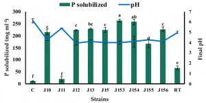
Bacteria screening identified nine best phosphate solubilizing strains as follows,

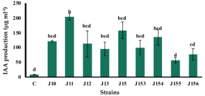
Indole acetic acid (IAA) stimulates plant growth,

Ammonia is a chemical compound having indirect plant health benefits, primarily by acting as metabolic inhibitor against phytopathogens. All tested strains were able to produce ammonia with various concentrations. The best one was Pseudomonas sp. J153.

As a conclusion, inoculation with P. moraviensis J12 and B. cereus J156 promote the highest rate of wheat seeds germination and seedlings growth.

L-SIGN is a receptor for authentic SARS-CoV-2 virus, using human liver sinusoidal endothelial cells (LSECs) with no ACE2 but L-SIGN

A group from Oklahoma Medical Research Foundation, Oklahoma, USA, etc. has reported that L-SIGN is a receptor for authentic SARS-CoV-2 infection.
https://insight.jci.org/articles/view/148999

To examine if L-SIGN interacts with SARS-CoV-2, lentivirus-based SARS-CoV-2 pseudo-typed virus (CoV-2–type virus) and HEK293T cell lines expressing L-SIGN-flag or ACE2-myc3 as positive control were established. L-SIGN is a C-type lectin which binds to high-mannose–type N-glycan and Ca2+ dependent manner. The RBD of the SARS-CoV-2 spike protein has an N-glycosylation site at Asn343. N-glycan–deficient spike/Fc, in which Asn343 was replaced with glutamine (Q) (SpikeN343Q/Fc) was developed, and the mannose moiety deficiency on SpikeN343Q/Fc were confirmed by mannose-specific GNL blotting. Thus, it was confirmed as shown below that SpikeN343Q/Fc binds to cells expressing ACE2 but not to cells expressing L-SIGN, indicating that L-SIGN recognizes an N-glycan structure on Asn343.

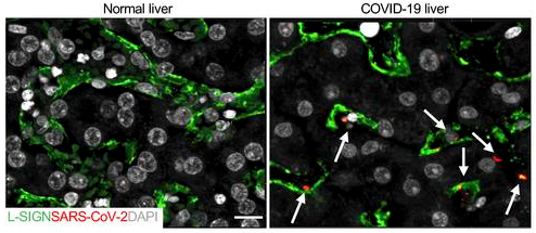
L-SIGN but not ACE2 is uniquely expressed on human liver sinusoidal endothelial cells (LSECs). To determine the clinical significance of the interaction between SARS-CoV-2 and L-SIGN, it was first examined if SARS-CoV-2 was present in LSECs from formalin-fixed paraffin-embedded liver autopsy samples from patients with COVID-19. In comparison with uninfected human liver autopsy samples, SARS-CoV-2 was detected with an anti-SARS-CoV-2 nucleocapsid antibody inside patient LSECs as shown below (LSECs were L-SIGN+ (green)and arrows mark SARS-CoV-2 (red)).

In addition, it was shown that SARS-CoV-2 infection was blocked with an antibody against human L-SIGN and also with mannan dose-dependently, suggesting these could be potential therapeutic options to treat severe COVID-19 infection.

Effectiveness of Tocilizumab and Dexamethasone in COVID-19 severe patients

A group from Jan Kochanowski University, Kielce, Poland, etc. has reported effectiveness of Tocilizumab and Dexamethasone in COVID-19 severe patients.
https://www.ncbi.nlm.nih.gov/pmc/articles/PMC8291861/

Comparing four groups (treated with Tocilizmab (TCZ), treated with TCZ and Dexamethason (DEX), treated with DEX, and no TCZ nor DEX), the death rate of 6.8% was significantly lower in patients receiving TCZ alone than other groups (19.6%–23.1%), particularly in patients with IL-6 concentration exceeding 100pg/mL, the death rate was significantly lower in patients receiving TCZ alone than other groups (5% vs 22.9%–51.7%, respectively).
The clinical improvements on day21 and day28 were also doubled in the case of TCZ alone (60% and 75%, respectively)comparing with that in that case of DEX alone (27.6% and 37.9%, respectively).

So, in patients with severe course of COVID-19, particularly those developing cytokine storm, administration of TCZ provides a significantly better effect than DEX regarding survival, clinical improvement, and hospital discharge rate. The combination of TCZ and DEX does not improve therapy effectiveness in patients with severe COVID-19 compared to the administration of TCZ alone.