Galectin-3 could be a GOOD prognostic marker against severe COVID-19

A group from University of Medicine “Aldo Moro”, Bari, Italy, etc. has reported on prognostic power of Galectin-3 against severe COVID-19.
https://www.ncbi.nlm.nih.gov/pmc/articles/PMC8332745/

This is the first study in scientific literature assessing the prognostic role of Galectin-3 in acute respiratory failure secondary to COVID-19 disease. Patients with higher serum levels of Galectin-3 tend to develop a more severe degree of ARDS with a worse prognosis. It is well known that SARS-COV2 infection can lead to the so called “cytokine storm” in some susceptible patients. For instance, our non-survivors group shows increased blood levels of various inflammation markers, which are frequently associated with negative outcomes in COVID-19 disease. Nevertheless, only IL-6, CRP and Galectin-3 remain statistically significant in our multivariate regression model. This finding is not surprising for IL-6 and CRP, which were previously reported as important prognostic markers in COVID-19 disease. On the contrary, this is the first study addressing this role for Galectin-3. Furthermore, among the explored parameters, Galectin-3 shows the best AUC curve in ROC analysis, showing good diagnostic power for severe ARDS (AUC 0.75, p = 0.001) using a cut-off value of 35.3 ng/ml.

In fact, patients with Galectin-3 serum levels above 35.3 ng/ml were not only more prone to develop severe ARDS, but also markedly at higher risk of ICU admission or death. The cohort size of this study was 156 patients.

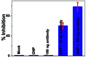
Anti-spike antibody attached gold nanoparticles (GNPs) have the capability to inhibit SARS-CoV-2 infection perfectly

A group from Jackson State University, USA, etc. has reported anti-spike antibody attached gold nanoparticles (GNPs) have the capability to inhibit SARS-CoV-2 infection perfectly.
https://www.ncbi.nlm.nih.gov/pmc/articles/PMC8323809/

Citrate coated original size of GNPs was 15 ± 2 nm, and the size of GNPs increased to ∼27 ± 6 nm by conjugating antibodies.

Pseudo SARS-CoV-2 (# C1110G, Montana Molecular, Bozeman, MT) was used as the model virus, and HEK293T cells were used as host cells, which express ACE2 known as infection receptor of SARS-CoV-2.

As shown below, anti-spike antibody attached gold nanoparticles block viral replication and virus spread in HEK293T cells. It was found that the inhibition efficiency was 100% for 100 ng/mL anti-spike antibody attached gold nanoparticles and 60% for 10 ng/mL anti-spike antibody attached GNPs. In contrast, our experimental data indicate that the inhibition efficiencies for PEG coated GNPs and 100 ng mL−1 antibody only were less than 1%.

< a href="https://www.emukk.com/WP/wp-content/uploads/2021/08/GNPantibody_SARS-CoV-2.png">

However, blog admin does not know if this method could be used in vivo.

B. amyloliquefaciens (Gram-positive Bacilli) applied singularly increased winter wheat crop biomass and Arbuscular Mycorrhizal fungal symbiosis

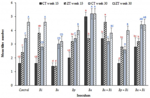
A group from University of Hertfordshire, Hatfield, UK has reported that Bacillus amyloliquefaciens inoculation singularly increased winter wheat crop biomass and arbuscular mycorrhizal (AM) fungal symbiosis.
https://www.ncbi.nlm.nih.gov/pmc/articles/PMC8309287/

Microbial associations between plant and soil are highly complex and provide a myriad of interactions with a wide range of plant and soil benefits including increased soil fertility and aggregation, improved plant immunity and defence, increased plant biomass and carbon sequestration. Arbuscular mycorrhizal (AM) fungi are one of the constituent organisms within the rhizosphere of an estimated 80% of terrestrial plants forming mutualistic biotrophic symbiosis with host plants. Mycorrhizal helper bacteria (MHB) stimulate mycorrhizae formation and further enhance plant-fungi symbiosis. The role of MHB and their interaction with AM fungi is poorly understood as is the extent of the tripartite rhizobacteria-AM fungi-host plant relationship. As MHBs, three Gram-positive Bacilli species were used in addition to (Ri) AM fungus R. intraradices in this experiment: (Bs) B. subtilis, (Bp) B. pumilis, and (Ba) B. amyloliquefaciens.

Soil cultivation by conventional tillage (CT) has been shown to directly influence the abundance and diversity of the soil microbiome. The hyphal networks of AM fungi are damaged by CT resulting in decreased root cortical arbuscules. However, the impact of tillage on rhizobacteria is relatively unknown. So, a series of experiments was done under two tillage regimes, CT and zero tillage (ZT).

As a conclusion, it was found that B. amyloliquefaciens (Gram-positive Bacilli) applied singularly increased winter wheat crop biomass and AM fungal symbiosis.

Four miRNAs that are remarkably decreased in the elderly and diabetic individuals can inhibit SARS-CoV-2 replication.

A group from School of Life Sciences, Nanjing University, Nanjing, China, etc. has reported four miRNAs (miR-7-5p, miR-24-3p, miR-145-5p and miR-223-3p) that are remarkably decreased in the elderly and diabetic individuals can inhibit SARS-CoV-2 replication.
https://www.nature.com/articles/s41392-021-00716-y

To comprehensively investigate the differences in circulating miRNA expression patterns in the serum between young and elderly people, age-related non-coding RNA expression profiles obtained by high-throughput sequencing from the NCBI Gene Expression Omnibus database were downloaded. A total of 13 samples (3 young, age <30 and 10 old, age >60) were analyzed, and differentially expressed miRNAs were screened and identified.

Quite interestingly, it was found that four miRNAs (miR-7-5p, miR-24-3p, miR-145-5p and miR-223-3p) through high-throughput sequencing and quantitative real-time PCR analysis, that are remarkably decreased in the elderly and diabetic groups.

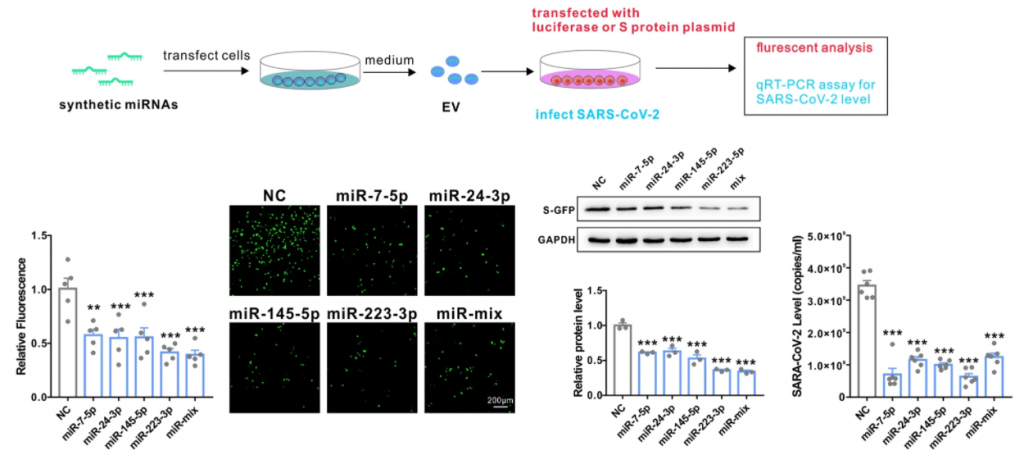
Then, it was demonstrated that these miRNAs, either in the exosome or in the free form, can directly inhibit S protein expression and SARS-CoV-2 replication. A figure below shows the case of exosome.

Mapping of SARS-CoV-2 spike glycoprotein-derived antigens presented by HLA class II on dendritic cells: how glycosylation is different from the original

A group from University of Oxford, UK, etc. has reported on mapping of SARS-CoV-2 spike glycoprotein-derived antigens presented by HLA class II on dendritic cells.
https://www.ncbi.nlm.nih.gov/pmc/articles/PMC8116342/

It is very curious for blog admin whether HLA-II bound glycopeptides presented as SARS-CoV-2-specific antigens on dendritic cells have the same glycosylation as phagocytized SARS-CoV-2 by dendritic cells.

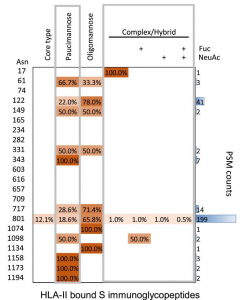
HLA-II-bound glycopeptides were identified from 14 N-linked glycosylation sites in Spike from total 22 glycosylation sites. HLA-II-bound peptides carried predominantly short paucimannosidic-type N-glycans while original Spike carried oligomannosidic- and GlcNAc-capped complex-type N-glycan structures at these sites. The paucimannosylation of the HLA-II-bound peptides comprised both core-fucosylated and fucosylated species. This reveals there is substantial trimming of glycan residues on the glycopeptides during antigen processing in dendritic cells.


The heatmap colors in a figure above indicate the relative frequency of each glycan composition present, and the total number of peptide spectral matches (PSM) is also shown (blue bars).

Monocytes and Macrophages in COVID-19: alveolar macrophages act as Trojan house carring SARS-CoV-2 and monocytes become immunosuppressive

This is a review paper written by a group from University of Bonn, Germany about monocytes and macrophages in COVID-19. Here is my summary of outlines discussed in the review paper.
https://www.ncbi.nlm.nih.gov/pmc/articles/PMC8335157/

Every day, the lung inhales thousands of liters of air containing high amounts of pathogens including viruses, bacteria, and fungi. To prevent infection and its resulting complications for the organism, a tight control by the immune system is needed. In the lung, macrophages are the most abundant immune cell type under homeostatic conditions. Based on their exact location, they can be separated in at least two different populations; the interstitial macrophages (IMs) and alveolar macrophages (AMs). AMs are the first defenders against pathogens entering the respiratory system. They originate from the yolk sac and populate the lung early after birth. AMs have proliferative capacity, thus can persist over the lifespan by self-renewal and are independent of replacement from the bone marrow.

Upon viral infection, AMs are responsible for early pathogen recognition, initiation and resolution of inflammation, as well as repair of tissue damage. AMs produce high levels of cellular mediators, including IL-1β, CCL3, CCL7 and CCL2, also known as monocyte chemotactic protein 1 (MCP1), which rapidly recruits CCR2-expressing bone marrow-derived monocytes into the lung. This is a necessary defense response, since viruses can either reduce the numbers of resident AMs dramatically or impair their phenotype. Furthermore, AMs are the main producers of type I IFN to trigger an antiviral response.

COVID-19 is characterized by a systemic increase of numerous cytokines, including IL-1α, IL-1β, IL-6, IL-7, tumor necrosis factor (TNF), type I and II IFN, and the inflammatory chemokines CCL2, CCL3 and CXCL10. Although the increased systemic cytokine response in COVID-19 is undisputed, the term “cytokine storm” in COVID-19 pathophysiology is a topic of debate, as TNF, IL-6, and IL-8 concentrations in COVID-19 are less strong compared to sepsis, acute respiratory distress syndrome unrelated to COVID-19, trauma, cardiac arrest, and cytokine release syndrome (CRS).
The systemic cytokine profile observed in patients suffering from severe COVID-19 does resemble those observed in macrophage activation syndrome (MAS), which led early on to the working hypothesis that dysregulated activation of mononuclear phagocytes (MNPs) compartment contributes to COVID-19-associated hyperinflammation.

SARS-CoV-2 infection of lung-resident MNPs might result either from phagocytosis of infected alveolar epithelial cells followed by viral escape from the lysosome or by direct infection. The induction of cytokine production in MNPs in COVID-19 can either be triggered via recognition of damage-associated molecular patterns (DAMPs) released from epithelial cells affected by SARS-CoV-2 by pattern recognition receptors (PRRs) or by direct recognition of viral pathogen-associated molecular patterns (PAMPs) via specific Toll-like receptors. Furthermore, C-type lectin receptors, including DC-SIGN, L-SIGN, etc., as well as Tweety family member 2 have been identified to interact with the SARS-CoV-2 spike protein inducing proinflammatory responses. It seems that AMs act as a Trojan horse, transferring the virus to adjacent lung regions, thereby slowly propagating SARS-CoV-2 infection and spreading hyperinflammation across the lung.

Most prominently and consistent across all studies, reduced HLA-DR expression on monocytes was reported in patients suffering from severe COVID-19. Decreased HLA-DR expression appeared to be strongly associated with COVID-19 disease severity. Downregulation of HLA-DR is a molecular feature often described for monocytic myeloid-derived suppressor cells (MDSC) which are observed in late-stage cancers and sepsis, and featured by immunosuppressive functions. In COVID-19, such HLA-DRlo monocytes are attracted to the lung by pro-inflammatory chemokines, resulting in a continuous accumulation of hyperactivated MNPs producing more pro-inflammatory mediators recruiting more inflammatory cells, including cytotoxic T cells and neutrophils, thus further exacerbating inflammation and tissue damage.

Infection susceptibility of various human endothelial cells by SARS-CoV-2

A group from Goethe University Frankfurt, Germany, etc. has reported on infection susceptibility of various human endothelial cells by SARS-CoV-2.
https://www.ncbi.nlm.nih.gov/pmc/articles/PMC8256413/

SARS-CoV-2 primarily invades alveolar epithelial cells and causes acute respiratory distress syndrome (ARDS). However, increasing evidence indicates that endothelial cell dysfunction and vascular events are major complications of the disease. Indeed, vascular inflammation, barrier defects leading to tissue edema, activation of disseminated intravascular coagulation and microthrombi were reported in moderate to severe COVID-19 cases. However, it is still unclear whether the endothelial dysfunction is caused by direct infection of endothelial cells or is mainly secondary caused by inflammation cascade due to cytokine release.

Various human endothelial cells from different vascular beds were incubated with SARS-CoV-2 to investigate infection susceptibility of SARS-CoV-2 to those endothelial cells. Evaluated cells are as follows;
Human umbilical vein endothelial cells (HUVEC),
human coronary artery endothelial cells (HCAEC),
human cardiac microvascular endothelial cells (HCMVEC), and
human lung microvascular endothelial cells (HLMVEC).

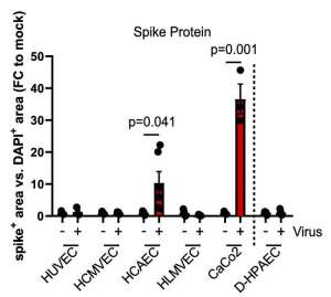
SARS-CoV-2 spike protein was only detected in HCAECs after the virus infection but not in the other endothelial cells tested. This is consistent with the fact the only HCAEC expressed the SARS-CoV-2 receptor angiotensin-converting enzyme 2 (ACE2) which is required for virus infection.

Human colon carcinoma cells (CaCo2) were used as positive control of the SARS-CoV-2 infection,

Although the spike protein was detected in HCAECs after SARS-CoV-2 infection, this does not necessarily mean infection of the endothelial cells. Therefore, the levels of double-stranded RNA, as a sign of viral RNA multiplication, and the presence of infectious virus in the cell supernatant, which would be indicative of viral replication, were evaluated further. However, neither double-strand RNA nor infectious virus in HCAEC supernatant could be detected 5 days after SARS-CoV-2 infection, suggesting that no new virus is generated by the tested endothelial cells and that the spike protein may originated from the virus that was originally taken up.

In conclusion, the massive endothelial dysfunction and microvascular thrombotic complications observed in patients suffering from COVID-19 is mainly secondarily caused by the inflammatory cascades mediated by the cytokine release syndrome.

Chitosan derivatives could be novel inhibitors against wild type and variants of SARS-CoV-2 virus infection

A group from Birla Institute of Technology and Sciences (BITS), Rajasthan, India, etc. has reported Chitosan derivatives could be novel inhibitors against wild type and variants of SARS-CoV-2 virus infection.
https://www.ncbi.nlm.nih.gov/pmc/articles/PMC8313795/

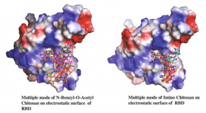
A library of chitosan derivatives was screened by site-specific docking at not only spike protein Receptor Binding Domain (RBD) of wild type SARS-CoV-2 but also on RBD of Alpha and Gamma SARS-CoV-2 variants. The obtained result was very interesting and ranks N-benzyl-O-acetyl-chitosan, Imino-chitosan, as a potent antiviral candidate due to its high binding affinity of the ligands (-6.4 to -6.7 kcal/mol) with SARS-CoV-2 spike protein RBD

Conformation models for both ligands in electrostatic surface are shown below,

A family of microsomal enzymes known as cytochrome P450 (CYP450) is responsible for xenobiotic metabolism. CYP3A4, CYP3A2, CYP2C9, CYP2C19, and CYP2D6 are all members of the CYP450 family, which are involved in metabolizing various drugs. The ideal scenario of a drug should be the non-inhibition of these enzymes. In this regard, findings of the test ligands suggested both Imino-chitosan and N-benzyl-o-acetyl-chitosan be the decent leads.

The toxicity profile of Imino-chitosan and N-benzyl-o-acetyl-chitosan was determined on the basis of their mutagenic and carcinogenic properties, using the in silico Ames test, carcinogenicity test in rats and mice, and HERG inhibition results. All the results of the toxicity profile were favorable indicating that the ligands can be potential drugs.

Wheat rhizosphere: High Zn wheat recruit more bacteria relevant to Zn mobilization in the rhizosphere

A group from Chinese Academy of Agricultural Sciences, Shenzhen, China, etc. has reported that high Zn wheat recruit more bacteria relevant to Zn mobilization in the rhizosphere.
https://www.ncbi.nlm.nih.gov/pmc/articles/PMC8261137/

Around 20% of the world population are suffering from zinc (Zn) deficiency, and the situation will become worse with the increase of atmosphere carbon dioxide. An effective solution to address human Zn deficiency is to increase the grain Zn concentration of staple food crops like wheat, namely, Zn biofortification. The target of wheat Zn biofortification is to increase the current grain Zn concentration of 20 ~ 30 mg/kg to above 40 mg/kg that is sufficient for human Zn nutrition.

Due to the poor mobility of Zn in soil, the absorption of Zn by plant roots mainly occurs in the rhizosphere, where the activities of roots and microorganisms can somewhat increase the amount of available Zn. In the calcareous soils distributed worldwide, Zn availability is restricted by alkaline environment and high carbonate content; and various root exudates like carboxylic acids, amino acids, and low-molecular-weight polypeptides can acidify the rhizosphere and solubilize the Zn immobilized in minerals. Besides, the microbes living on root exudates can also produce organic acids, siderophores, and exopolysaccharides that can mobilize micronutrients in rhizosphere soil.

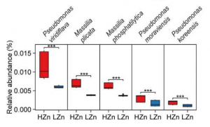
Comparisons of the relative abundances of rhizosphere-enriched species between High Zn (HZn) and Low Zn (LZn) wheat found that 30 species were significantly enriched in HZn with the abundance ratio of HZn to LZn >1.5 and two species were enriched in LZn with the abundance ratio of HZn to LZn <0.5. Of the 32 species, three HZn-enriched species belong to the reported soil Zn-mobilizing genus Pseudomonas, which is one of the 38 previously published Zn-mobilizing microbial species or genera. Nearly half of the previously reported Zn-mobilizing microbes were enriched in wheat rhizosphere, but most of them showed no significant difference between HZn and LZn. Differently, the 32 HZn- or LZn-enriched species were also enriched in wheat rhizosphere, and they exhibited significant differences between HZn and LZn, such as much higher abundances of Pseudomonas and Massilia species in HZn than those in LZn. Besides, genome functional annotation of the 32 species showed that 28 HZn-enriched and two LZn-enriched species possess the functional genes involved in soil Zn mobilization. Therefore, the previously reported Zn-mobilizing microbes can promote the Zn uptake of all wheat plants, while the identified 30 HZn- or LZn-enriched species may be the potential soil Zn-mobilizing microbes contributing to the variations of shoot Zn uptake and grain Zn concentration among cultivars.

It would be true that the HZn wheat recruit more bacteria relevant to soil Zn mobilization in the rhizosphere, suggesting that the difference between Hzn wheat and LZn wheat cultivars comes from the difference in microbiome of the rhizosphere. However, how HZn wheat recruit Zn mobilization bacteria is still unknown.

Good Serum cytokine and chemokine markers associated with effective immune response to SARS-CoV-2 in mRNA vaccination

A group from National Cancer Institute, Frederick, USA, etc. has reported good Serum cytokine and chemokine markers associated with effective immune response to SARS-CoV-2 in mRNA vaccination.
https://www.ncbi.nlm.nih.gov/pmc/articles/PMC8299183/

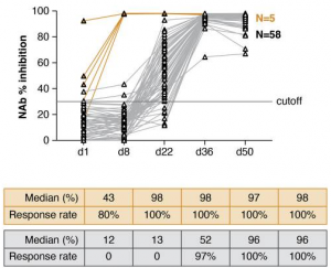
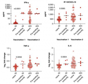
A cohort of 63 health-care workers received the BNT162b2 mRNA vaccine and was monitored for the development of neutralizing antibodies. There were two types of recipients in this cohort. One is individuals without any SARS-CoV-2 infection before the vaccination (58 recipients), and the other was individuals with pre-existing SARS-CoV-2 infection (6 recipients). The 58 recipients showed immune responses first detected 3 weeks after the 1st dose (day 22), which was followed by a significant increase after the 2nd dose by day 36. The neutralizing ability was measured using a surrogate virus neutralization test, and an assay that reached a median of 96% inhibition after the 2nd dose, In contrast, the 5 recipients with pre-existing SARS-CoV-2 immunity showed antibody responses to SARS-CoV-2 at the day of vaccination, followed by an immediate strong anamnestic response after the 1st dose (day 8). The antibody responses did not further increase upon the 2nd vaccination and remained significantly higher than those in the SARS-CoV-2-naive vaccine recipients.

This study highlights important associations of several immunoregulatory molecules induced by vaccination with innate and adaptive immune responses elicited by an mRNA-based vaccine. The early cytokine/chemokine signature featuring IL-15, IFN-γ, and IP-10/CXCL10 may be used to monitor effective vaccination and as a guide to optimize the efficacy of mRNA vaccination strategies.