Seroprevalence of anti-SARS-CoV-2 IgG antibodies in COVID-19 patients 

A group from Juntendo University Faculty of Medicine has reported seroprevalence of IgG and IgM antibodies in COVID-19 patients, although the cohort size was small including only 34 patients.
https://www.ncbi.nlm.nih.gov/pmc/articles/PMC8023454/

Using a chemiluminescent microparticle immunoassay (CMIA)-based SARS-CoV-2 IgG test (cat. # 06R90, Abbott),
Severe/Critical cases:within a week after symptom onset=40%、1~2 weeks=88%、after two weeks=100%,
Mild/Moderate cases:within a week after symptom onset=0%、1~2 weeks=38%、after two weeks=100%.

Using an IC IgG antibody assay using the Anti-SARS-CoV-2 Rapid Test (cat. # RTA0203, AutoBio),
Severe/Critical cases:within a week after symptom onset=60%、1~2 weeks=63%、after two weeks=100%,
Mild/Moderate cases:within a week after symptom onset=17%、1~2 weeks=63%、after two weeks=100%.

In this study, IgG titers remained at significantly elevated levels for 2 months, regardless of disease severity. These results indicate that IgG serologic tests could be used as a complementary test to PCR to diagnose COVID-19 from 14 days after symptom onset. However, since this cohort is so small without including asymptomatic individuals, a larger scale cohort is needed to conclude final answer.
     

Difference in site-specific glycosylation of SARS-CoV-2 spike proteins (all recombinants) among several laboratories

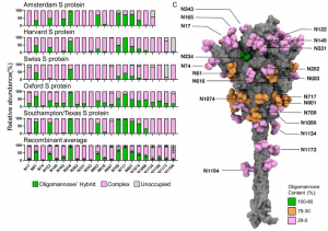
A group from University of Southampton, etc. has compared site-specific glycosylation of SARS-CoV-2 spike proteins (all recombinants) among five laboratories.
The cells used for Sproten expression were as follows.
HEK293: Amsterdam, Harvard,
HEK293F: Southampton/Texas,
HEK293T: Oxford,
CHO: Swiss,
It is celarly shown that site-specific glycosylation changes considerablly with reflecting differences in cells and culture conditions.
Fundamentally speaking, it is a mixture of oligo mannose and complex type N-glycans.

Blog admin is interested in how much these differences cause difference in the infectivity, how glycosylation changes with SARS-CoV-2 mutations, and how much the glycosylation changes due to mutations affect the infectivity.
https://www.biorxiv.org/content/10.1101/2021.03.08.433764v1.full

Characteristics of  SARS-CoV-2 neutralizing antibodies targeting NTD Spike protein: Effects of Glycans and Variants

A group from Columbia University, etc. has reported about characteristics of SARS-CoV-2 neutralizing antibodies targeting NTD Spike protein.
https://www.ncbi.nlm.nih.gov/pmc/articles/PMC7953435/

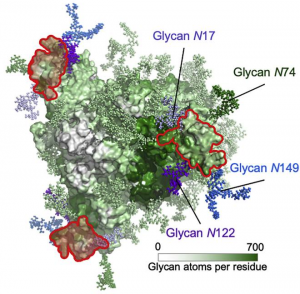
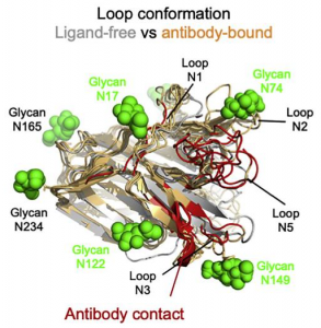
Totally 17 NTD antibodies have been already published. It was found that these antibodies target a specific area (called Supersite) on NTD of the spike protein. The NTD supersite was located at the periphery of the spike protein, distal from the 3-fold axis, and facing away from the viral membrane. This surface was surrounded by four N-glycans, N17, N74, N122, and N149, and nominally “glycan free,” although there are some glycan coverage because of the dynamic motional fluctuation of glycans. And also, the supersite had strong positive electrostatic potential, while recognizing antibodies had complementary strong electronegative potential.

The SARS-CoV-2 variants, particularly B.1.1.7 and B.1.351, are concerning due to increased infectivity, and these variants escape most NTD-directed neutralizing antibodies. B.1.1.7 includes NTD deletion mutations D69-70 and D144, and strain B.1.351 includes NTD mutations D242-244 and R246I. The mutated positions including 144, 242-244, and 246 are all within the NTD supersite. While the deletion at 69-70 is outside of the supersite, it forms part of the hairpin N2 loop of NTD. So, its deletion could significantly impact the conformation of the NTD supersite.

A new pathway was discovered in activation of the complement system of the innate immunity: MASP-1 and MASP-3 have dual functions as enzymes and as PRMs 

In the lectin pathway as one of innate immunities, pattern recognition molecules (PRMs) (i.e., mannose-binding lectin (MBL), ficolins, and collectin-10/-11) bind to pathogens recognizing specific molecular structures on the membranes, make complexes with MAPSs, activate complements sequentially, and finally form Membrane Attack Complex (MAC) on the membranes to kill pathogens.

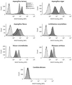
However, a group from University of Copenhagen, etc. has reported a new pathway in which MAPSs directly bind to pathogens and activate the complement system using Aspergillus fumigatus as a pathogen. This means that a new pathway in the complement system was discovered showing MASP-1 and MASP-3 have dual functions as enzymes and as PRMs
https://www.karger.com/Article/FullText/514546

A cocktail monoclonal antibody (REGN-COV2) is unaffected by B.1.1.7, B.1.351, P.1 variants, however, the vaccine Pfizer BNT162b2 shows markedly reduced inhibition against B.1.351, P.1 variants 

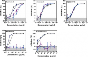
A group from German Primate Center, Göttingen, etc. has reported on the effectiveness of major monoclonal antibodies for COVID-19 (Casirivimab, Bamlanivimab, Imdevimab)against SARS-CoV-2 variants, that of a cocktail monoclonal antibody (REGN-COV2: consisting of Casirivimab and Imdevimab), and also that of Pfizer BNT162b2 vaccine against those variants.
https://www.ncbi.nlm.nih.gov/pmc/articles/PMC7980144/

A cocktail monoclonal antibody (REGN-COV2)efficiently inhibited infection mediated by the S proteins of all variants (B.1.1.7, B.1.351, P.1). However, infection mediated by the S proteins of the B.1.351 and P.1 variant was completely resistant to REGN10989 and Bamlanivimab.

On the other hand, the Pfizer BNT162b2 vaccine is based on an mRNA that encodes for the SARS-CoV-2 S protein and is said to be highly protective against COVID-19. All sera from 15 donors immunized twice with BNT162b2 efficiently inhibited entry driven by the WT S protein and inhibition of entry driven by the S protein of the B.1.1.7 variant was only slightly reduced. However, 12 out of 15 sera showed a markedly reduced inhibition of entry driven by the S proteins of the B.1.351 and P.1 variants


Diagnostic accuracy of COVID-19 Chest CT images with using Deep Learning methodology

A group from Sejong University, Seoul, etc. has reported on the accuracy of COVID-19 diagnosis in chest CT images with applying Deep Learning.
https://journals.plos.org/plosone/article?id=10.1371/journal.pone.0249450

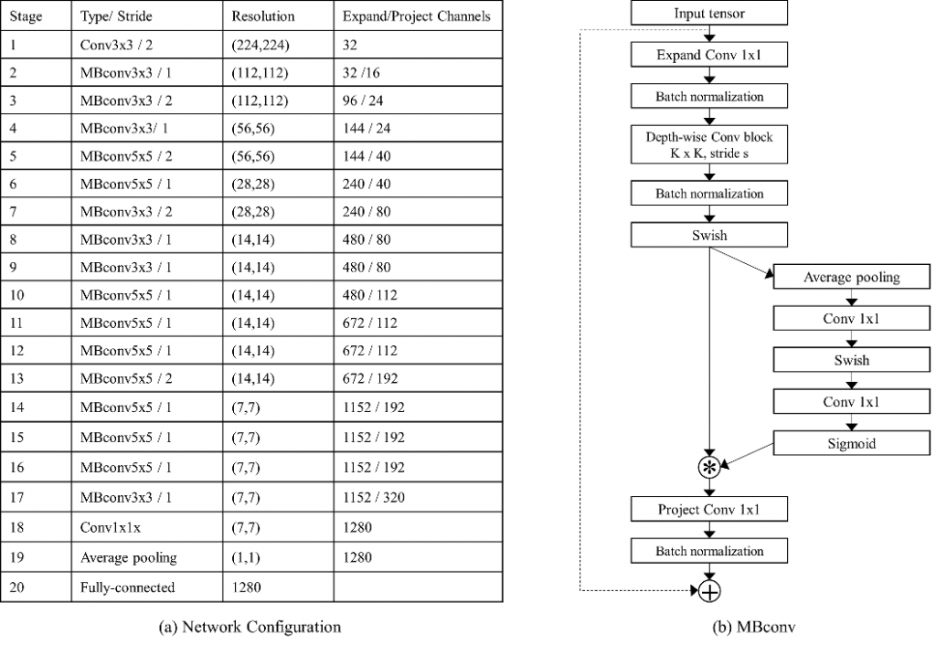
The neural network using in Deep Learning was composed of 20 stages, and convolution and pooling functions were incorporated. The resolution of the input images were (224 x 224, and the sizes of convolution were (3 x 3)and(5 x 5). The obtained overall accuracy was 99.83%(sensitivity=0.9286, specificity=0.99). In the future, it will be accelerate to adopt Deep Learning in diagnostic applications.

Using SARS-CoV-2 nanoparticles as a vaccine, a broadly neutralizing antibody strong to SARS-CoV-2 variants could be induced 

A group from Scripps Research Institute has reported that a broadly neutralizing antibody strong to SARS-CoV-2 variants could be induced by using SARS-CoV-2 nanoparticles as a vaccine.
https://www.ncbi.nlm.nih.gov/pmc/articles/PMC8010731/

The main stream of the current vaccines is using platforms which deliver the recombinant SARS-CoV-2 spike as an antigen such as mRNA-encapsulated liposomes (e.g., BNT162b2 and mRNA-1273), adenovirus vectors (e.g., ChAdOx1 nCoV-19 [AZD1222], CTII-nCoV, Sputnik V, and Ad26.COV2.S). There vaccines are compatible with B.1.1.7 variant, but a notable loss of efficacy was reported for B.1.351 and P.1 variants. Therefore, it is required to develop vaccines that can elicit a broadly neutralizing antibody (bNAb) response to SARS-CoV-2 variants. For this purpose, those vaccines have to induce long-lived germinal center (GC) reactions to activate precursor B cells, stimulate affinity maturation, and form long-term immune memory.

The SARS-CoV-2 spike protein is a trimer of S1-S2 heterodimers. The S1 subunit contains a receptor-binding domain (RBD) to initiate infection. The S2 subunit consists of a fusion peptide (FP) and heptad repeat regions 1 and 2 (HR1 and HR2). Authors designed an HR2-deleted glycine-capped spike (S2GΔHR2) to increase the spike stability, and assembled those spikes on a nanoparticle platform called SApNP using I3–01v9 60-mers as a linker (S2GΔHR2-10GS-I3-01v9-LD7-PADRE (I3-01v9-L7P)). The I3–01v9-LP7 presents 20 stabilized spikes.

Using S2GΔHR2-10GS-I3-01v9-L7P as a vaccine, a bNAb were induced showing compatible titers for B.1.1.7, B.1.351, and P.1 variants. Compared with the soluble spike, this nanoparticle showed 6-fold longer retention, 4-fold greater presentation on follicular dendritic cell dendrites, and 5-fold higher germinal center reactions in lymph node follicles. The reason of this effect is not clear yet, but would be related to the size effect.

The effect of Transgenic Expression of Human C-Type Lectin CLEC18A in Aedes aegypti on suppressing Dengu Virus infectivity and proliferation

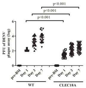
A group from National Health Research Institutes, Miaoli, Taiwan, etc. has established a transgenic Aedes aegypti line that expresses human CLEC18A, and has confirmed suppression of dengue virus (DENV) infection and proliferation in Aedes aegypti.
https://www.frontiersin.org/articles/10.3389/fimmu.2021.640367/full

A human C-type lectin protein CLEC18A binds to glycoprotein of dengue virus, induces type-I interferon secretion, making CLEC18A one of players in innate immune responses to DENV infection (detailed glycan binding specificity of CLEC18A is not clear, but usually C-type lectins bind to high mannose or Gal/GalNAc). This study suggests a possibility that could suppress the spread of DENV infection and proliferation by using transgenic mosquitoes.

Immune Memory (CD4+T-cell reaction) in mild COVID-19 patients and Cross-rectivity of CD4+T-cells against SARS-CoV-2 in unexposed donors

A group from National Institute of Immunology, New Delhi, etc. has reported on immune memory (CD4+T-cell reaction) in mild COVID-19 patients and cross-reactivity of CD4+T-cells against SARS-CoV-2 in unexposed donors
https://www.frontiersin.org/articles/10.3389/fimmu.2021.636768/full

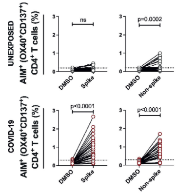
In mild COVID-19 patients, it was reported that the immune memory (i.e, CD4+T-cell reactions to SARS-CoV-2) continues for about 5 months (median about 3 months). On the other hand, in SARS-CoV-2 unexposed donors, there was no immune reaction to SARS-CoV-2 spike protein, but the CD4+ T-cells specific to SARS-CoV-2 N protein were observed in 66% of the donors. In SARS-CoV-2 exposed patients, the CD4+ T-cells targeted SARS-CoV-2 spike protein rather than SARS-CoV-2 N protein.
Whether the cross-reactive CD4+ T cells are contributing to suppressing SARS-CoV-2 infection and further less severe outcome needs to be addressed in the prospective cohort before and after COVID-19.

Proteinuria would be a good Biomarker for the new coronavirus (COVID-19) severity 

A group from Université Côte d’Azur, Nice, etc. has reported that Proteinuria would be a good Biomarker for the new coronavirus (COVID-19) severity.
https://www.ncbi.nlm.nih.gov/pmc/articles/PMC7985082/

Significant proteinuria (>0.3 g/g) was related to
higher prevalence of ICU admission [OR = 4.72, IC95 (1.16–23.21), p = 0.03],
acute respiratory distress syndrome (ARDS) [OR = 6.89, IC95 (1.41–53.01, p = 0.02)],
longer hospital stay [19 days (9–31) versus 7 days (5–11), p = 0.001].