Chitinase 3-like-1 could be a good Therapeutic Target for the new coronavirus (COVID-19)

A group from Brown University, etc. has reported that Chitinase 3-like-1 (CHI3L1/YKL-40) could be a good therapeutic target for the new coronavirus (COVID-19).
https://pubmed.ncbi.nlm.nih.gov/33442679/

In the case of COVID-19, majority is asymptomatic or mild, but 10-20% of patients require hospitalization. Especially, it is so interesting that the severity of COVID-19 is deeply related to aging and comorbid disorders (such as diabetes, hypertension, obesity and metabolic syndrome, cardiovascular disease and chronic lung diseases like COPD and asthma). It is know that CHI3L1 is secreted from a spectrum of cells in response to a variety of injury and inflammation, and regulates innate and adaptive immunity and also protects apoptosis. What’s even more interesting is that CHI3L1 increases with aging and also with risk factors of COVID-19 (i.e., presence of comorbid disorders). Taking these things into consideration, authors focused on the relationship between CHI3L1 and angiotensin-converting enzyme 2 (ACE2), transmembrane protease serine 2 (TMPRSS2), cathepsin L (CTSL).

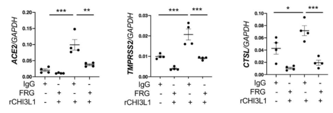
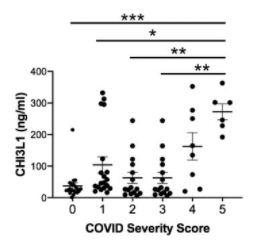
In an in vivo experiment using mice, it was found that CHI3L1 stimulates expression of pulmonary ACE2, TMPRSS2, and CTSL. In other experiments using Calu-3 lung epithelial cells, the expression of ACE2, TMPRSS2, CTSL increased monotonically with CHI3L1 dose, and also it was found that a monoclonal antibody for CHI3L1 (FRG) strongly inhibits the expression of ACE2, TMPRSS2, and CTSL. In actual COVID-19 patients hospitalized, it was also indicated that there is a significant correlation between severity and CHI3L1.

These evidences suggest strongly that CHI3L1 could be a good therapeutic target for COVID-19. Let’s anticipate the future research. 

IgM plays a major role unexpectedly in preventing the new coronavirus (SARS-CoV-2) infection

A group from University de Monterey, etc. has reported an evaluation result of SARS-CoV-2 neutralization activities of IgM, IgA, and IgG using plasma samples from 25 convalescent patients with the new coronavirus (COVID-19).
https://pubmed.ncbi.nlm.nih.gov/33596407/

IgM, IgA, and IgG were selectively depleted using isotype-specific ligands immobilized on beads, respectively. A comparison of the SARS-CoV-2 pseudoviral inhibitory dilution (ID50) for each plasma is shown below. In the IgM-depleted plasma, neutralization activity was greatly reduced by 5.5 times, the secondary one was the IgG-depleted plasma with the reduction of 4.5 times, and the reduction of IgA-depleted plasma was 2.4 times. Considering that IgM is only 5% of total immunoglobulin, the neutralization activity of IgM is very curious.

Q677 mutations in clade 20G (lineage B.1.2) of the new coronavirus (SARS-CoV-2) are increasing rapidly since late 2020

A group from University of Bern, Switzerland, etc. has reported a sharp increase of Q677 mutations (Q677H and Q677P) in clade 20G (lineage B.1.2) of the new coronavirus (SARS-CoV-2) in the USA. Since this mutation is present in the proximity of the furin cleavage site, it may have an effect on infectivity, but detailed research will be a future issue.
https://pubmed.ncbi.nlm.nih.gov/33594385/

Excessive complement activation occurs in kidneys of patients with the new coronavirus (COVID-19)

A group from Friedrich-Alexander-University (FAU) Erlangen-Nürnberg, etc. has reported excessive complement activation in kidneys of patients with the new coronavirus (COVID-19).
https://www.ncbi.nlm.nih.gov/pmc/articles/PMC7878379/

COVID-19 causes severe acute respiratory syndrome (ARDS), and the tissue damage is not restricted to lungs but also develop cardiac and kidney injury. In the case of kidneys, the glomerules and tubules seem to be damaged. Excessive activation of complements develops excessive membrane attack complex (MAC) resulting in damage of the glomerular and tubular tissues.
The table below shows a comparison of the level of complement expression among COVID-19 and typical kidney diseases comparing with a control.


ATI: acute tubular injury
HUS: hemolytic uremic syndrome
DIC: disseminated intravascular coagulation

The functions of glycans and lectins in myogenesis: Galectin-1 promotes muscle development

A group from University of Melbourne, etc. has investigated changes in glycan modification in myogenesis over time, while studying the functions of Galectins.
https://www.mcponline.org/article/S1535-9476(20)35144-6/fulltext

In myogenesis, there was a tendency that terminal di-Gal modification down regulated, α2-6Sia modification up regulated, α2-3Sia modification down regulated inversely, and paucimannose up regulated. These changes were thought to be related to intercellular signaling in myogenesis, but the specific signal paths were still unknown. On the other hand, as for galectins, the expression of Galectin-1 increased and the expression of Galectin-3 decreased.
Using newborn mice, an empty multiple cloning site (MCS) was injected with AAV6 into the left foot (as a control), and a galectin-1 gene (LGALS1) was injected with AAV6 into the right foot, and the difference between them was compared after 42 days. it was found that muscle mass was significantly increased with the injection of LGALS1. So, it is clear that galectin-1 is able to promote muscle development.
In the figure below, 14-3-3 protein was referenced as a comparison of LGALS1. 14-3-3 protein is related to intercellular signaling, and recognizes Ser/Thr residues in a specific domain of target protein with a phosphorylation-dependent way, and extracts the physiological function of the phosphorylation state.

On the effects of O-glycan modification around furin cleavage site of the new coronavirus (SARS-CoV-2), the infectivity of UK variant (B.1.1.7)

A group of NIH has reported the effects of O-glycan modification around the furin cleavage site of the new coronavirus (SARS-CoV-2) to the increased infectivity of the UK variant (B.1.1.7).
https://www.ncbi.nlm.nih.gov/pmc/articles/PMC7872346/

At the S1/S2 boundary of SARS-CoV-2 S-protein, an unique sequence of 4 amino acids (PRRA) is inserted forming the furin cleavage site as shown below.


S673, T676 are the positions where O-glycans are modified, as shown above. The presence of proline at 681 was deeply related to O-glycan modification at those positions. The authors also found that GALNT1 is the most active one glycosylating the furin proximal region among glycosyltransferase GALNT family. Furthermore, addition of O-glycans was shown to significantly suppress the cleavage at the furin cleavage site into S1 and S2. The UK variant (B.1.1.7) has a mutation called P681H, which replaces proline with histidine. Based on the above mentioned results, it is inferred that the non-presence of proline at 681 in UK variant suppresses O-glycan modification, resulting in increased cleavage efficiency and increased infectability.

IgG of the new coronavirus (SARS-CoV-2) was found in bovine whey milked in November 2018 and August 2019

A group from Juntendo University etc. has found antibodies (IgG) against the S-protein of the new coronavirus (SARS-CoV-2) from bovine milk whey.

https://www.sciencedirect.com/science/article/pii/S0958694621000303?via%3Dihub

The bovine milk was prepared from New Zealand cows in November 2018 and August 2019, and they discovered the presence of IgG for RBD present in the S-protein of SARS-CoV-2 from the bovine milk whey. The genome sequence of the S-protein is shown below, and the epitope mapping of the IgG shows that RBD is the one. Therefore, IgG enriched from milk whey may show neutralize activity against SARS-CoV-2.
However, this bovine milk whey was milked before the spread of COVID-19, and it is difficult to think that the cows were infected with SARS-CoV-2, and it might have been infected with an unknown coronavirus that shares immunogenicity of SRAS-CoV-2.


Glycan modification changes in gastric epithelial cells infected with Helicobacter pylori

A group from Chinese Center for Disease Control and Prevention, Beijing etc. has infected gastric epithelial cells (GES-1) with Helicobacter pylori isolated from patients with different diseases, and investigated changes in glycan modification with lectin microarrays.
https://www.mdpi.com/2076-0817/10/2/168/htm

Patient’s disease states from which H. pylori bacteria were isolated were as follows:
Chronic gastritis (YN4-62),
Duodenal ulcer disease (M84, P164),
Gastric cancer (HLJ011, HLJ030),

The trend is characterized by the increase in α1-2Fuc and α2-3Sia modifications. GES-1 which was not infected with H. pylori, was used as a control, to compare changes in glycan expression levels. Red indicates increased ones.

Lactoferrin functions as an antiviral drug for the new coronavirus (SARS-CoV-2)

Lactoferrin is an iron-binding glycoprotein with a molecular weight of 80,000 contained in milk. It is found in mammalian milk (especially colostrum), including human, but is also found in tears, saliva, pancreatic fluid and other exocrine fluids and neutrophils in adults. Lactoferrin is also known to maintain and improve health, including immune regulation, antiviral function,
lactobacillus bifidus proliferation, iron absorption regulation associated with iron binding ability, anti-inflammatory action, and lipid metabolism improvement action.

A group from University of Arizona, Tucson etc. has reported that lactoferrin actually shows antiviral effects against SARS-CoV-2, and what is the mechanism behind it.
https://www.tandfonline.com/doi/full/10.1080/22221751.2021.1888660

Using ACE2 overexpressed 293 T cells, Vero E6 cells, and Calu-3 cells, they show the inhibitory activity of lactoferrin on infection of SARS-CoV-2 in vitro. They have also verified using a molecular docking method that the mechanism of infection inhibition is blocking SARS-CoV-2 binding to heparan sulfate, which is a co-receptor of ACE2. Heparan sulfate has a negative charge and is thought to bind to a positively charged region (17 to 41 rest regions) present in the NTD of the SARS-CoV-2 S-protein. In the figures below, BLF=Bovine lactoferrin and HLF=Human lactoferrin. The lower right hand side figure shows a molecular docking image of heparin dp4 with bovine lactoferricin.

Why SARS-CoV-2 (20I/501Y. V1) UK Variant does increase its infectability?

A group from Anschutz Medical Center, University of Colorado etc. has analyzed why does the SARS-CoV-2 (20I/501Y. V1) UK variant increase its infectability? using a molecular docking method.
https://www.biorxiv.org/content/10.1101/2021.02.02.428884v1.full

Comparing with the wild type N501, RBD of the mutant Y501 binds to ACE2 about 10 times more tightly, because a new hydrogen bond is formed between RBD and ACE2 (bold dashed line in the figure below), and there is a ring-ring interaction (light dashed line in the figure below).

Powered by WordPress |Copyright © 2020 Emukk. All rights reserved