Archive 22/2/19

The most effective inoculation method of plant-growth-promoting bacteria: different between Gran-positive bacteria and Gram-negative bacteria

A group of Department of Agronomy and Horticulture and Center for Plant Science Innovation, University of Nebraska – Lincoln, Lincoln, NE, USA, etc. has reported about optimal inoculation methods of plant growth-promoting bacteria.
https://www.ncbi.nlm.nih.gov/pmc/articles/PMC8826558/

The inoculation method of plant growth-promoting bacteria is an important factor that can affect the colonization of the inoculant in the host plant rhizosphere and impact its downstream effect on plant growth. Seed inoculation is the most widely used on a commercial scale since it is suited to agricultural production and requires less inoculant than the other two under field conditions. To enhance the survival of the bacteria coated on the seeds, a carrier such as peat slurry or a film coat consisting of alginate polymers are often mixed with bacteria during the coating process as a layer protecting inoculants from environmental stresses such as desiccation and temperature perturbations. Soil drench or in-furrow inoculation, on the other hand, is performed by applying the inoculants in soil before or after planting. It has several advantages over seed inoculation as it prevents the inoculants from being inhibited by the chemicals coated on seeds and can be used to deliver inoculants at higher density without being constrained by seed size. Foliar spray and root dipping are two of the most commonly used methods for plant inoculation.

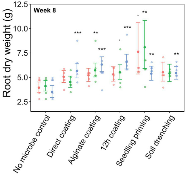
In this report, several inoculation methods ”i.e, seedling priming, soil drench, and three seed coating methods (direct seed coating, alginate seed coating, and 12-h coating)” were compared for their efficacy of delivering three different bacterial strains to sorghum under sterile and field conditions.

Three bacteria isolated from field-grown sorghum were used in this study, with Chitinophaga pinensis (Gram-negative) originating from the root endosphere while Caulobacter rhizosphaerae (Gram-negative) and Terrabacter sp. (Gram-positive) were from the soil.

Under Sterile Greenhouse Conditions:
Greater root growth-promotion was detected when inoculating C. rhizosphaerae and C. pinensis with the seedling priming compared to other inoculation methods. In fact, root growth-promotion from C. rhizosphaerae was only detectable with seedling priming despite this effect being marginally significant. For C. pinensis, significant root growth-promotion was also observed with alginate coating and marginally significant for 12 h coating. No significant root growth-promotion was measured when inoculating C. rhizosphaerae and C. pinensis with soil drench method. Among the three bacteria used, only C. pinensis and Terrabacter sp. exhibited significant shoot growth enhancement. Significant shoot growth-promotion from C. pinensis was measured when inoculated with seedling priming, alginate, and 12 h coating methods. Significant shoot growth-promotion was also observed when Terrabacter sp. was inoculated with the same seed coating methods but not the seedling priming.
Under Field Condition:
Two inoculation methods suitable for field planting, alginate and 12 h coating, were tested under field condition. C. rhizosphaerae and C. pinensis were detected in the rhizosphere up to 12 weeks after inoculation in the field, DNA copy numbers in the rhizosphere of the inoculated plants were lower as compared to that of the greenhouse experiment and not significantly different from the uninoculated control, and Terrabacter sp. was not detected in either of the sampling timepoints. This may have been because the bacterial isolate concentration used to coat the seeds (103–104 CFU per seed) was too low to facilitate their establishment in sorghum rhizosphere under non-sterile conditions where there is competition from the natural microbial communities.

Simply coating seeds with a bacterial suspension was suitable for the inoculation and successful colonization of Gram-positive bacteria in the greenhouse, whereas the field results were inconclusive. For Gram-negative bacteria direct inoculation using seedling priming led to higher colonization efficiency than seed coating.
C-Mannosyl tryptophan (CMW) could be a Good in vitro diagnostic marker for Ovarian cancer

A group from Department of Obstetrics and Gynecology, Wakayama Medical University, Wakayama 641-0012, Japan, etc. has reported on diagnostic application of C-mannosyl Tryptophan.
https://www.ncbi.nlm.nih.gov/pmc/articles/PMC6924205/

C-Mannosyl tryptophan (CMW) is a glycosylated amino acid first isolated from human urine with a unique glycan structure in which an α-mannose is bound to the indole C2 carbon of a Trp residue through a C-C linkage. CMW was also identified in human ribonuclease 2 (RNase2) as a post-translational modification.

In medical field, it was first reported that blood CMW is elevated in patients with renal dysfunction, including renal diseases associated with type 2 diabetes. In cancer biology, it was recently reported that C-mannosylation of R-spondin 2 activates Wnt/β-catenin signaling and migration activity in various human tumor cells. This study suggested that C-mannosylation of R-spondin 2 is involved in the promotion of cancer progression. Furthermore, spondin 2 (mindin), a substrate protein for C-mannosylation, is increased in the blood of ovarian cancer patients. These studies suggest that protein C-mannosylation and CMW may be involved in the pathophysiological processes of cancer progression.

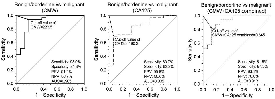
Plasma CMW in ovarian cancer was significantly higher in the malignant tumor group than in the borderline and benign tumor groups compared with healthy controls. Receiver operating characteristic curve analysis of plasma CMW distinguished malignant tumors from borderline/benign tumors (AUC=0.905). Discrimination performance was greater than that of conventional cancer antigen CA125 (AUC=0.835), and CMW + CA125 combined achieved even greater discrimination (AUC=0.913, 81.8% sensitivity, 87.5% specificity).

CMW in biological samples was analyzed and quantified by chromatographic assay. The samples were injected into an UPLC system, and CMW was quantified by measuring the fluorescence (excitation at 285 nm/emission at 350 nm).

Biocontrol activity of nonpathogenic Fusarium oxysporum strains against pathogenic Fusarium wild type strains

A group from National Agriculture and Food Research Organization, Tsu, Japan, etc. has reported on biocontrol activity of nonpathogenic Fusarium oxysporum strains against pathogenic Fusarium wild type strains.
https://www.ncbi.nlm.nih.gov/pmc/articles/PMC8828976/

The idea of using nonpathogenic F. oxysporum to control Fusarium diseases came from studies of soils naturally suppressive to Fusarium wilt disease.

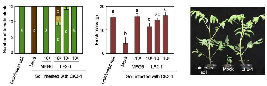
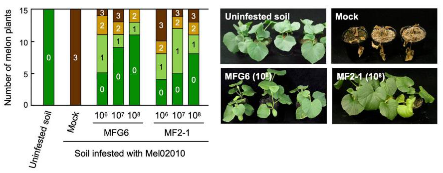
In this report, the following strains were used to demonstrate biocontrol activities of nonpathogenic mutants of F. oxysporum against pathogenic fusarium wild types.
Pathogenic Fusarium wild type strains: F. oxysporum f. sp. melonis strain Mel020120, and F. oxysporum f. sp. lycopersici strain CK3-1
Nonpathogenic Fusarium strains: F. oxysporum strain MFG6, ΔFOW2 Mel02010 MF2-1, and ΔFOW2 CK3-1 LF2-1

As shown below, biocontrol Activities of nonpathogenic Fusarium strains against the pathogenic F. oxysporum wild type strains were clearly demonstrated.

Pre-inoculation of roots with the nonpathogenic strains are quite effective. Actually, it was shown separately that conidial germination and hyphal elongation of the pathogenic fusarium wild-type strain were markedly inhibited on the root surface pre-inoculated with the nonpathogenic strains.

Glycosylation of IgG changes under the influence of cytokines (IFN-γ, IL21, IL-17A etc.)

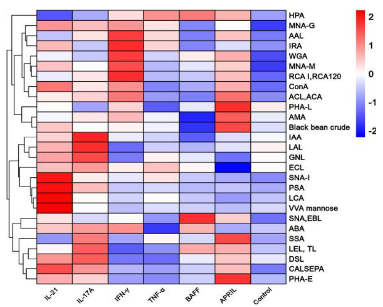
A group from Department of Endocrinology, Peking University First Hospital, Beijing, China, etc. has reported that the galactose level of IgG was increased by IFN-γ stimulation (p<0.05), and the sialylation of IgG was increased by IL-21 and IL-17A (p<0.05). This data was taken by using lectin microarrays. https://www.ncbi.nlm.nih.gov/pmc/articles/PMC8818798/

But, what does this mean?

655Y mutation, present in the SARS-CoV-2 gamma and omicron variants, is a key determinant of SARS-CoV-2 infection and transmission

A group from Department of Microbiology, Icahn School of Medicine at Mount Sinai, New York, NY 10029, USA, etc. has reported that the 655Y mutation, present in the SARS-CoV-2 gamma and now omicron variants, is a key determinant of SARS-CoV-2 infection and transmission.
https://www.ncbi.nlm.nih.gov/pmc/articles/PMC8776496/

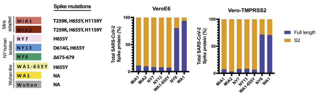
In order to evaluate the effect of H655Y mutation, five types of variants (MiA1, MiA2, NY7, NY13, and WA1-655Y) containing H655Y mutant, and two types of variants as controls (WA1 and NY6) not containing H655Y mutation were prepared.
Differences in replication and S processing of this panel of viruses were assessed by comparing growth in both Vero E6 and Vero-TMPRSS2 cells. More than 90% of the total S protein from these 655Y variants corresponded to the S2 cleaved form of the spike. In contrast, NY6 and WA1 controls showed poor cleavage efficiency.

In addition, human pneumocyte-like cells were infected with a representative panel of viruses containing the 655Y (NY7, NY13, WA1-655Y, NY6, and WA1) to assess viral growth and S protein processing. WA1-655Y demonstrated higher replication efficiency in our human airway epithelial system compared to WA1 wild type. Moreover, all isolates encoding the 655Y spike mutation exhibited enhanced spike cleavage. This demonstrates that the S:655Y polymorphism plays a crucial role in SARS-CoV-2 S protein processing and cell entry in human pneumocyte-like cells.

As a reference, it should be also noted that the spike mutations P681H in alpha variant and P681R harbored by kappa and delta variants are known to bring about enhanced Spike cleavage.

Effects of Arbuscular mycorrhizal fungi (AMF) inoculation on Maize plants

A group from Guangxi Colleges and Universities Key Laboratory of Crop Cultivation and Tillage, College of Agriculture, Guangxi University, Nanning, Guangxi, China, etc. has reported effects of Arbuscular mycorrhizal fungi (AMF) inoculation on Maize plants.
https://www.ncbi.nlm.nih.gov/pmc/articles/PMC8817564/

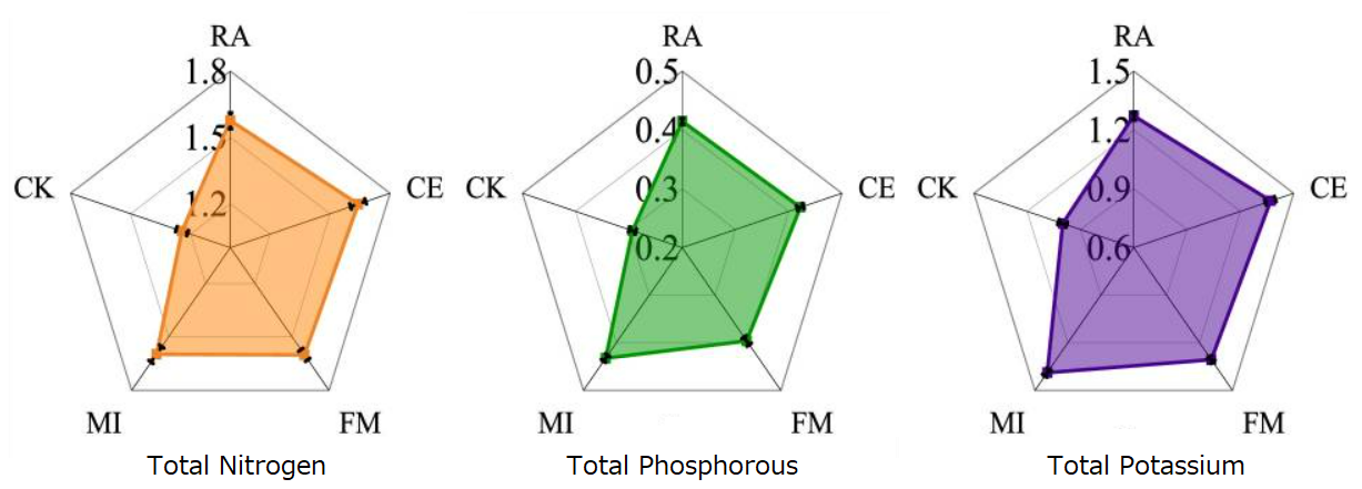
Five treatments were designed:
maize plants inoculated with Rhizophagus aggreratus were recorded as RA,
those inoculated with Claroideoglomus etunicatum were recorded as CE,
those inoculated with Funneliformis mosseae were recorded as FM,
those inoculated with three species of AMF were recorded as MI, and
those without AMF were recorded as CK.

The total N, P and K levels of the maize plants (g plant-1)were measured and analyzed during the harvest period.
It was clearly shown that the accumulation of N, P and K in MI was the highest, and the accumulation capacity of N, P and K in maize symbiosis with AMF was significantly higher than that in CK as shown below.

Different AMF inoculations had different effects on maize root exudates at different growth stages. In this experiment, 10 kinds of organic acids were isolated from maize root exudates.
From the total level of organic acids secretion in the whole maize growth period, the followings were found,
FM promoted the secretion of p-hydroxybenzoic, p-coumaric and caffeic acid,
CE promoted the secretion of syringic acid,
RA promoted the secretion of chlorogenic and succinic acid, and
the levels of protocatechuic, vanillic, citric and ferulic acid were lower than those of CK.

Some studies have shown that p-hydroxybenzoic acid can reduce the number of Fusarium and the occurrence of Fusarium wilt, vanillic acid can inhibit soil-borne pathogens and reduce soil-borne diseases, vanillic acid can inhibit the growth of Fusarium wilt, caffeic acid plays an important role in inhibiting soil-borne pathogens, caffeic acid can directly inhibit the growth of Ralstonia solanacearum, and ferulic acid has strong allelopathy and can inhibit the growth of plant roots.

So, AMF may alleviate soil-borne diseases by increasing the levels of p-hydroxybenzoic, vanillic and caffeic acid in plant root exudates. The role of these organic acids in maize growth and their impact on maize growth after AMF symbiosis must be further studied.

The severity rate of SARS-CoV-2 Omicron is lower than that of Delta, but is almost the same as that of ancestral Wuhan

A group from Division of Infectious Diseases, Department of Medicine, Johns Hopkins University School of Medicine, Baltimore, MD, USA, etc. has reported about inpatient clinical outcome of SARS-CoV-2 variants including Omicron.
https://www.ncbi.nlm.nih.gov/pmc/articles/PMC8820675/

Although unvaccinated patients hospitalized with Omicron variant had a lower risk of developing severe disease or death compared to those with Delta variant, the risk of severe disease or death was similar compared to SARS-CoV-2 lineages that circulated earlier in the pandemic. Among vaccinated patients, there was no difference in the risk of developing severe illness between Delta and Omicron variants once patients were hospitalized.

In concrete terms,

Among unvaccinated patients,
The percentages of developing severe disease or death within 14 days of hospitalization were (27%) ancestral, (31%) Alpha, (32%) Delta, (26%) Omicron, and (26%) other variants.
The relative risk of developing severe disease or death within 14 days for Delta variant compared to ancestral lineages was 1.34. Compared to Delta variant, the 14-day relative risk of severe disease or death for Omicron was 0.78 and compared to ancestral lineages was 1.04 (almost the same).

Among vaccinated patients,
The adjusted hazard ratio developing the 14-day risk of severe disease or death of inpatients who were vaccinated was 0.46 (almost half) compared to unvaccinated inpatients. Severe outcomes were less common in vaccinated patients, but there was no difference between Delta and Omicron infections.

Rhizosphere microbiota of Fritillaria ussuriensis (in the lily family Liliaceae) at different health levels

A group from College of Forestry, Northeast Forestry University, Harbin, China, etc. has reported about changes in rhizosphere microbiota of Fritillaria ussuriensis at different health levels.
https://www.ncbi.nlm.nih.gov/pmc/articles/PMC8796711/

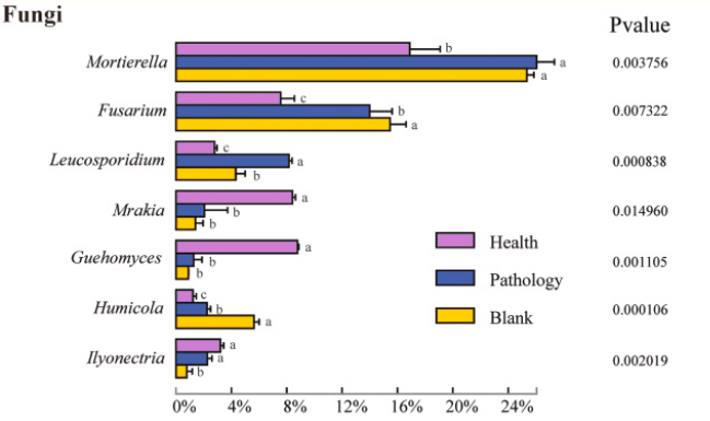
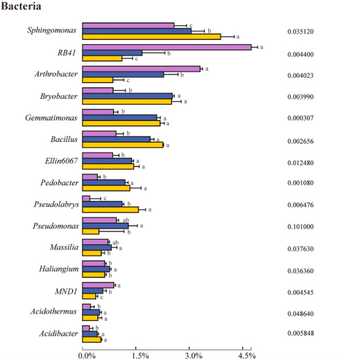
The difference in the composition of the soil microbial community at different health levels of Fritillaria ussuriensis, the top 7 fungal and 15 bacterial genera with high abundances were selected from the fungal and bacterial communities in healthy(H), pathologic(P), and blank(B) samples.

The difference in the relative abundance at the genus level indicated that Mortierella, Fusarium, Leucosporidium, Mrakia, Guehomyces, Humicola, and Ilyonectria were members of the fungal genera with higher abundances, among which Mortierella exhibiting the highest abundance (22.86%). Compared with the healthy sample, the abundance of Mortierella increased significantly in diseased samples (P and B). The abundances of Fusarium and Humicola increased significantly with increasing the severity of disease (H →P →B), with the blank sample having the highest abundance (15.49% and 5.60%, respectively). On the contrary, the abundances of Mrakia and Guehomyces decreased significantly, with the highest abundance in the healthy sample and more abundance and variation in the bacterial community compared with the fungal community.

In the case of rhizobacteria, the highest abundances of RB41 (4.74%) and Arthrobacter (3.30%) were found in the healthy sample. With increasing the severity of disease (H →P →B), the abundance decreased significantly. The relative abundances of Sphingomonas, Bryobacter, Gemmatimonas, Bacillus, Ellin6067, Pedobacter, Acidothermus, and Acidibacter in diseased samples (P and B) were significantly higher than that in healthy samples. Furthermore, the highest abundances of Bryobacter, Acidibacter, Pseudomonas, Massilia, and Haliangium were found in pathologic samples.

Glycan profiling analysis of serum NRG1 in papillary thyroid cancer with using Lectin Microarrays

A group from Department of Head and Neck Surgery, Fudan University Shanghai Cancer Center, Shanghai, China, etc. has reported on glycan profiling analysis of serum NRG1 in papillary thyroid cancer with using Lectin Microarrays.
https://www.ncbi.nlm.nih.gov/pmc/articles/PMC8798758/

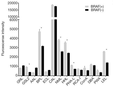
This study is evaluating if BRAF V600E mutant status in papillary thyroid cancer (PTC) patients could be detected by analyzing multiple glycan profiles of serum NRG1 through lectin microarray assays.

It has been reported that NRG1 overexpression was related in various cancers. NRG1 protein can bind to ERBB3 or ERBB4 protein, activating ERBB receptor tyrosine kinases, followed by the initiation of signal cascades, including PI3K/AKT pathways. NRG1 can be secreted to serum as reported.

lectin microarrays were used to analyze the different glycan profiles of NRG1 from sera of BRAF(+) PTC and BRAF wild type(-) PTC patients. Lectin microarray is a rapid and sensitive high-throughput technique to get glycan profiles, allowing researchers to directly analyze glycoproteins without liberation of glycans from the core substrate.

It was found that LEL, BPL and NML tended to have bound to NRG1 in BRAF(+) PTC patients compared to BRAF(-) controls.

Rhizosphere Biofilm: Characteristics of Mucigel exuded from plant roots and EPS exuded from Rhizobacteria

A group from Division of Biogeochemistry of Agroecosystems, Georg-August University of Göttingen, Göttingen, Germany, etc. has reported on Biofilm Matrix in the Plant Rhizosphere.
https://www.ncbi.nlm.nih.gov/pmc/articles/PMC8792611/

Roots exude a diverse set of compounds into the rhizosphere, including mucigel, amino acids, and secondary metabolites, which regulate rhizosphere functions. Mucigel is a gelatinous high-molecular-weight substance produced by almost all plants. The mucigel backbone is know to be built of polysaccharides, but proteins, minerals, and lipids are also part of the biogel. Rhizobacteria also exude extracellular polymeric substances (EPS). EPS are mainly composed of polysaccharides, but also contain proteins, nucleic acids, lipids, and minerals, and forms biofilms on root surfaces functioning as comfortable place to live. Thus, the polysaccharides were the major chemical constituent of both biogels (77.4% and 74.6% for mucigel and EPS, respectively) and did not significantly differ between them.

The polysaccharide backbone did not significantly differ in proportion between mucigel and EPS, namely galactose (mucilage = 23.8%; EPS = 22.8%), fucose (13.9%; 9.9%), glucose (16.7%; 28.7%), rhamnose (12.4%; 15%), xylose (13.4%; 8.1%), and glucuronic acid (8%; 12.8%). In contrast, mannose (3.9%; 18.6%) was significantly higher in EPS than in Mucigel (nearly fivefold higher), whereas arabinose (16.3%; 4.8%) and galacturonic acid (27.3%; 7.8%) had higher proportions (3.4-fold and 3.5-fold higher, respectively) in Mucigel than in EPS.

Alginate is an anionic polysaccharide found in EPS, consisting of only uronic acids such as glucuronic acid, galacturonic acid, and mannuronic acid. Alginate participates in the formation of microcolonies at the beginning of the biofilm formation process, increases EPS hydration, and assists in trapping cations such as Ca2+, Zn2+, Cd2+, and Ni2+. The high proportion of galacturonic acid in mucigel is likely one of the major reasons for the higher water absorption capacity of mucigel than EPS.

Mucigel can also be decomposed and consumed by microorganisms. Enzymatic release of highly abundant sugars in mucigel such as galactose, fucose, and arabinose can feed microorganisms residing in the mucigel. The presence of endogenous glycosyl hydrolase enzymes in mucilage augments this claim. It seems that microorganisms utilize mucigel as an energy source, with average times of 7–15 days for the consumption of 50% of the mucigel carbon added to the soil. The high protein content of mucigel leads to a C:N ratio of approximately 16:1, which is approximately double the C:N ratio of microorganisms. Thus, considering that 50% of the C is utilized via catabolism and oxidized to gain energy, mucigel has the ideal composition to function as a sole energy, C and N source for microorganisms. Consequently, microorganisms solely need to be supplied with mineral nutrients (P, K, Ca, Mg, etc.)—a common nutrients shared with their mucigel-providing plants.

Powered by WordPress |Copyright © 2020 Emukk. All rights reserved