Archive 21/4/18

Differences in SARS-CoV-2 infectivity between M1 alveoli macrophages and M2 macrophages 

A group from Peking Union Medical College, etc. has reported on differences in SARS-CoV-2 infectivity between M1 macrophages and M2 macrophages including alveolar epithelial type II (AT2) cells.
https://www.nature.com/articles/s41421-021-00258-1

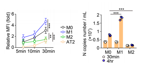
Generally speaking, M1 macrophages are inflammatory and M2 macrophages are anti-inflammatory. Comparing AT2, M1 macrophages, and M2 macrophages, SARS-CoV-2 infection and its replication was higher in macrophages than AT2, M1 macrophages took up SARS-CoV-w2 with higher efficiency than M2 macrophages, and the viral loads increased in an exponential fashion in M1 macrophages but in a flat fashion in M2 macrophages. It is considered that ACE2 could be the receptor that mediates the infection with SARS-CoV-2, and ACE2 has been reported to be expressed also by macrophages. However, there were no difference in the infectivity between ACE2-overexpressing macrophages and ACE2-knockeddown macrophages. So, virus takeup through phagocytosis could initiate SARS-CoBV-2 infection.
Authors related the difference in the infectivity between M1 and M2 to difference in cell softness, endosomal, and lysosomal pH.
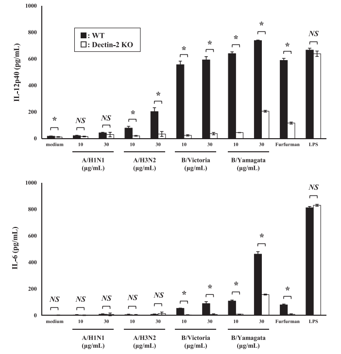
Dectin-2 is related to Infection of Influenza virus and the induction of inflammatory responses

A group from Niigata University, etc. has reported that Dectin-2 is related to Infection of Influenza virus and the induction of inflammatory responses.
https://www.jstage.jst.go.jp/article/biomedres/42/2/42_53/_pdf/-char/en

Although the importance of relationship between hemagglutinin (HA) and sialic acids has been emphasized, agents targeting HA and sialic acids are not effective in suppressing severe influenza unless they are administered within 48 h after symptoms begin. Antigen-presenting cells (APCs), such as dendritic cells (DCs) and macrophages, recognize pathogen-associated molecular patterns (PAMPs) and damage-associated molecular patterns (DAMPs) of dead cells or damaged tissues, and activate inflammatory immune responses. Toll-like receptors (TLRs), Nod-like receptors (NLRs), and C-type lectin receptors (CLRs) are known as such pattern recognition receptors.

Since HA is strongly high mannosylated, there must be a signal path activating inflammatory immune responses through the interaction between C-type Lectins and glycosylated. C-type Lectin family includes DC-SIGN, Dectin-1, Dectin-2, Mincle, etc. Authors have found that Dectin-2 expressed on BM-DCs recognizes glycosylated HA from the type A and type B strains, and induce production of inflammatory cytokines. Dectin-2 recognized high mannose polysaccharides.
A figure below shows that production of inflammatory cytokines is greatly reduce by knocking out Dectin-2.

Characteristics of neutralizing antibodies obtained from SARS-CoV-2-infected donors

A group from King’s College London, etc. has reported on characteristics of neutralizing antibodies obtained from SARS-CoV-2-infected three donors.
https://www.ncbi.nlm.nih.gov/pmc/articles/PMC8015430/pdf/main.pdf

Authors have isolated total 107 SARS-CoV-2 Spike-specific mAbs from three SARS-CoV-2-infected donors, P003 (hospitalized and spent time in ICU), P054 (symptomatic but no hospitalization), and P008 (SARS-CoV-2 infected, but asymptomatic). In total 107 mAbs, 35.5% of the Spike reactive mAbs were RBD-specific, 32.7% were NTD specific, 0.9% bound S1 only, and 30.8% mAbs only bound Spike. 43.9% of total 107 mAbs showed neutralizing activity, and the majority about 70% of the neutralizing Abs (nAbs) targeted RBD and about 20% of nAbs targeted NTD.

Overall, highly potent nAbs targeting RBD and NTD had similar titers, IC50 ranged from 2.3 to 488ng/mL.
RBD-specific mAb P008_108 showed the most potent anti-SARS-CoV-2 neutralizing activity, IC50 of 2.3 ng/mL, and P008_056 neutralized SARS-CoV-2 with an IC50 of 14 ng/mL making this one of the most potent NTD nAbs reported thus far.
It is very curious to know that the nAbs with higher neutralizing activity were obtained from asymptomatic donor.

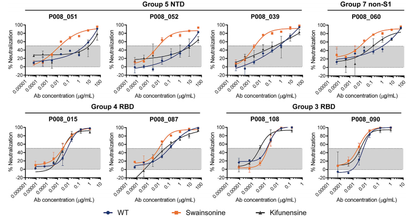
Since NTD is heavily glycosylated than RBD, the effect of glycosylation onto nAbs were studied by using glycosidase inhibitors such as kifunensine and swainsonine (see figure below). These results suggest that glycan structures can affect nAb epitope recognition either through modulating the conformation of Spike or altering the accessibility of nAb epitopes.

IL-33 expression in COVID-19 covalescent individuals correlates with seropositivity of SARS-CoV-2 specific IgG

A group from Max Planck Institute of Immunobiology and Epigenetics, etc. has found that IL-33 expression in COVID-19 covalescent individuals correlates with seropositivity of SARS-CoV-2 specific IgG.
https://www.nature.com/articles/s41467-021-22449-w

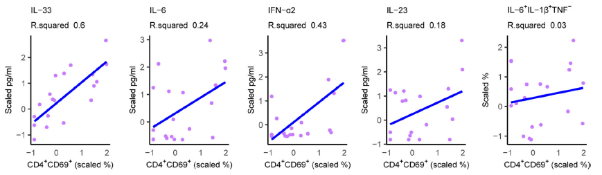
Authors purified PBMCs from COVID-19 covalescent 20 individuals and examined their composition using conventional CD markers, and monitored cytokine production with activation following SARS-CoV-2 Spike stimulation. It was found that IL-33 was most strongly correlated with CD4+CD69+ T-cells among cytokines investigated here, IL-33, IL-6, IFN-ɑ2, and IL-23. IL-33 has a high homology amino acid sequence with IL-1β and IL-18 and belongs to the IL-1 family, but its detailed characteristics are still unknow. Authors have investigated the source of IL-33 in PBMCs by staining for intracellular IL-33 in conjunction with cell-specific markers, and found that it was most highly expressed in CD14+ monocytes. However, there were no differences in the frequency of IL-33+ cells in PBMC from seropositive vs. seronegative individuals, suggesting that the emergence of COVID-19-specific T cells that are capable of eliciting the release of bioactive IL-33 from other cells that constitutively produce this cytokine. These results suggest that IL-33 might be deeply linked to COVID-19 pathogenesis and immunity.

A polyphenol from green tea (GCG) could inhibit SARS-CoV-2 replication effectively

A group from Fudan University, etc. has shown that Gallocatechin gallate (GCG), a polyphenol from green tea, inhibits SARS-CoV-2 replication efficiently.
https://www.nature.com/articles/s41467-021-22297-8

SARS-CoV-2 N protein is a structural protein binding to RNA, and form a shell embracing SARS-CoV-2 RNA. This is a typical example of so called Liquid-Liquid Phase Separation (LLPS) which is a mechanism in organizing macromolecules such as proteins and RNAs into membrane-less oil droplet like organelles.
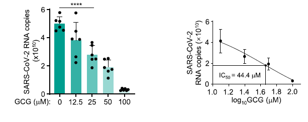
Authors has shown that GCG could could inhibit N protein LLPS in the context of SARS-CoV-2 infection effectively. Since, the amino acid sequence shares ~90% homology among corona viruses, targeting N protein by GCG could be a novel drug candidate not only for SARS-CoV-2 but also for new coronaviruses in the future.

Effects of glycosylation and disulfide bonding of SARS-CoV-2 Spike protein

A group from Harvard Medical School, etc. has reported on the effects of glycosylation and disulfide bonding of SARS-CoV-2 Spike protein onto infectivity and susceptibility to antibody inhibition.
https://pubmed.ncbi.nlm.nih.gov/33821278/

There are 22 N-glycosylation sites, 10 O-glycosylation sites and 15 disulfide bonding in the spike protein.
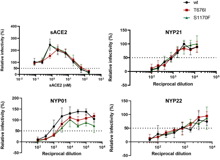
In this paper, it was shown experimentally how the mutations at O-glycosylation sites(676, 1170)affect binding to ACE2 and titer of sera from convalescing SARS-CoV-2-infected individuals. Let me introduce that point for this blog readers. The evaluated mutations were T676I and S1170F. As shown below, it seems that these mutations do not affect so much.

A key why children do not develop severe COVID-19 is in strong innate immune response in nasopharyngeal mucosa

In the case of influenza or respiratory syncytial virus, children are often more severe than adults. However, in the case of COVID-19, children infected with SARS-CoV-2 have a milder clinical course with significantly less morbidity and mortality than adults.
Several hypotheses have been proposed to explain why children are more protected than adults, for example, expression of angiotensin-converting enzyme 2 (ACE2) is lower in children, resulting in lower viral loads, presence of antibodies to common cold coronaviruses (229E, NL63, HKU1)that might provide partial protection is stronger in children, and a more robust innate response in children in early course of infection that mitigates against a vigorous adaptive response.

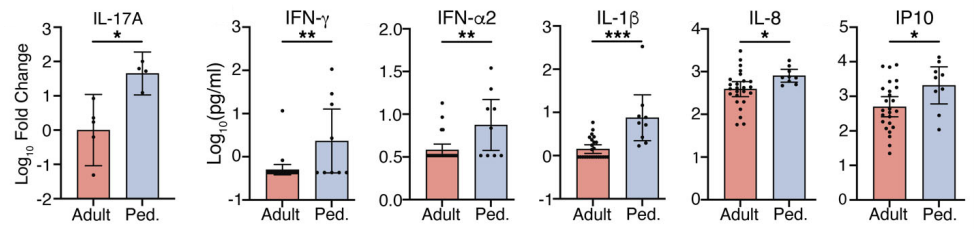
A group from Albert Einstein College of Medicine, etc. has discovered that nasopharyngeal mucosal immune response is stronger in children. It is speculated that the immune response is strengthened in nasopharyngeal mucosa because children have more frequent respiratory infections than adults. Measured cytokines in nasopharyngeal swab fluid (IFN-γ, IFN-α2, IL-1β, IL-8) was significantly upregulated in children than adults. So, the robust mucosal immune response lowers viral loads and gives protection against advancing in severity.
https://insight.jci.org/articles/view/148694

IL-1R7 antibody would be effective in COVID-19 to suppress cytokine storms

A group from University of Colorado Denver Anschutz Medical Campus has reported that IL-1R7 antibody would be effective in COVID-19 to suppress cytokine storms.
https://www.jbc.org/article/S0021-9258(21)00416-6/fulltext

Excessive inflammation observed in macrophage activation syndrome (MAS) results in severe diseases with high mortality. The cytokine storms observed in COVID-19 patients would be a typical example similar to MAS.
Interleukin-18 (IL-18), a proinflammatory cytokine belonging to the IL-1 family, is elevated in both MAS and COVID-19 patients, and its level is known to correlate with the severity of COVID-19 symptoms

IL-18 binds its specific receptor IL-1 Receptor 5 (IL-1R5, also known as IL-18 Receptor alpha chain), leading to the recruitment of the co-receptor, IL-1 Receptor 7 (IL-1R7, also known as IL-18 Receptor beta chain).

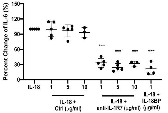
Authors found that the anti-IL-1R7 antibody significantly suppressed IL-18-mediated NFκB activation, IL-18-stimulated IFNγ production, and IL-6 production in human cell lines.

Seroprevalence of anti-SARS-CoV-2 IgG antibodies in COVID-19 patients 

A group from Juntendo University Faculty of Medicine has reported seroprevalence of IgG and IgM antibodies in COVID-19 patients, although the cohort size was small including only 34 patients.
https://www.ncbi.nlm.nih.gov/pmc/articles/PMC8023454/

Using a chemiluminescent microparticle immunoassay (CMIA)-based SARS-CoV-2 IgG test (cat. # 06R90, Abbott),
Severe/Critical cases:within a week after symptom onset=40%、1~2 weeks=88%、after two weeks=100%,
Mild/Moderate cases:within a week after symptom onset=0%、1~2 weeks=38%、after two weeks=100%.

Using an IC IgG antibody assay using the Anti-SARS-CoV-2 Rapid Test (cat. # RTA0203, AutoBio),
Severe/Critical cases:within a week after symptom onset=60%、1~2 weeks=63%、after two weeks=100%,
Mild/Moderate cases:within a week after symptom onset=17%、1~2 weeks=63%、after two weeks=100%.

In this study, IgG titers remained at significantly elevated levels for 2 months, regardless of disease severity. These results indicate that IgG serologic tests could be used as a complementary test to PCR to diagnose COVID-19 from 14 days after symptom onset. However, since this cohort is so small without including asymptomatic individuals, a larger scale cohort is needed to conclude final answer.
     

Difference in site-specific glycosylation of SARS-CoV-2 spike proteins (all recombinants) among several laboratories

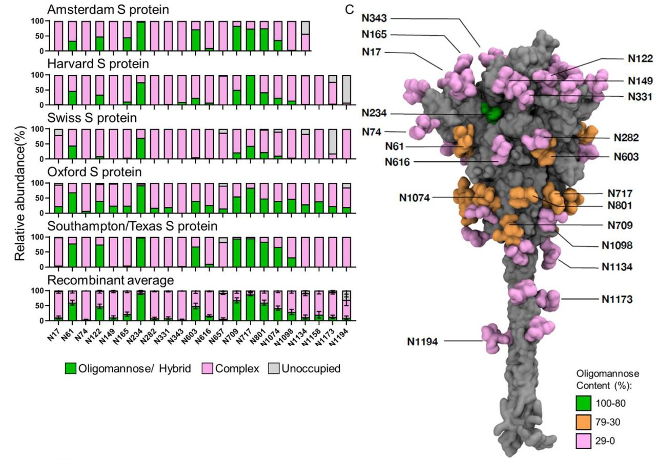
A group from University of Southampton, etc. has compared site-specific glycosylation of SARS-CoV-2 spike proteins (all recombinants) among five laboratories.
The cells used for Sproten expression were as follows.
HEK293: Amsterdam, Harvard,
HEK293F: Southampton/Texas,
HEK293T: Oxford,
CHO: Swiss,
It is celarly shown that site-specific glycosylation changes considerablly with reflecting differences in cells and culture conditions.
Fundamentally speaking, it is a mixture of oligo mannose and complex type N-glycans.

Blog admin is interested in how much these differences cause difference in the infectivity, how glycosylation changes with SARS-CoV-2 mutations, and how much the glycosylation changes due to mutations affect the infectivity.
https://www.biorxiv.org/content/10.1101/2021.03.08.433764v1.full

Powered by WordPress |Copyright © 2020 Emukk. All rights reserved