Archive 21/5/16

Characteristics of the lung microbiome of COVID-19 patients 

A group from IRCCS Azienda Ospedaliero-Universitaria di Bologna, Italy has reported on differences in lung microbiome between COVID-19 patients and COVID-19 negative persons.
https://www.nature.com/articles/s41598-021-89516-6

Several indexes have been developed to discuss about similarity among groups and characteristics of each group.
Similarity Percentage analysis highlighted that the lung microbiome of COVID-19 patients was characterized by a higher relative abundance of Pseudomonas spp. compared to COVID-19-negative persons. On the other hand, the lung microbiome of COVID-19-negative persons was mainly characterized by the enrichment of lung commensal bacteria (such as Haemophilus influenzae, Veillonella dispar, Granulicatella spp., Porphyromonas spp., and Streptococcus spp).

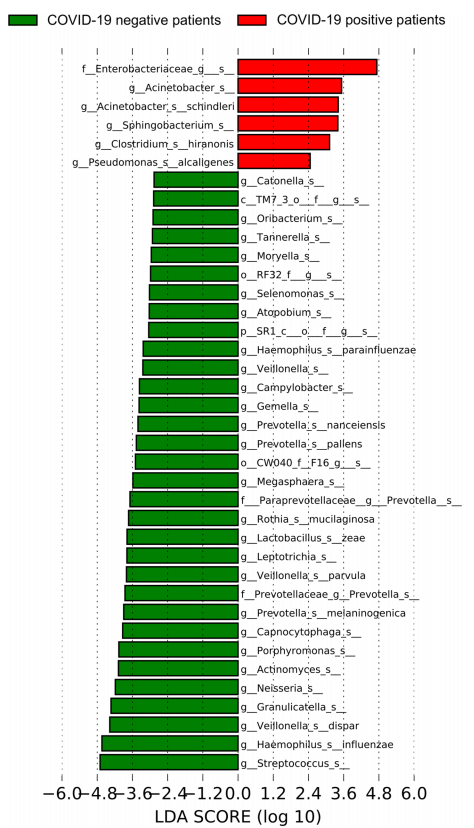
The linear discriminant analysis (LDA) highlighted that the lung microbiome of COVID-19 patients was characterized by the presence of Pseudomonas alcaligenes, Sphingobacterium spp., Clostridium hiranonis, Acinetobacter schindleri, Enterobacteriaceae of unknown genus and Acinetobacter spp.

A common feature between these two analysis indexes was Pseudimonas spp. This means that enrichment of opportunistic gram-negative pathogens frequently associated with multidrug resistance increase in COVID-19 patients and the lung commensal bacteria decrease inversely.
Changes in the expression levels of ACE2 and TMPRSS2 with agonists for Toll-like receptors (TLRs)

A group from Hokkaido University, etc. has reported on changes in the expression levels of ACE2 and TMPRSS2 with agonists for Toll-like receptors (TLRs) and with Fluticasone Propionate (FP)
https://www.ncbi.nlm.nih.gov/pmc/articles/PMC8107375/

It is said that SARS-CoV-2 cell entry depends on two proteins present on the surface of host cells, angiotensin-converting enzyme 2 (ACE2) and transmembrane protease serine 2 (TMPRSS2). Authors has investigated effects of activation of innate immune signal path onto the expression levels of ACE2 and TMPRSS2, and also if there is any effect of FP administration on that.

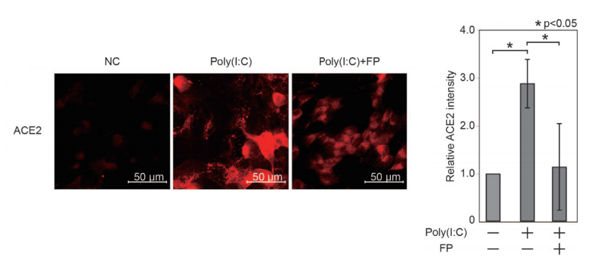
As sample cells, primary human nasal epithelial cells (HNECs) were collected from the nasal mucosa. HNECs were stimulated by Poly(I:C), which is a double-stranded RNA (dsRNA) often used as a agonist for TLRs in a model of viral infection.

ACE2 and TMPRSS2 protein expression levels were evaluated by using immunofluorescent analysis. The ACE2 intensity in HNECs was significantly promoted by Poly(I:C) (a 2.884±0.505-fold change vs. untreated cells, p=0.003), but there were no significate changes in the TMPRSS2 expression levels. And, this Poly(I:C)-induced change in ACE2 was significantly suppressed by FP (a 0.405±0.312-fold change vs. Poly(I:C)-treated cells, p=0.044).

Further studies are required to evaluate whether FP suppresses SARS-CoV-2 viral cell entry in vivo.

Passive immunity of infants born to mothers infected with SARS-CoV-2 

A group from Stanford University School of Medicine, etc. has reported about passive immunity of infants born to mothers infected with SARS-CoV-2.
https://pubmed.ncbi.nlm.nih.gov/33972953/

An important aspect of immunity against infectious pathogens in young infants relies on effective maternal antibody production, transfer of maternal antibodies across the placenta to the fetus, and persistence of passive immunity in the infant. Authors have investigated SARS-CoV-2 antibody transplacental transfer ratios with respect to the timing of maternal infection during gestation, antibody response to SARS-CoV-2 infection in the newborns, and persistence of passively- and actively-acquired SARS-CoV-2 antibodies in infants.

The study enrolled 145 mothers with SARS-CoV-2 infection and 147 of their infants. There was a significant positive correlation between IgG levels in the 125 paired maternal and cord blood samples (Rs=0.93, p<0·0001). Transplacental IgG transfer ratios were calculated in 77 IgG positive mother-infant dyads, and the median transfer ratio was 1.0 (95% CI 0.86-1.09). The transfer ratio was significantly higher in the mothers who were severe-critically symptomatic (n=4) compared to mothers who were asymptomatic (n=23) (1.6 vs. 1.0, p=0.003) or mild-moderately symptomatic (n=50) (1.6 vs. 0.9, p=0.002). The transfer ratios based on time elapsed from the first maternal positive PCR to delivery were 0.6 (<60 days, n=22), 1.2 (60-180 days, n=27), and 0.9 (>180 days, n=5). These studies demonstrate that cross-placental SARS-CoV-2 IgG transfer occurs throughout gestation, and a higher transfer efficiency is achieved when infection onset is more than two months prior to delivery. These findings have important implications in determining optimal timing of vaccination in pregnant mothers and infants. 

Gal-9 could be a quite good diagnostic marker specificity/sensitivity (95%) for COVID-19 

A group from University of Alberta, Canada, etc. has reported that Gal-9 could be a quite good diagnostic marker for COVID-19.
https://mbio.asm.org/content/12/3/e00384-21.long

Innate immune cells recognize and respond to a wide variety of pathogens. For example, activated macrophages/monocytes secrete excessive amounts of cytokines including IFN-γ, IL-1, IL-6, TNF-α, and IL-18. Similarly, neutrophils produce neutrophil extracellular traps and NK cells prolong the antigenic stimulation due to diminished/skewed cytolytic functions, both of which can amplify cytokine production. Overall, it appears that the viral burden, accompanied by the dysregulated innate immune response due to underlying disease, and aging may ignite the cytokine storm.

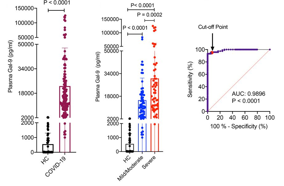
Galectins are involved in many biological functions such as development, signal transduction, and immune responses. This study was performed in a cohort of 120 SARS-CoV-2-infected individuals and 59 healthy controls (HCs) to understand how galectins, especially focusing on Gal-9, could be positioned in COVID-19.

Significantly higher levels of Gal-9 in the plasma of COVID-19 patients were observed comparing to HCs (ranging between 0 and 2,042 pg/ml). The plasma Gal-9 concentrations were substantially greater in severe cases (ranging between 1,950 and 125,510 pg/ml) comparing to those with mild/moderate cases (ranging between 1,000 and 83,717 pg/ml). From a ROC curve, quite high specificity/sensitivity (95%) to discriminate COVID-19 from either HCs or patients with HIV and virus-associated cancers with a cutoff value of 2,042 pg/ml. It is quite interesting to know that the plasma Gal-9 concentration in COVID-19 patients surpasses the detectable levels reported in other conditions such as HIV, dengue fever, influenza, and virus-associated solid tumors.

Through a series of experiments to identify what cells secrete Gal-9 and to understand how Gal-9 activates innate immune cells contributing to the cytokine storm, the following model was proposed as a conclusion. The damaged lung epithelial cells following SARS-CoV-2 infection release Gal-9, which activates alveolar macrophages, resulting in the secretion of proinflammatory cytokines and Gal-9 from the activated or apoptotic cells. Subsequently, Gal-9 activates monocytes and other immune cells, orchestrating another wave of proinflammatory cytokines and Gal-9 release, and exacerbates the cytokine storm further.

A positive result from a Phase 3 clinical trial of Lenzilumab against COVID-19 

A group from Mayo Clinic, etc. has reported a promising result of a phase 3 clinical trial of Lenzilumab against COVID-19.
https://pubmed.ncbi.nlm.nih.gov/33972949/

In COVID-19, it has been known that high levels of granulocyte-macrophage colony-stimulating factor (GM-CSF)-secreting T-cells are associated with disease severity, myeloid cell trafficking to the lungs, and ICU admission. GM-CSF is a kind of cytokine which activates differentiation of myeloid cells, leading to elevations of downstream inflammatory chemokines (MCP-1, IL-8, IP-10) and cytokines (IL-6, IL-1).

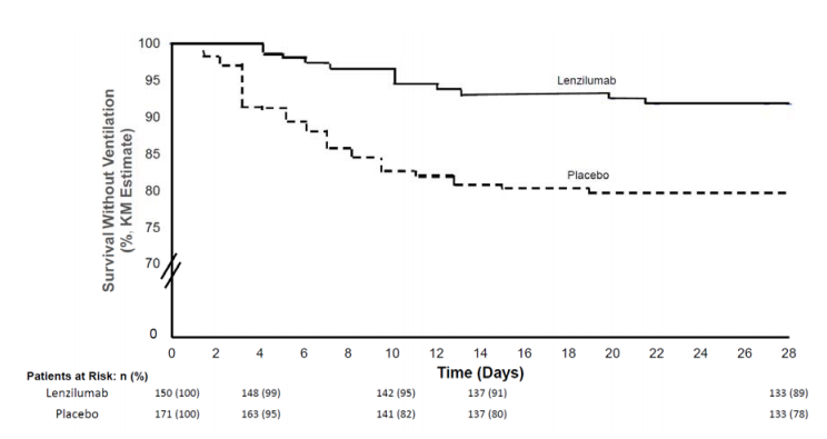
Lenzilumab is a novel anti-human GM-CSF monoclonal antibody that directly binds GM-CSF and prevents signaling through its receptor. It has high binding affinity (25 pM) for glycosylated human GM-CSF and a slow off-rate. A phase 3 randomized, double-blind, placebo-controlled clinical trial was designed to demonstrate the effectiveness of lenzilumab. The dose of lenzilumab was 600mg and administered 8hours apart for 28 days.

Lenzilumab could improve the likelihood of survival without ventilation (SWOV) through Day 28 by 54%. It should be noted that the improvement in SWOV was most evident in patients with CRP<150 mg/L who are less than 85 years of age.

Characteristics of antibody responses in COVID-19 asymptomatic individuals 

A group from National Institute of Metrology, Beijing, China, etc. has reported characteristics of antibody responses in COVID-19 asymptomatic individuals.
https://www.nature.com/articles/s41392-021-00596-2

Over 40% of individuals undergo asymptomatic infection without showing any symptoms. It is considered that these asymptomatic individuals can efficiently transmit viral infection accounting for more than 30% of virus infection

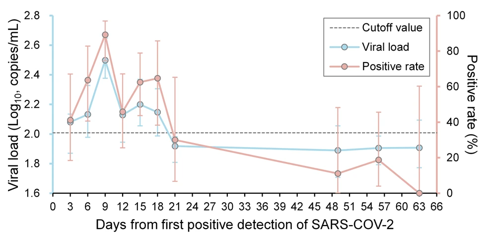
A total of 143 asymptomatic individuals with SARS-COV-2 were recruited in the study. The viral loads of SARS-CoV-2 based on N gene in saliva peaked at Day 9 of the first detection (315.1 copies/mL, 95% CI 238.1–417.1), followed by gradual decrease and from Day 21 detection was below the cut-off value (102 copies/mL) in a figure below. The positive rate of SARS-CoV-2 detection showed the same trend with the viral loads.

It is found that the antibody responses are short-lived lasting only about 69 days.

Increased risk of SARS-CoV-2 infection in Diabetes Mellitus patients would be related to the increased expression levels of ACE2 

A group from University of Campania L. Vanvitelli, Naples, Italy has reported the relationship between expression levels of ACE2 and increased risks of SARS-CoV-2 infection to cardiomyocytes due to Diabetes Mellitus
https://cardiab.biomedcentral.com/articles/10.1186/s12933-021-01286-7

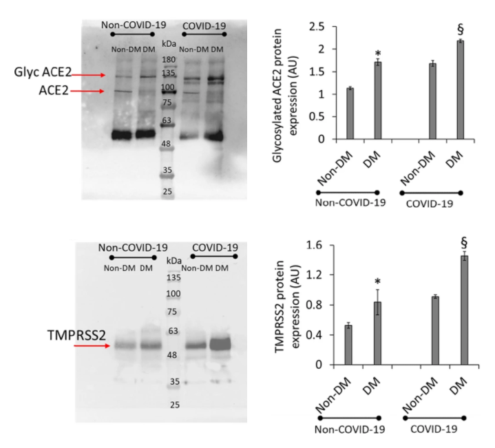
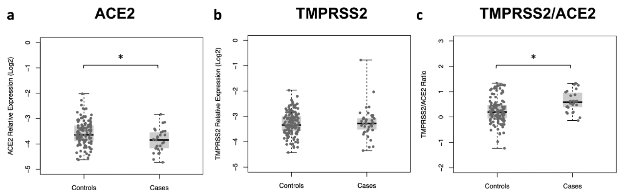
Comparing diabetes mellitus (DM) patients and healthy individuals, the expression levels of ACE2 and TMPRSS2 were higher in DM patients than non-DM individuals significantly (see a figure below).
Further, comparing COVID-19 patients and non-COVID-19 individuals, the expression levels of ACE2 and TMPRSS2 were much higher in COVID-19 patients.
As a result, the increased risk of SARS-CoV-2 infection in DM patients would be related to the increased expression levels of ACE2.

What relationship is there among the expression levels of ACE2, TMPRSS2, and severity of COVID-19?

A group from Universidade Federal do Rio de Janeiro, etc. has reported the relationship among the expression levels of ACE2, TMPRSS2, and severity of COVID-19.
https://www.nature.com/articles/s41598-021-88944-8

ACE2 and TMPRSS2 are known to be key players on SARS-CoV-2 entry into host cells. However, it is still unclear whether expression levels of these factors could reflect disease severity. The angiotensin-converting enzyme 2 (ACE2) has been described as the entry receptor for SARS-CoV-2 and the transmembrane serine protease 2 (TMPRSS2) as an important priming enzyme required during this process.

A case–control study was carried out including 213 individuals with SARS-CoV-2 infection confirmed by RT-qPCR in nasopharyngeal samples.
Expression of both genes of ACE2 and TMPRSS2 was positively correlated to age (r = 0.20; p = 0.03 and r = 0.21; p = 0.01 for ACE2 and TMPRSS2, respectively), while no association with sex was observed.
Transcription levels of ACE2 were significantly lower among cases than in controls, while similar levels of TMPRSS2 were observed in both groups. However, TMPRSS2/ACE2 ratios were significantly higher among cases than those of controls. From blog admin’s point of view, It is interesting but quite difficult to understand that the expression levels of ACE2 showed a protective effect for the infection, no significant association between the levels of TMPRSS2 and the infection, but TMPRSS2/ACE2 ratio could be a risk factor on COVID-19 severity, suggesting that other infection paths would be behind the ACE2/TMPRSS2 infection path.


Vaccine effectiveness of Pfizer-BioNTech or Moderna vaccine against COVID-19

CDC COVID-19 Response Team has reported about the vaccine effectiveness of Pfizer-BioNTech or Moderna vaccine.
https://www.cdc.gov/mmwr/volumes/70/wr/pdfs/mm7018e1-H.pdf

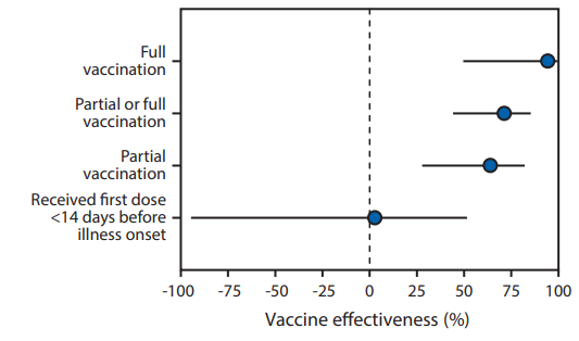
The team has reported that adjusted vaccine effectiveness for full vaccination using Pfizer-BioNTech or Moderna vaccine was 94% (95% CI = 49%–99%), and that for partial vaccination was 64% (95% CI = 28%–82%).
In this study, the vaccine effectiveness was assessed among 417 adults (including 187 case-patients and 230 controls) aged ≥65 years who admitted with COVID-19–like illness and confirmed by RT-PCR during January 1, 2021–March 26, 2021. Case-patients were those who received one or more positive test results for SARS-CoV-2. Patients meeting eligibility criteria who received negative SARS-CoV-2 RT-PCR test results served as controls. Participants in this study were considered to have received COVID-19 vaccine doses based on official CDC vaccination record card etc. and by plausible self-report.

It should be noted that there was no significant effect for receiving the first dose of a 2-dose COVID-19 vaccine series within 14 days before illness onset (adjusted vaccine effectiveness = 3%, 95% CI = −94%–51%)

Using lectin–glycan interactions to combat COVID-19 reviewing past instances against enveloped viruses

A group from Universidade CEUMA, Brazil, etc. has reviewed past instances using lectin–glycan interactions to combat enveloped viruses, and mentioned about challenge to SARS-CoV-2.
https://academic.oup.com/glycob/article/31/4/358/5934657

The presence of glycoproteins in the viral envelope opens a wide range of possibilities for application of lectins to fight COVID-19. Lectins could be leading molecules for the development of new antiviral drugs due to their ability to inhibit viral entry into the host cell by binding to glycans expressed on he virus envelop.

The overall view of glycosylation profiles of SARS-CoV-2 is as follows. There are 22 N-glycosylation sites and 6 O-glycosylation sites on the spike protein.  The oligomannose-type glycans are predominant in two sites (N234 and N709). Complex-type glycans are predominantly expressed in 14 amino acid residues (N17, N74, N149, N165, N282, N331, N343, N616, N657, N1098, N1134, N1158, N1173 and N1194), while six sites show a mixture of oligomannose- and complex-type glycans (N61, N122, N603, N717, N801 and N1074). The most common configuration of oligomannose-type glycans was Man5GlcNAc2. The short O-glycans such as Tn and core 1  structures are mainly expressed in 6 sites (T73, T76, T478, T676, T678, T1076).

Typical lectins evaluated as antiviral drugs are explained in the review paper (FRAIL, GRFT, Cyanovirin-N, BanLec, MVN, and Avaren). All of these lectins have binding specificity to oligo and high mannose, probably because envelopes of targeted viruses in the past (e.g., HIV, HCV, influenza, and Ebora) were heavily mannosylated. In that sense, we can say that glycosylation profile of SARS-CoV-2 is somewhat different from other enveloped viruses, and therefore, there might be more suitable lectins for SARS-CoV-2 besides those lectins.

Anyhow, the major limitation in using lectins as therapeutic drugs is the possible binding of the lectin in unwanted glycosylated targets. For instance, the administration of lectins could result in the agglutination and proliferation of cells. Therefore, it is so important to modify lectins with using protein engineering to suppress such side effects and to extract only the antiviral property against SARS-CoV-2. Probably, the key word would be making fusion proteins, e.g., fusion to Fab, fusion to Fc, PEGylation, and so forth. 

Powered by WordPress |Copyright © 2020 Emukk. All rights reserved