Archive 21/5/22

Characteristics of cross-reactive serum and memory B-cells between SARS-CoV-2 and endemic HCoVs 

A group from Scripps Research Institute has reported about characteristics of cross-reactive serum and memory B-cells between SARS-CoV-2 and endemic HCoVs.
https://www.nature.com/articles/s41467-021-23074-3

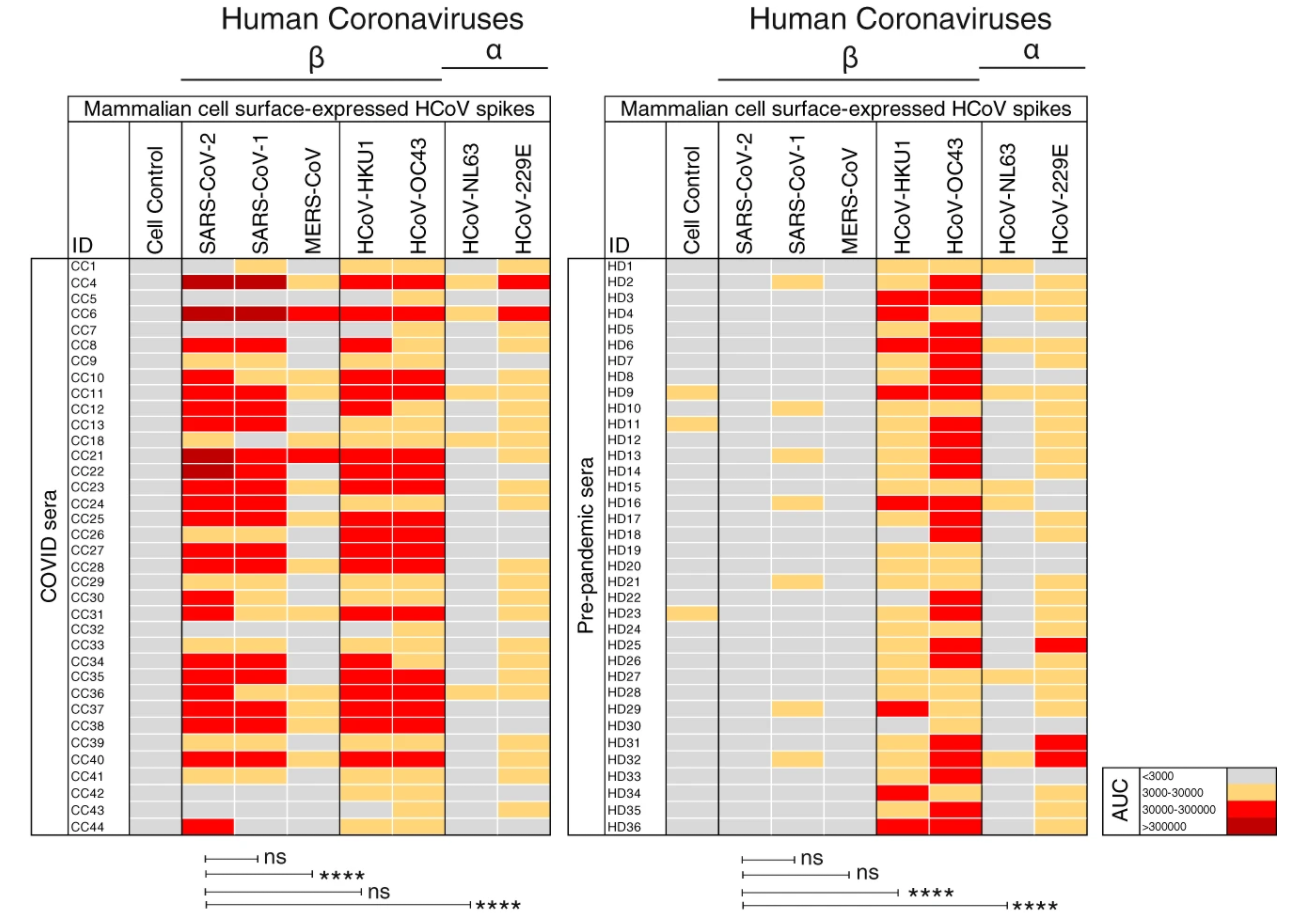
COVID-19 convalescent sera from 36 donors showed strong reactivity to the SARS-CoV-2 spike in the vast majority of infected donors, somewhat lower reactivity with the SARS-CoV-1 spike and much lower reactivity with the MERS-CoV spike. COVID-19 sera also exhibited strong cross-reactivity with endemic HCoV spikes, especially with the HCoV-HKU1 and HCoV-OC43 β-HCoVs. The α-HCoV-derived HCoV-NL63 spike was least reactive among the four endemic HCoVs. From a cohort of 36 HIV seropositive but otherwise healthy human donors whose samples were collected SARS-CoV-2 pre-pandemic. The sera showed almost no reactivity to SARS-CoV-2/CoV-1 and MERS-CoV spikes but showed strong binding to the endemic HCoV spikes, especially against the HCoV-HKU1 and HCoV-OC43 β-HCoVs (see a figure below).

In sera from SARS-CoV-2 pre-pandemic cohort, there was no evidence of pre-existing SARS-CoV-2 S-protein reactive antibodies that resulted from endemic HCoV infections. A recent study has, however, reported the presence of SARS-CoV-2 S-protein reactive antibodies in a small fraction of pre-pandemic human sera from children and adolescents as explained in the following paper.
Preexisting immunity to SARS-CoV-2 before the pandemic

The SARS-CoV-2/HKU1-CoV cross-reactive mAbs failed to bind any of the S1 subunit domains or subdomains, suggesting targeting to the more conserved S2 subunit

Interestingly, SARS-CoV-2 infection boosted titers to endemic HCoV-HKU1 S protein, but not for other HCoVs, suggesting that SARS-CoV-2 infection activated cross-reactive endemic HCoV-HKU1 S-protein-specific B cells.
Alternative host receptors (AXL, L-SIGN and DC-SIGN) for SARS-CoV-2 to ACE2

Blog admin completely agree with the following author’s opinion about alternative host receptors for SARS-CoV-2 to ACE2.
A review paper from a group of Shandong University of Traditional Chinese Medicine, China,
https://www.sciencedirect.com/science/article/pii/S2211383521001726?via%3Dihub

Although the role of ACE2 as a receptor for SARS-CoV-2 is clear, extensive studies have demonstrated that the expression of ACE2 is tissue- and cell-type specific and SARS-CoV-2 appears to infect tissues that are negative for ACE2. For example, ACE2 expression in human lung and respiratory tract is extremely low and limited in the epithelium, however, it’s well studied that SARS-CoV-2 preferentially infects cells of the respiratory tract, and SARS-CoV-2 can successfully infect human H522 lung adenocarcinoma cells despite complete absence of ACE2. The existence of alternative host receptors for SARS-CoV-2 entry was therefore speculated. Interestingly, Wang et al. recently identified the tyrosine-protein kinase receptor UFO (AXL) as a candidate receptor that promoting SARS-CoV-2 infection of the human respiratory system. Based on their study, the NTD rather than RBD of SARS-CoV-2 Spike is responsible for AXL recognition, highlighting the importance of NTD during SARS-CoV-2 infection. Meanwhile, Amraei et al. demonstrated thatCD209L/L-SIGN and CD209/DC-SIGN may also serve as alternative receptors for SARS-CoV-2 in disease-relevant cell types, including the vascular system.

The following papers are cited in this review paper regarding AXL and L-SIGN/DC-SIGN.
Regarding AXL by Wang et al.
Regarding L-SIGN/DC-SIGN by Amraei et al. 

SARS-CoV-2 has a Ganglioside binding domain in its NTD 

A group from Aix-Marseille Université, France has reported that there is a ganglioside binding domain in SARS-CoV-2 NTD.
https://www.ncbi.nlm.nih.gov/pmc/articles/PMC7547605/

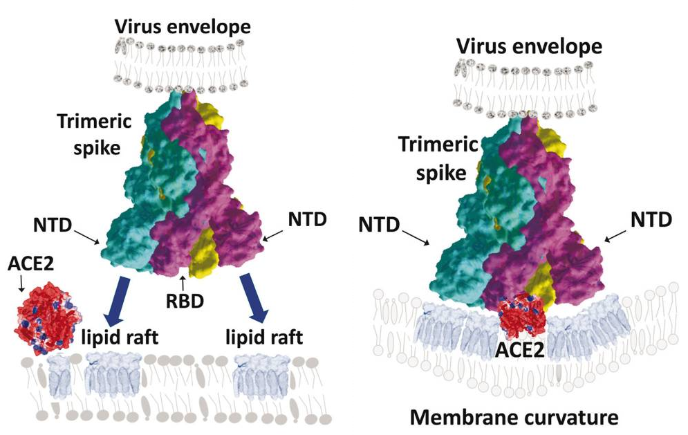
Residues 111-162 would be a ganglioside binding domain, and the binding could be inhibited by azithromycin which is a sugar-containing macrolide antibiotic which has some ganglioside mimicry properties, and also by 4A8 antibody that has been characterized in convalescent Covid-19 patients recognizes a discontinuous epitope of the NTD.

The figure blow shows an idea that the NTD binding to gangliosides induces the coalescence of lipid rafts together with a local modulation of membrane curvature that may facilitate the recruitment of the ACE-2 receptor.

POCT biosensor (named RAPID) for SARS-CoV-2 based on electrochemical reading with quite high sensitivity and high detection speed

A group from Perelman School of Medicine, University of Pennsylvania, etc. has reported a POCT biosensor (named RAPID) for SARS-CoV-2 based on electrochemical reading with quite high sensitivity and high detection speed.
https://pubmed.ncbi.nlm.nih.gov/33997767/

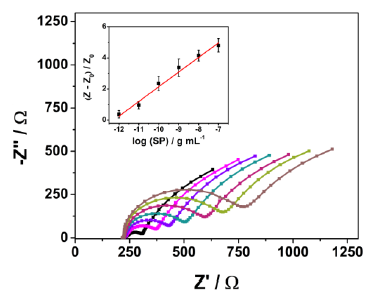
ACE2 was used as a probe detecting SARS-CoV-2 Spike protein. Enzyme immobilization on the electrode was done by cross-linking ACE2 using the bifunctional chemical cross-linker glutaraldehyde. BSA was used to block the electrode’s surface after immobilization of ACE2. Nafion was added to increase the sensitivity further.
The binding between two molecules (ACE2 and SARS-CoV-2 Spike protein) causes a change in interfacial electron transfer kinetics between the redox probe, ferricyanide/ferrocyanide in solution and the conducting electrode sites. This electrochemical change is then detectable by monitoring the charge-transfer resistance.

As a result, a linear concentration range from 10 fg/mL to 100 ng/mL was obtained (R2 = 0.993) and limits of detection (LOD) and quantification (LOQ) were calculated as 2.18 fg/mL on signal to noise ratios (S/N=3). With RAPID, result is obtained in 4 minutes (2 minutes of sample incubation + 2 minutes to perform the EIS analysis)..

In blinded tests using 139 nasopharyngeal swab samples, 109 of which were COVID-19 positive and 30 COVID-19 negative as determined by RT-qPCR, RAPID demonstrated high sensitivity, specificity and accuracy for nasopharyngeal (83.5%, 100% and 87.1%, respectively) and saliva (100%, 86.5% and 90.0%, respectively) samples.

Pomegranate peel extract (PPE) would be promising as an anti-SARS-CoV-2 drug 

A group from Arterra Bioscience SPA, Naples, Italy, etc. has reported that pomegranate peel extract (PPE) would be a promising source of novel anti-SARS-CoV-2 drugs.
https://www.frontiersin.org/articles/10.3389/fchem.2021.638187/full

Two major extracts from pomegranate peel are Punicalagin and Ellagitannins.

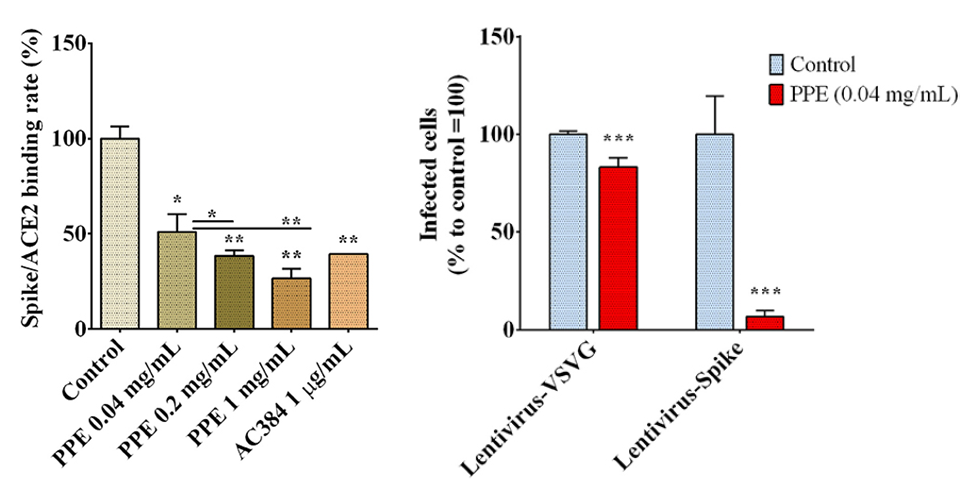
SARS-CoV-2 Spike/ACE2 binding was inhibited significantly in the presence of PPE as shown below. AC384 was used as a control, a monoclonal antibody that inhibited the binding between Spike and ACE2 by specifically recognizing ACE2 itself.

Infection experiments were done with using human kidney-2 cells (HK-2). The infection rate of lentivirus carrying SARS-CoV-2 Spike protein into was greatly reduced in the presence of PPE, and a lentivirus carrying vesicular stomatitis virus G (VSVG) protein was used as a control.

These in vitro experiments are quite reassuring to go to the next stage of the development of effective and innovative therapies using PPE.

Serum glycan biomarkers for Intracerebral hemorrhage (ICH) : ConA showed the highest specificity

A group from The First Affiliated Hospital of Anhui Medical University, Hefei, China, etc. has reported serum glycan biomarkers for Intracerebral hemorrhage (ICH).
https://www.ncbi.nlm.nih.gov/pmc/articles/PMC8105815/

The area under curve (AUC) were yielded as
0.93 for ConA (P<0.01), 0.95 for PNA (P<0.01), 0.67 for VVA (P=0.04), 0.92 for AAL (P<0.01), 0.86 for LTL (P<0.01), 0.84 for AIL (P<0.01). The sensitivity/specificity were as follows, 75.0%/95.8% for ConA, 100.0%/64.7% for PNA, 75.0%/58.3% for VVA, 100.0%/72.9% for AAL, 87.5%/79.2% for LTL, A68.8%/83.3% for AIL. As a result, α-Man/α-Glc (ConA) exhibited the best specificity. It is worthy to note that α-Man/α-Glc may be a potential biomarker with diagnostic and prognostic value in ICH and ConA will be a good probe for that.

Effects of B.1.427/B.1.429 SARS-CoV-2 variants onto infectivity and neutralization 

A group from University of California, San Francisco has reported effects of B.1.427/B.1.429 SARS-CoV-2 variants onto infectivity and neutralization.
https://pubmed.ncbi.nlm.nih.gov/33991487/

Famous SARS-CoV-2 variants are B.1.1.7(originally detected in UK), B.1.351(South Africa), and P.1(Brazil) variants. The B.1.1.7 variant is characterized by N501Y mutation, and the B.1.351 and P.1 carry E484K, K417N/K417T in addition to N501Y mutations.
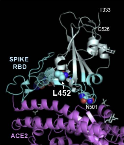
B.1.427/B.1.429 variants appeared in California from September, 2020 are characterized by S13I, W152C, L452R mutations in the spike protein coding domain, and L452R is in RBD.

These variants showed 18.6%–24% increased transmissibility relative to wild-type circulating strains, and antibody neutralization assays revealed 4.0- to 6.7-fold decrease in neutralizing titer from convalescent patients and 2.0-fold decreases in neutralizing titers from vaccine recipients, respectively

The reason of this change could be due to structural changes in the RBD which are caused by formation of a hydrophobic patch on the surface of spike RBD by L452 together with F490 and L492.

A unique glycosylation pattern in relapse-prone breast cancer comparing with non-relapsed cancer : TJA-II binding β-GalNAc

A group from Juntendo University School of Medicine, etc. has found a unique glycosylation pattern in relapse-prone breast cancer comparing with non-relapsed cancer with using lectin microarrays.
https://journals.plos.org/plosone/article?id=10.1371/journal.pone.0250747

Breast cancer is one of the most common malignant tumors among women. Triple-negative breast cancer (TNBC) is a relapse-prone breast cancer subset that accounts for about 15–20% of all breast cancers and is defined by tumors lacking estrogen receptor expression, progesterone receptor expression, and increased expression of human epidermal growth factor receptor-2 (HER2). An adjuvant therapy, cytotoxic chemotherapy is the only available treatment option for TNBC patients, because they do not respond to hormone or anti-HER2 treatment.

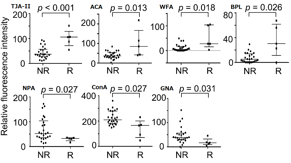
Authors found that TJA-II, which has binding specificity to Fucα1-2Gal and/or β-GalNAc glycans, shows higher intensity in cell extracts from surgical specimens of relapsed TNBC patients than in non-relapsed patients, and histochemical TJA-II staining of adjacent sections confirmed this difference. Since two more lectins, WFA and BLP, showed similar tendency to TJA-II, some glycoproteins having β-linked terminal GalNAc are to be in the background of this finding. The next stage is definitely to find out new target molecules for the effective therapy of the relapse-prone TNBC subset.

Alterations in the dendritic cells (DCs) signal paths after α2-3Sia binding: α2-3Sia induces anti-inflammatory actions

A group from Vrije Universiteit Amsterdam, Netherlands has studied alterations in the dendritic cells (DCs) signal paths after α2-3Sia binding.
https://www.frontiersin.org/articles/10.3389/fimmu.2021.673454/full

DCs possess the extraordinary capacity to recognize various pathogens with its pattern recognition receptors (PPRs) and present those antigens with MHC to tell the information to T-cells.
Sialic acids are increasingly attracting attention with their role in the immune regulation of cancer. During cancer progression, tumor cells often highly increase their sialic acid expression to create an immunosuppressive tumor microenvironment. Sialic acids are also advantageous for pathogens to evade from immune attacks. Bacteria obtain sialic acids by de novo synthesis or from an environmental source, and thereby can hide and escape from immune surveillance.

Authors analyzed alterations in the DC phosphoproteins, Kinase signatures, and JAK-STAT signaling pathway due to α2-3Sia stimulation in the presence of Lipopolysaccharide (LPS). LPS is often used as a model antigen (as an endotoxin) to activate immunity.

Overview of the result was summarized as follows.
Phosphorylation was enhanced upon α2-3Sia stimulation, while simultaneous α2-3sia and LPS stimulation resulted in less phosphorylation, indicating that recognition of a2-3Sia by DC alters TLR 4 triggering and DC signaling. α2-3Sia stimulated DCs compared to control with simultaneous α2-3sia and LPS stimulation showed decreased scoring of kinases ERK, AKT1, PKCB, GSK3, PKCD, PAK1, PKA, GSK3, GRK, IκB, and RAF1, and those affected kinase signatures were involved in the chemokine signal pathway. α2-3sia stimulation affected the JAK-STAT signaling by lowering the phosphorylation status of STAT3 and STAT5A.

Although the things are so complicated, these changes result in downregulation of IL-12 (inflammatory cytokine) pathway and inversely upregulation of IL-10 (anti-inflammatory cytokine) pathway, leading allover features to the direction of suppressing inflammation.
 

Using natural biomolecules (polysaccharides and lectins) extracted from Algae as antiviral therapeutics 

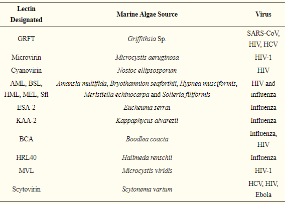
There have been so many examples in using natural biomolecules extracted from Algae as antiviral therapeutics. A group from Zhengzhou University, China, etc. has summarized as a review paper.
https://www.mdpi.com/1420-3049/26/8/2134/htm

Carrageenan, Gaalactan, Chitosan, Agar, Fucoidan, Laminaran are so famous as algae-derived antiviral polysaccharides, and you might have heard about it once. The antiviral mechanisms of these polysaccharides underlie several processes, including the inhibition of viral absorption, the inhibition of virus transcription and replication, and the improvement of host antiviral immune responses. Since polysaccharides are water-soluble and vary safe, it would be quite easy to use as internal medicines. If you are interested in these polysaccharide in more detail, please refer to the original paper and its references.

As you know, since the viral envelop proteins are heavily glycosylated, lectins could be also used to inhibit virus adsorption onto host cells. There have been a number of such examples as shown below.


A past blog article summarizing how lectin-glycan interactions are to be used to combat COVID-19 would be a good reference from a view point of antiviral therapeutics. It would be a big challenge how side effects of using lectins as antiviral ones could be controlled.

Powered by WordPress |Copyright © 2020 Emukk. All rights reserved