A group from University of Alberta, Canada, etc. has reported that Gal-9 could be a quite good diagnostic marker for COVID-19.
https://mbio.asm.org/content/12/3/e00384-21.long
Innate immune cells recognize and respond to a wide variety of pathogens. For example, activated macrophages/monocytes secrete excessive amounts of cytokines including IFN-γ, IL-1, IL-6, TNF-α, and IL-18. Similarly, neutrophils produce neutrophil extracellular traps and NK cells prolong the antigenic stimulation due to diminished/skewed cytolytic functions, both of which can amplify cytokine production. Overall, it appears that the viral burden, accompanied by the dysregulated innate immune response due to underlying disease, and aging may ignite the cytokine storm.
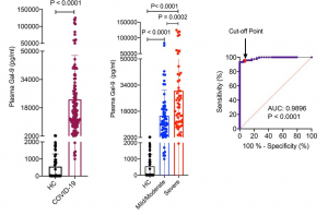
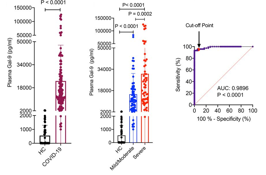
Galectins are involved in many biological functions such as development, signal transduction, and immune responses. This study was performed in a cohort of 120 SARS-CoV-2-infected individuals and 59 healthy controls (HCs) to understand how galectins, especially focusing on Gal-9, could be positioned in COVID-19.
Significantly higher levels of Gal-9 in the plasma of COVID-19 patients were observed comparing to HCs (ranging between 0 and 2,042 pg/ml). The plasma Gal-9 concentrations were substantially greater in severe cases (ranging between 1,950 and 125,510 pg/ml) comparing to those with mild/moderate cases (ranging between 1,000 and 83,717 pg/ml). From a ROC curve, quite high specificity/sensitivity (95%) to discriminate COVID-19 from either HCs or patients with HIV and virus-associated cancers with a cutoff value of 2,042 pg/ml. It is quite interesting to know that the plasma Gal-9 concentration in COVID-19 patients surpasses the detectable levels reported in other conditions such as HIV, dengue fever, influenza, and virus-associated solid tumors.
Through a series of experiments to identify what cells secrete Gal-9 and to understand how Gal-9 activates innate immune cells contributing to the cytokine storm, the following model was proposed as a conclusion. The damaged lung epithelial cells following SARS-CoV-2 infection release Gal-9, which activates alveolar macrophages, resulting in the secretion of proinflammatory cytokines and Gal-9 from the activated or apoptotic cells. Subsequently, Gal-9 activates monocytes and other immune cells, orchestrating another wave of proinflammatory cytokines and Gal-9 release, and exacerbates the cytokine storm further.