Effects of glycosylation (Oligomannose and bi-antennary complex N-glycan) onto binding between SARS-CoV-2 RBD and ACE2 

A group from Los Alamos National Laboratory, etc. has reported on effects of glycosylation onto binding between SARS-CoV-2 RBD and ACE2 with using molecular dynamics simulations.
https://www.mdpi.com/1999-4915/13/5/927

Two cases were evaluated: one is that six oligomannose (MAN9) glycans (magenta) are bound to ACE2 (at Asn53, Asn90, Asn103, Asn322, Asn432, Asn546) and one FA2 glycan (blue) is bound to RBD (at Asn343) and the other is that the six ACE2 glycans are replaced by bi-antennary complex N-glycan (FA2)

The result is as follows. When ACE2 glycans are MAN9, the binding energy decreased by 14.7%, and when ACE2 glycans are FA2, the binding energy increased by 9.1%.

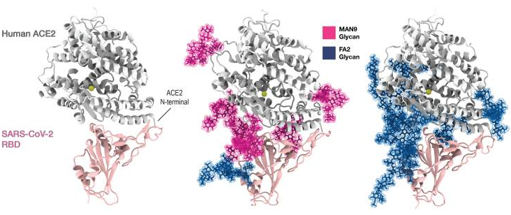
These simulation results seem not to contradict with the other experimental result.

In addition, it was shown that N501Y would introduce additional stabilizing interactions with Y41 and K353 of ACE2, which will increase the binding affinity between RBD and ACE2. The same results from different groups are introduced in the past blog.


N-glycan Binding specificity of Siglecs: from a study using glycan arrays 

A group from Georgia State University, etc. has reported N-glycan binding specificity of siglecs using glycan arrays.
https://www.ncbi.nlm.nih.gov/pmc/articles/PMC8116747/

Siglec-3, -9, and -10 prefer the α1-3 branching structure when Siaα2-6Galβ1-4GlcNAc terminal epitope serves as the binding ligand but prefer the opposite α1-6 branching structure when Siaα2-3Galβ1-4GlcNAc epitope serves as the ligand. Interestingly, Siglec-10 exhibited strong affinity to Neu5Gc-containing N-glycans

Characteristics of cytokine storms caused by highly pathogenic virus infections including SARS-CoV-2

Highly pathogenic virus infections usually trigger cytokine storms, which may have adverse effects on vital organs and result in high mortalities.
Sichuan Agricultural University, China has investigated characteristics of cytokine storms caused by the following viruses: three coronaviruses (SARS-CoV, MERS-CoV, and SARS-CoV-2), three influenza viruses (2009H1N1, H5N1 and H7N9), Ebola virus, HIV, dengue virus, Zika virus, West Nile virus, HBV, HCV, and enterovirus, and has found three characteristics in the cytokine storms.
https://www.frontiersin.org/articles/10.3389/fimmu.2021.659419/full

IP-10, L-6, IL-8, and IL-17 Are the Most Increased Cytokines

Variations of IFN-γ and IL-4 Determine the Maximum Amplitude of All Cytokines
The two cytokines IL-4 and IFN-γ play major roles in the generation and regulation of immune responses. Central in this respect are their mutually antagonistic functions. IFN-γ plays a key role in the inhibition of Th2-cell differentiation and Th1-cell stabilization; IL-4 promotes Th2-cell differentiation and stability and inhibits Th1-cell differentiation. The amplification factor should be the ratio of IFN-γ FC/IL-4 FC (if IFN-γ FC/IL-4 FC < 1, the amplification factor should be the ratio of IL-4 FC/IFN-γ FC). Correlation between [IFN-γ FC × (IFN-γ FC/IL-4 FC) (if IFN-γ FC/IL-4 FC > 1)] or [IFN-γ FC × (IL-4 FC/IFN-γ FC) (if IFN-γ FC/IL-4 FC < 1)] and the max FC was calculated and a very high correlation coefficient R2 = 0.988 was obtained.

viral load is weakly positive-correlated with IFN-γ FC, and virus clearance rate is negatively correlated with IFN-γ FC

Pathogenesis suggested from autopsies where the direct cause of death was determined as COVID-19 associated ARDS: Macrophages are directly infected with SARS-CoV-2

A group from Karolinska University Laboratory, etc. has reported on their new findings from 12 patients autopsies where the direct cause of death was determined as COVID-19 associated ARDS.
https://www.ncbi.nlm.nih.gov/pmc/articles/PMC8141733/

ARDS caused by COVID-19 appears to be the massive consolidation of the lung parenchyma. The process is initiated by virus replication in the pneumocytes leading to the desquamation of the alveolar epithelia and various levels of breach in the barriers between alveolar capillaries and the intra-alveolar space. With the increasing severity of this breach, edema fluid, plasma and eventually whole blood is leaking out into the alveolar space. Initiation of the coagulation cascade leads to the accumulation of fibrin filaments and/or intra-alveolar coagulated blood. Importantly only a minority of pneumocytes showed lytic virus replication but most pneumocytes in the affected area showed pronounced cytopathic effects in form of cytoplasmic swelling, vesicular degeneration and nuclear atypia.

Interestingly, the virus was replicating in the pneumocytes and macrophages but not in bronchial epithelium, endothelium, pericytes or stromal cells, suggesting that phagocytic cells consuming virus carrying cellular debris might themselves become infected, although the detailed mechanism is unknown.

The lung consolidation was accompanied with massive accumulation of CD163+ macrophages and immature myeloid elements, extensive proliferative response both in the epithelial and stromal components as well as exuberant neo-angiogenesis. The endothelial damage leading to the lung consolidation was considered to be caused by massive bystander effect, beyond the direct virus induced cytopathic damage, possibly induced by soluble factors such as ORF3a, inducing apoptosis in non-infected cells, released from the infected pneumocytes.
 

C-type lectins expressed on myeloid cells induce proinflammatory responses through the interaction with SARS-CoV-2 

A group from New York University Grossman School of Medicine, etc. has reported that C-type lectins expressed on myeloid cells induce proinflammatory responses through the interaction with SARS-CoV-2.
https://www.ncbi.nlm.nih.gov/pmc/articles/PMC8106883/

It was investigated that the binding of human immunoglobulin Fc-tagged SARS-CoV-2 S, S1, and RBD recombinant proteins to HEK293T cells transfected with individual cDNA including five C-type lectins (DC-SIGN, L-SIGN, LSECtin, ASGR1, and CLEC10A) and Tweety family member 2 (TTYH2). It was clearly shown that the ectopic expression of the myeloid cell receptors in HEK293T cells supported SARS-CoV-2 engagement independently from ACE2. The binding of DC-SIGN, L-SIGN, ASGR1, and CLEC10A was not affected by soluble ACE2-His, but that of ACE2 and LSECtin was completely blocked by soluble ACE2-His, and also the binding of DC-SIGN and L-SIGN was completely inhibited by mannan.

Furthermore, GFP-encoding HIV-based lentivirus pseudotyped with SARS-CoV-2 S protein (SARS-CoV-2 pseudovirus) was used to evaluate binding to those myeloid cell receptors. In the presence of the SARS-CoV-2 pseudovirus, robust GFP signals were observed in HEK293T expressing ectopic DC-SIGN or L-SIGN as well as expressing ectopic ACE2. The low level of SARS-CoV-2 pseudovirus engagement through LSECtin, ASGR1, CLEC10A, and TTYH2 was enhanced by co-expression of furin and/or TMPRSS2, which mediate S protein cleavage for viral entry. Consistent with our S protein mannan blockade results, pseudovirus-derived GFP signals through DC-SIGN and L-SIGN were inhibited by mannan treatment. These data suggested that these myeloid cell receptors can engage with the SARS-CoV-2 S protein on the pseudotyped virus.

Unbelievable discovery that RNA is glycosylated 

A group from Stanford University, etc. has reported that small noncoding RNAs are the third scaffold of glycosylation.
https://pubmed.ncbi.nlm.nih.gov/34004145/

Existence of glycosylated RNAs (named GlycoRNAs) was confirmed with the following protocol. Authors metabolically labeled cells with peracetylated N-azidoacetylmannosamine (Ac4ManNAz), and RNA was extracted with warm TRIzol, then ethanol precipitated, desalted via silica columns, stripped of protein contamination via high concentration proteinase K digestion, and repurified over silica columns. After RNA purification, Ac4ManNAz was conjugated to DBCO-biotin, visualized with streptavidin-IR800 (Strep), and imaged on an infrared scanner.

The RNA scaffolds for glycosylation was found to be small noncoding ones. Most of glycoRNAs existed on cell surface, and GlycoRNAs were modified with complex-type N-glycans with at least one terminal sialic acid residue. It was found that these GlycoRNAs directly bind to Siglecs.

The framework in which glycobiology is presently understood excludes RNA as a substrate for N-glycosylation. This discovery of glycoRNA suggests the current view is incomplete and points to a new axis of RNA glycobiology, including as of yet undiscovered biosynthetic and trafficking mechanisms. Further, it highlights the possibility that cell surface glycoconjugates, which mediate and regulate important inter-cellular interactions. 

An infection mechanism of SARS-CoV-2 infection through C-type lectin receptors (CLRs) 

A group from Univ. Grenoble Alpes, France, etc. has reported a mechanism of SARS-CoV-2 infection through C-type lectin receptors (CLRs).
https://journals.plos.org/plospathogens/article/authors?id=10.1371/journal.ppat.1009576

Authors studies molecular interaction between various CLRs (DC-SIGN, L-SIGN, MGL, and Langerin) with SARS-CoV-2 spike protein using SPR. DC-SIGN, L-SIGN, and MGL showed affinities in the μMrange in terms of Kd, from around 2 to 10 μM, and that of Langerin was speculated to be weaker at least one order of magnitude.

In order to understand the roles of CLRs in SARS-CoV-2 infection, infection experiments were performed using monocyte-derived dendritic cells (MDDCs), monocyte-derived macrophages (M2-MDM) and Vero E6 cells. Interestingly, VSV/SARS-CoV-2 pseudotyped virus did not infect MDDCs or M2-MDMs, despite DC-SIGN expression. In the case of VSV/EBOV-GP, however, direct infection was observed and DC-SIGN-mediated cis-infection was clearly blocked with anti-DC/L-SIGN in the case of MDDCs (92.5% inhibition of infection), followed by M2-MDM (68.4% inhibition). VSV/VSV-G also showed strong infectivity to both MDDC and M2-MDM. However, this infection was DC-SIGN independent, since anti-DC-SIGN antibodies did not impact the infection level.

To study the potential function of DC/L-SIGN in SARS-CoV-2 trans-infection, MDDCs were incubated with VSV/SARS-CoV-2 and placed onto susceptible Vero E6 cells which is the reference ACE2+ cell line for SARS-CoV-2 cell culture. Interestingly, DC-SIGN promoted efficient SARS-CoV-2 transfer from MDDC to Vero E6. An anti-DC-SIGN antibody could reduce substantially the infectivity observed (98% inhibition). These experiments confirmed that the role of DC-SIGN and L-SIGN in the process of SARS-CoV-2 could be trans-infection.

Characteristics of cross-reactive serum and memory B-cells between SARS-CoV-2 and endemic HCoVs 

A group from Scripps Research Institute has reported about characteristics of cross-reactive serum and memory B-cells between SARS-CoV-2 and endemic HCoVs.
https://www.nature.com/articles/s41467-021-23074-3

COVID-19 convalescent sera from 36 donors showed strong reactivity to the SARS-CoV-2 spike in the vast majority of infected donors, somewhat lower reactivity with the SARS-CoV-1 spike and much lower reactivity with the MERS-CoV spike. COVID-19 sera also exhibited strong cross-reactivity with endemic HCoV spikes, especially with the HCoV-HKU1 and HCoV-OC43 β-HCoVs. The α-HCoV-derived HCoV-NL63 spike was least reactive among the four endemic HCoVs. From a cohort of 36 HIV seropositive but otherwise healthy human donors whose samples were collected SARS-CoV-2 pre-pandemic. The sera showed almost no reactivity to SARS-CoV-2/CoV-1 and MERS-CoV spikes but showed strong binding to the endemic HCoV spikes, especially against the HCoV-HKU1 and HCoV-OC43 β-HCoVs (see a figure below).

In sera from SARS-CoV-2 pre-pandemic cohort, there was no evidence of pre-existing SARS-CoV-2 S-protein reactive antibodies that resulted from endemic HCoV infections. A recent study has, however, reported the presence of SARS-CoV-2 S-protein reactive antibodies in a small fraction of pre-pandemic human sera from children and adolescents as explained in the following paper.
Preexisting immunity to SARS-CoV-2 before the pandemic

The SARS-CoV-2/HKU1-CoV cross-reactive mAbs failed to bind any of the S1 subunit domains or subdomains, suggesting targeting to the more conserved S2 subunit

Interestingly, SARS-CoV-2 infection boosted titers to endemic HCoV-HKU1 S protein, but not for other HCoVs, suggesting that SARS-CoV-2 infection activated cross-reactive endemic HCoV-HKU1 S-protein-specific B cells.

Alternative host receptors (AXL, L-SIGN and DC-SIGN) for SARS-CoV-2 to ACE2

Blog admin completely agree with the following author’s opinion about alternative host receptors for SARS-CoV-2 to ACE2.
A review paper from a group of Shandong University of Traditional Chinese Medicine, China,
https://www.sciencedirect.com/science/article/pii/S2211383521001726?via%3Dihub

Although the role of ACE2 as a receptor for SARS-CoV-2 is clear, extensive studies have demonstrated that the expression of ACE2 is tissue- and cell-type specific and SARS-CoV-2 appears to infect tissues that are negative for ACE2. For example, ACE2 expression in human lung and respiratory tract is extremely low and limited in the epithelium, however, it’s well studied that SARS-CoV-2 preferentially infects cells of the respiratory tract, and SARS-CoV-2 can successfully infect human H522 lung adenocarcinoma cells despite complete absence of ACE2. The existence of alternative host receptors for SARS-CoV-2 entry was therefore speculated. Interestingly, Wang et al. recently identified the tyrosine-protein kinase receptor UFO (AXL) as a candidate receptor that promoting SARS-CoV-2 infection of the human respiratory system. Based on their study, the NTD rather than RBD of SARS-CoV-2 Spike is responsible for AXL recognition, highlighting the importance of NTD during SARS-CoV-2 infection. Meanwhile, Amraei et al. demonstrated thatCD209L/L-SIGN and CD209/DC-SIGN may also serve as alternative receptors for SARS-CoV-2 in disease-relevant cell types, including the vascular system.

The following papers are cited in this review paper regarding AXL and L-SIGN/DC-SIGN.
Regarding AXL by Wang et al.
Regarding L-SIGN/DC-SIGN by Amraei et al. 

SARS-CoV-2 has a Ganglioside binding domain in its NTD 

A group from Aix-Marseille Université, France has reported that there is a ganglioside binding domain in SARS-CoV-2 NTD.
https://www.ncbi.nlm.nih.gov/pmc/articles/PMC7547605/

Residues 111-162 would be a ganglioside binding domain, and the binding could be inhibited by azithromycin which is a sugar-containing macrolide antibiotic which has some ganglioside mimicry properties, and also by 4A8 antibody that has been characterized in convalescent Covid-19 patients recognizes a discontinuous epitope of the NTD.

The figure blow shows an idea that the NTD binding to gangliosides induces the coalescence of lipid rafts together with a local modulation of membrane curvature that may facilitate the recruitment of the ACE-2 receptor.

Powered by WordPress |Copyright © 2020 Emukk. All rights reserved