Relationship between serum uric acid level and COVID-19 severity 

A group from Cliniques universitaires Saint-Luc, Belgium, etc. has reported relationship between serum uric acid level and COVID-19 severity.
https://www.ncbi.nlm.nih.gov/pmc/articles/PMC8201458/

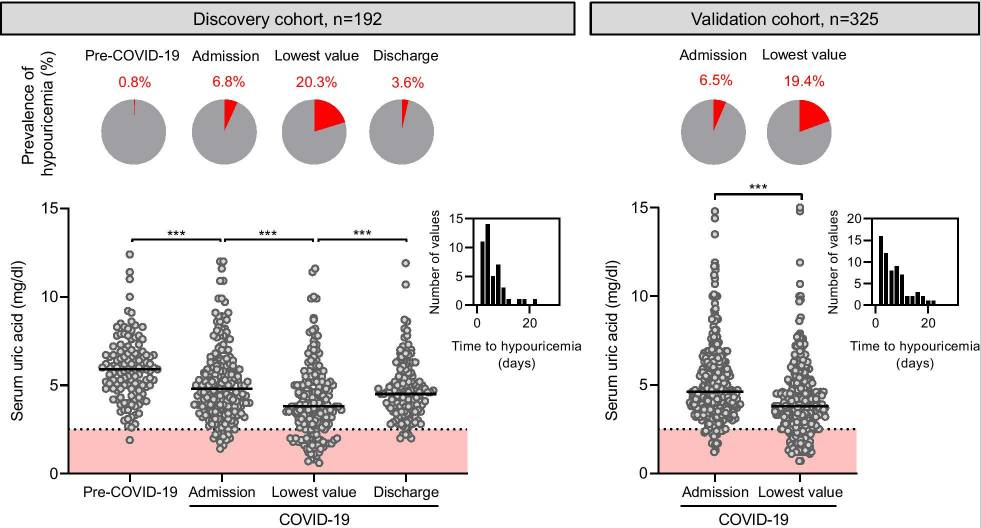
The prevalence of hypouricemia is approximately 0.3% in the general ambulatory population. In this study, 20% of the patients hospitalized for SARS-CoV-2 infection developed hypouricemia, a proportion that increased to 77% among patients requiring mechanical ventilation.

Uric acid is the end-product of purine metabolism in humans and is generated in the liver. The kidney is an important regulator of circulating uric acid levels as it excretes most of total body uric acid. Serum urate is freely filtered by the glomeruli followed by a complex balance of reabsorption and secretion in the kidney proximal tubule. Although the molecular mechanisms of urate transport in the proximal tubule are still incompletely understood, URAT1 (SLC22A12) is the main apical transporter mediating urate reabsorption in the brush border of the proximal tubule. In a small subset of kidney samples from patients who died from COVID-19, it was shown that life-threatening SARS-CoV-2 infection is associated with a significant (~ 70%) decrease in the expression of the apical urate transporter URAT1 in the kidney proximal tubule, contributing to the impaired tubular absorption of urate. The mechanisms linking hypouricemia and progression to severe disease requiring mechanical ventilation in patients with COVID-19 remains speculative and may be diverse

Anyhow, it seems that serum uric acid could be used as a reliable biomarker to identify patients at risk of life-threatening COVID-19.

Whether HIV antibodies could neutralize SARS-CoV-2 or not 

A group from University of British Columbia, Canada has reported whether HIV antibodies could neutralize SARS-CoV-2 or not.
https://www.nature.com/articles/s41598-021-91746-7

Cross-reactive interactions of three HIV antibodies (2G12, PGT128, PGT126) in the presence of methyl α-d-mannopyranoside, a stabilized mannose analogue, were evaluated using an ELISA assay. Disruption of cross-reactivity was observed for all these antibodies with increasing concentrations of methyl α-d-mannopyranoside, demonstrating that the binding of these antibodies to SARS-CoV-2 were via glycan sensitivity of these interactions.

HEK293-T cells stably overexpressing ACE-2 were incubated with SARS-CoV-2 S pseudo-typed virus harbouring a luciferase reporter gene, in the presence of serial dilutions of three HIV antibodies. Luciferase activities in cellular lysates were determined 48 h post-infection (RLU: relative luciferase units). However, no neutralization capabilities were detected for these antibodies over a wide range of concentrations, while VH-FC ab8 demonstrated potent neutralization, a positive control antibody VH-FC ab8 which targets the SARS-CoV-2 RBD.

But, the blog admin thinks that the infection to immune cells (i.e., macrophages) expressing C-type lectins might be neutralized by these HIV antibodies? 

Whether ABO blood group or secretor status was associated with COVID-19 severity 

A group from Bristol Institute for Transfusion Sciences (BITS), NHSBT, UK, etc. has investigated whether ABO blood group or secretor status was associated with COVID-19 severity.
https://onlinelibrary.wiley.com/doi/10.1002/jha2.180

A number of studies carried out in several countries have supported an association between ABO type and SARS-CoV-2 susceptibility to infection and/or outcomes. The reasons for the association of severe COVID-19 with blood type A are unknown, but it has been suggested that this could be caused by O group patients having anti-A type antibodies, that A type glycans could function as co-receptors for SARS-CoV-2 or due to the known effects of blood groups on thrombosis risk due to von Willebrand factor (VWF) levels.

The result obtained here is that the association of blood group A with hospitalization was (RR = 1.24, CI 95% [1.05, 1.47], p = 0.0111), and blood group A with cardiovascular complications was greatly increased (RR = 2.56, CI 95% [1.43, 4.55], p = 0.0011), but group A non-secretors were significantly less likely to be hospitalized than secretors. Why the difference between group A and O was so large in COVID-19 related cardiovascular complications was considered to be due to VWF related origins.

As is known, in the presence of active FUT2 A, B, H, and Lewis b antigens can be expressed on mucosal surfaces. Individuals with this phenotype are known as secretors. In individuals lacking active FUT2, known as non-secretors, only the Lewis a antigen can be expressed.     

New urinary biomarkers for cardiovascular events originating from Type 2 diabetes: High mannose is the one

A group from Okayama University Graduate School of Medicine, Dentistry and Pharmaceutical Sciences, etc. has reported new urinary biomarkers for cardiovascular events originating from Type 2 diabetes.
https://www.frontiersin.org/articles/10.3389/fcvm.2021.668059/full

Targeting cardiovascular events (CVE) as one of primary outcomes originating from the diabetes, urinary glycomes in 680 patients with type 2 diabetes were evaluated with using Lectin microarrays.

During approximately a 5-year follow-up period, 62 patients reached the endpoint. Cox proportional hazards analysis revealed that urinary glycan signals binding to two lectins were significantly associated with the outcome after adjustment for known indicators of CVE and for false discovery rate, as well as increased model fitness. Hazard ratios for these lectins (+1 SD for the glycan index) were UDA (recognizing glycan: mixture of Man5 to Man9): 1.78 (95% CI: 1.24–2.55, P = 0.002) and Calsepa [High-Man (Man2–6)]: 1.56 (1.19–2.04, P = 0.001). Common glycan binding to these lectins was high-mannose type of N-glycans.

The urinary excretion of high-mannose glycan may be a valuable biomarker for improving prediction of CVE in patients with type 2 diabetes. It is still unclear what kinds of mechanisms are underlaying such abnormal N-glycosylation.

HOMER3 with sialyl-T and sialyl-Tn antigen could be a therapeutic target for bladder cancer 

A group from Portuguese Institute of Oncology, etc. has reported that HOMER3 with sialyl-T and sialyl-Tn antigen could be a therapeutic target for bladder cancer (BC).
https://www.ncbi.nlm.nih.gov/pmc/articles/PMC8188679/

A total of 10 major glycans structures were identified for both BC cell lines: 5637 and T24, which present similar O-glycomes. Notably, both cell lines expressed high levels of mono- and di-sialylated T antigens (sialyl-T), also exhibiting neutral core 1 and sialylated and/or fucosylated core 2 structures

Based on glycomics analysis, samples were first enriched for membrane proteins by differential ultracentrifugation of whole cell protein extracts, digested with sialidase and enriched for T antigen expressing glycoproteins by PNA affinity pulldowns. The glycoproteins were identified by conventional bottom-up proteomics after trypsin digestion and analysis by nanoLC-CID-MS/MS. As a result, over 900 glycoproteins were identified as potentially expressing sialyl-T.

Among these, GLUT1(SLC2A1) and HOMER3 ranked first, suggesting potential for targeted therapeutics. GLUT1 is a pivotal membrane glucose transporter frequently overexpressed in more aggressive bladder tumours. HOMER3 has been implicated in neuronal signaling, T-cell activation and trafficking of beta amyloid peptides. HOMER3 at the cell membrane co-localizes in the same tumour area with sialyl-Tn and sialyl-T antigens in BC. According to immunohistochemistry, HOMER3 was diffusely expressed in wide tumour areas, both at the cytoplasm and cell membranes. These areas co-localized with very high sialyl-Tn and/or sialyl-T expressions areas.

Only two parameters are good enough to predict need for hospitalization with COVID-19: IL-6 and CRP 

Through case studies with over 80,000 samples, it was concluded that having both IL-6 ≥10 pg/mL and hs-CRP ≥10.0 mg/L very accurately predicts the need for hospitalization in COVID-19 positive patients. Only two parameters are good enough to predict.

In detail, Median IL-6 levels were the same in controls and outpatients but were about 75-fold higher in inpatients as compared to other groups (P<0.0001). Median hs-CRP levels were very similar in control subjects and outpatients but were about 80-fold higher in inpatients as compared to other groups (P<0.0001).

This is a report from a group of Boston Heart Diagnostics/Eurofins Scientific Network, USA, etc.

https://www.ncbi.nlm.nih.gov/pmc/articles/PMC8191995/

Cell-to-cell transmission could be a major factor in SRAS-CoV-2 spread:ACE2 is not so important in this case

A group from The Ohio State University, etc. has reported that cell-to-cell transmission could be a major factor in SRAS-CoV-2 infection spread.
https://www.ncbi.nlm.nih.gov/pmc/articles/PMC8183005/

ACE2 is the primary receptor of both SARS-CoV-2 and SARS-CoV, mediating viral entry into host cells. The virus infection through the binding between SARS-CoV-2 Spike and ACE2 increases linearly with increasing ACE2 expression on host cells as shown below for both SARS-CoV-2 and SARS-CoV. However, interestingly, with a relatively low dose of ACE2, SARS-CoV-2 cell-to-cell transmission reached ~70% of its maximal and started to decrease with increasing further as shown below. This pattern of SARS-CoV-2 cell-to-cell transmission was different from that of cell-free infection, suggesting that the cell-to-cell transmission happens independently from ACE2 mediated infection.

Interestingly, it was shown that endosomal entry pathways were also involved in cell-to-cell transmission of SARS-CoV-2 and SARS-CoV. This is evidenced as shown below by the inhibitory effect of drugs that specifically target the endosomal entry pathway of these viruses, including the CatL inhibitor III, which blocks cleavage of the viral glycoprotein, as well as BafA1, which neutralizes endosomal pH.

Natural evolution of neutralizing antibodies after one year from SARS-CoV-2 infection 

A group from Rockefeller University, etc. has reported on naturally enhanced neutralizing antibodies after one year from SARS-CoV-2 infection.
https://www.biorxiv.org/content/10.1101/2021.05.07.443175v2.full

Convalescent participants who had not been vaccinated maintained most of their anti-RBD IgG titers between 6 and 12 months. Vaccination increased the anti-RBD plasma antibody levels, with IgG titers increasing by nearly 30-fold compared to unvaccinated individuals (in a figure below, Vac means vaccinated individuals).

Plasma neutralizing activity in 63 participants was measured using an HIV-1 pseudotyped with the SARS-CoV-2 spike protein. 12 months after infection, the geometric mean half-maximal neutralizing titer (NT50) for the 37 individuals that had not been vaccinated was 75, which was not significantly different from the same individuals at 6.2 months. In contrast, the vaccinated individuals showed a geometric mean NT50 of 3,684, which was nearly 50-fold greater than unvaccinated individuals.

To determine whether there was an increase in neutralization breadth over time, the neutralizing activity of the 60 antibodies was assayed against a panel of RBD mutants covering residues associated with circulating variants of concern: R346S, K417N, N440K, A475V, E484K and N501Y. Increased activity was evident against K417N, N440K, A475V, E484K and N501Y suggesting that evolution of the antibody repertoire results in acquisition of neutralization breadth over time.

Sea is a reservoir of lectins

Septifer virgatus, Sponge, Turban shell, Capitulus mitella, Sea cucumber, Star fish, Sea hare, and Sea urchin……….
Our journey continues to discover new lectins.

Human surfactant protein D would have therapeutic potential against SARS-CoV-2 infection 

A group from National Cheng Kung University, Tainan, etc. has reported that human surfactant protein D would have therapeutic potential against SARS-CoV-2 infection.
https://www.ncbi.nlm.nih.gov/pmc/articles/PMC8161545/

Human surfactant protein D (SP-D), a collagen-containing C-type lectin and a member of the collectin family, is known to be involved in pulmonary surfactant homeostasis and immunity. The ability of recombinant fragment of human SP-D(rfhSP-D) to inhibit infection of SARS-CoV-2 was examined using pseudotyped lentiviral particles expressing SARS-CoV-2 S1 protein.
It was found that rfhSP-D bound SARS-CoV-2 S1 protein in a dose-dependent manner; this interaction was inhibited by maltose and EDTA. No rfhSP-D binding was observed in the absence of RBD in this assay.

Inhibition of rfhSP-D binding to S protein by EDTA or maltose suggested that rfhSP-D bound to the carbohydrate moieties on S protein of SARS-CoV-2. A luciferase reporter assay with pseudotyped lentiviral particles expressing SARS-CoV-2 S1 protein was used to evaluate infectivity. Approximately 0.5 RLU fold reduction was seen with rfhSP-D (5 or 10 µg/ml) treatment comparing to untreated sample (1 RLU fold; ACE2 overexpressing HEK293T cells + SARS-CoV-2). A significantly reduced luminescent signal following rfhSP-D treatment indicated that the interaction of rfhSP-D with SARS-CoV-2-S1 restricted the binding and entry of the virus.

These results highlight the therapeutic potential of rfhSP-D in SARS-CoV-2 infection. 

Powered by WordPress |Copyright © 2020 Emukk. All rights reserved