A new pathway was discovered in activation of the complement system of the innate immunity: MASP-1 and MASP-3 have dual functions as enzymes and as PRMs 

In the lectin pathway as one of innate immunities, pattern recognition molecules (PRMs) (i.e., mannose-binding lectin (MBL), ficolins, and collectin-10/-11) bind to pathogens recognizing specific molecular structures on the membranes, make complexes with MAPSs, activate complements sequentially, and finally form Membrane Attack Complex (MAC) on the membranes to kill pathogens.

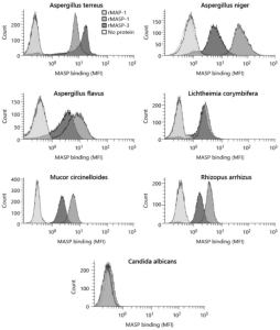
However, a group from University of Copenhagen, etc. has reported a new pathway in which MAPSs directly bind to pathogens and activate the complement system using Aspergillus fumigatus as a pathogen. This means that a new pathway in the complement system was discovered showing MASP-1 and MASP-3 have dual functions as enzymes and as PRMs
https://www.karger.com/Article/FullText/514546

Immune Memory (CD4+T-cell reaction) in mild COVID-19 patients and Cross-rectivity of CD4+T-cells against SARS-CoV-2 in unexposed donors

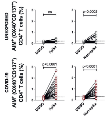
A group from National Institute of Immunology, New Delhi, etc. has reported on immune memory (CD4+T-cell reaction) in mild COVID-19 patients and cross-reactivity of CD4+T-cells against SARS-CoV-2 in unexposed donors
https://www.frontiersin.org/articles/10.3389/fimmu.2021.636768/full

In mild COVID-19 patients, it was reported that the immune memory (i.e, CD4+T-cell reactions to SARS-CoV-2) continues for about 5 months (median about 3 months). On the other hand, in SARS-CoV-2 unexposed donors, there was no immune reaction to SARS-CoV-2 spike protein, but the CD4+ T-cells specific to SARS-CoV-2 N protein were observed in 66% of the donors. In SARS-CoV-2 exposed patients, the CD4+ T-cells targeted SARS-CoV-2 spike protein rather than SARS-CoV-2 N protein.
Whether the cross-reactive CD4+ T cells are contributing to suppressing SARS-CoV-2 infection and further less severe outcome needs to be addressed in the prospective cohort before and after COVID-19.

Proteinuria would be a good Biomarker for the new coronavirus (COVID-19) severity 

A group from Université Côte d’Azur, Nice, etc. has reported that Proteinuria would be a good Biomarker for the new coronavirus (COVID-19) severity.
https://www.ncbi.nlm.nih.gov/pmc/articles/PMC7985082/

Significant proteinuria (>0.3 g/g) was related to
higher prevalence of ICU admission [OR = 4.72, IC95 (1.16–23.21), p = 0.03],
acute respiratory distress syndrome (ARDS) [OR = 6.89, IC95 (1.41–53.01, p = 0.02)],
longer hospital stay [19 days (9–31) versus 7 days (5–11), p = 0.001].

Cell surface N-glycan of Candida albicans (mannose core) works anti-inflammatory in sepsis

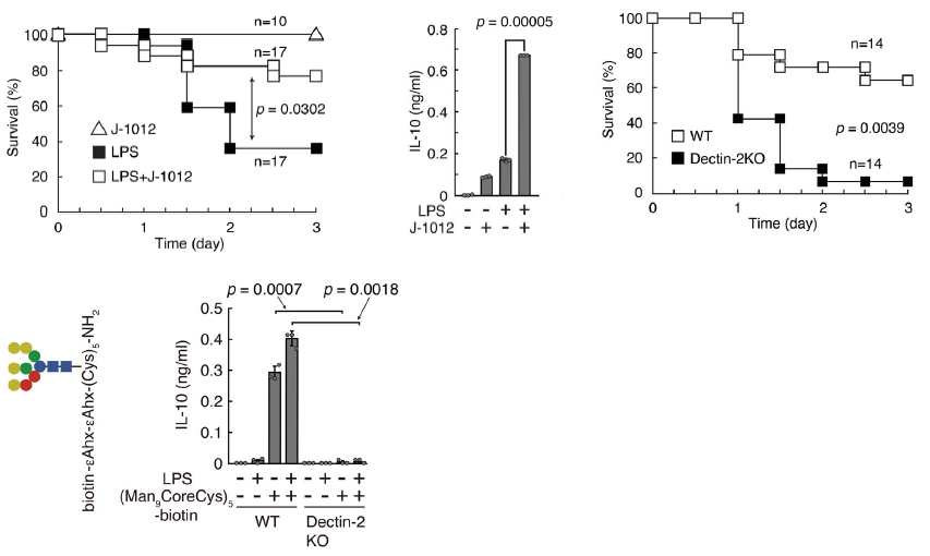
A group from Kyoto University has reported that the cell surface N-glycan (mannose core) of Candida albicans works anti-inflammatory in sepsis.
https://www.nature.com/articles/s42003-021-01870-3

Sepsis was induced by lipopolysaccharide(LPS)in mouse, and it was demonstrated that purified mannoprotein of Candida albicans stratn J-1020 works anti-inflammatory. Injection of the extracted mannoprotein from J-1020 improved survival greatly. It was found that anti-inflammatory cytokine IL-10 was up-regulated by the injection of J-1020 at this time. It was also confirmed that these phenomena were induced by the interaction between mannoprotein of J-1020 and C-type lectin Dectin-2. When Dectin-2 was knocked out, the survival decreased greatly, and J-1012 mannoprotein did not affect IL-10 production. Further, it was suggested the mannose core of Candida albicans N-glycan is recognized by Dectin-2, leading to suppression of the early onset of septic responses.

The amount of serum IgG in SARS-CoV-2 positive patients is inversely correlated with age at around 20 years old as a boundary

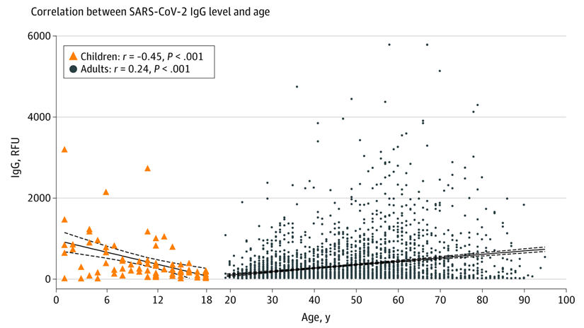
A group from Weill Cornell Medicine, New York has reported on the relationship between serum IgG and Age with using serum samples gathered from April 2020 to June 2020.
https://jamanetwork.com/journals/jamanetworkopen/fullarticle/2777743

The correlation diagram between serum IgG and Age using 85 SARS-CoV-2 positive pediatric and 3648 positive adult patient samples is shown below. The result is quite interesting. It is clearly found that the amount of serum IgG is inversely correlated with age at around 20 years old as a boundary. It remains unclear why patients aged 19 to 25 years exhibited lower levels of SARS-CoV-2 IgG antibodies than children and older adults.

Site-specific O-glycan analysis of the new coronavirus (SARS-CoV-2)

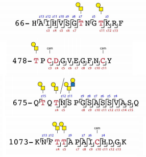
A group from University of Copenhagen, etc. has reported on site-specific O-glycan analysis of the new coronavirus (SARS-CoV-2).
https://www.biorxiv.org/content/10.1101/2021.02.03.429627v2

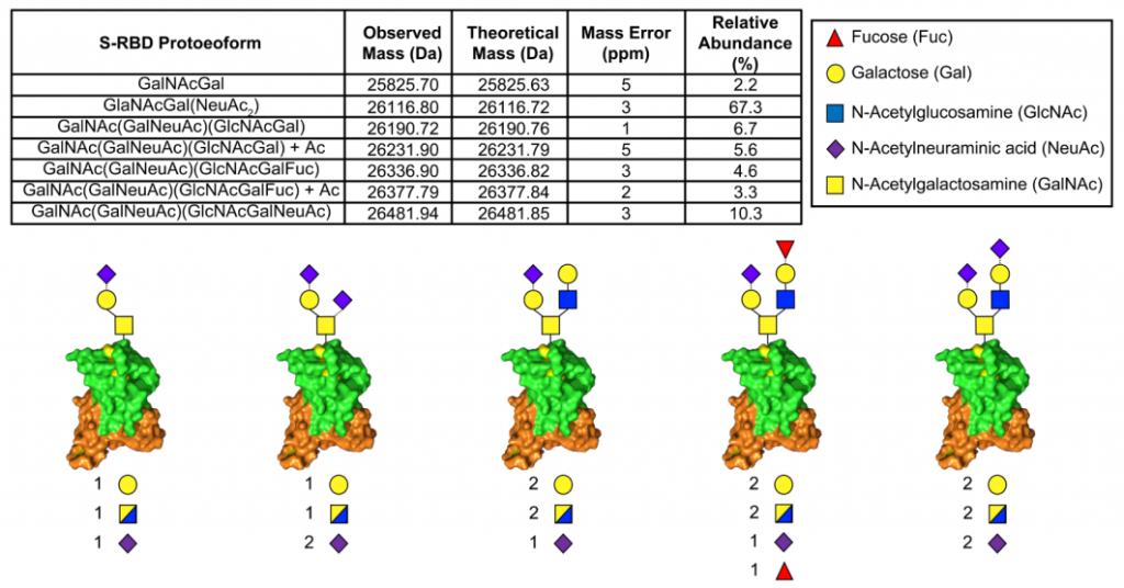
Authors used SARS-CoV-2 Spike proteins expressed in Drosophia and HEK 293 F cells. N-glycans were removed by PNGase F, and then Sialic acids were also removed by neuraminidase. Glycan analysis was done by using ETD/HCD LC-MS/MS. O-glycan modification in HEK 293 F is shown blow. Tn-antigen seems to be more expressed than Tn-antigen. There is no information about sialylation of T- and Tn-antigens.

O-glycan analysis of the new coronavirus (SARS-CoV-2)

A group from University of Wisconsin-Madison, etc. has reported on the analysis of O-glycans expressed on the new coronavirus (SARS-CoV-2).
https://www.biorxiv.org/content/10.1101/2021.02.28.433291v1

In order to analyze O-glycans, N-glycans on SARS-CoV-2 spike protein expressed with HEK293 were removed by PNGase F treatment. Authors combined ion mobility spectrometry (TIMS) and ultrahigh-resolution Fourier transform ion cyclotron resonance (FTICR) MS analysis.
As a result, it was found that the majority of the O-glycan modification was Sialyl-T-antigen.

The low level of anti-Tn is related to COVID-19 status

A group from Université de Nantes, etc. has reported on the relationship between anti-Tn and COVID-19 status.
https://www.frontiersin.org/articles/10.3389/fmicb.2021.641460/full

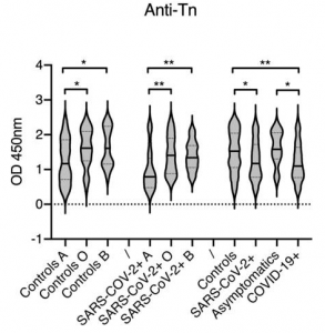
Taking into consideration that Tn-antigen (α-GalNAc) has a structural similarity to blood A-antigen (GalNAcα1-3(Fucα1-2)Galβ1-4GlcNAc), it is easily understood that the level of anti-Tn in bood group A individuals is lower than those in blood groups B and O individuals. The fact that the level of anti-Tn is lower in SARS-CoV-2 infected and developed COVID-19 individuals than controls and asymptomatic individuals would mean that the lower the level of anti-Tn is, the higher the COVID-19 developing possibility is. This does not conflict with the existing report saying that blood group A individuals are prone to develop COVID-19 than blood group O individuals.
https://www.nejm.org/doi/pdf/10.1056/NEJMoa2020283?articleTools=true
In additon, there are almost no changes in anti-T, anti-core-3, anti-Lec, anti-GlcNAcLac, and anti-Gb3.

Quantitative and Comparative Urine proteome analysis of the new coronavirus (COVID-19) 

A group from Beijing Proteome Research Center, etc. has reported results of quantitative and comparative urine proteome analysis from healthy individuals, mild and severe COVID-19 positive patients.
https://pubmed.ncbi.nlm.nih.gov/33688631/

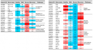
A total number of 2656 proteins was identified from healthy control samples. There were 1008 proteins being commonly identified and quantified among healthy, mild and sever cases. However, 211 and 63 proteins were uniquely expressed in COVID-19 patients and recovery samples.

Some proteins increased in COVID-19, but some proteins decrease inversely in COVID-19.
Typical examples are listed below. However, from a view point of this blog Admin, there is some question if urine proteome can be used as a diagnostic tool, because it seems that urine proteome can not discriminate severity of COVID-19 from moderate, mild, to sever. But, it would definitely contribute to elucidation of COVID-19 pathology. The fact that the urine proteome of recovered patients is not the same as healthy individuals would suggest that the aftereffects are remaining for a longer time.

IGLV1-40: 14 times higher in the severe COVID-19
FKBP1A: 13 times higher in the severe COVID-19
LUNA: 37 times higher in the severe COVID-19
PARK7: 7 times higher in the severe COVID-19

CD9: 1/12 times lower in the sever COVID-19
EGF: 1/25 times lower in the sever COVID-19
MME: 1/18 times lower in the sever COVID-19
CUBN: 1/19 times lower in the sever COVID-19

Immune modulations in Pancreatic Ductal Adenocarcinoma (PDAC) through aberrant glycan modification, especially upregulated Sialylation, and its interaction with Siglec-7, Siglec-9 

A group from Amsterdam UMC, etc. has reported on mechanisms of immune modulations in pancreatic ductal adenocarcinoma (PDAC) through aberrant glycan modification, especially upregulated α2-3 sialic acids.
https://www.nature.com/articles/s41467-021-21550-4.pdf

PDAC is surrounded by dense fibrotic stroma and suppressive immune cells, mostly of the myeloid lineage such as dendric cells and macrophages (called TME). It has been known that aberrant glycosylation changes in PDAC are upregulation of sialyl Lewisx, sialyl Lewisa, truncated O-glycans, increased branched and fucosylated N-glycans, and are related to tumor cell proliferation, invasion, metastasis, and inflammation. However, their detailed link to the immune cell function in PDAC is still unknown.

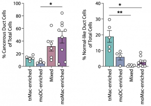
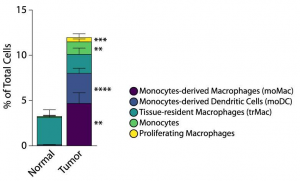
Authors have deep dived into the link between the aberrant sialylation and immune cells, and further its relationship with survival rate of PDAC. Composition of myeloid linkage cells composing TMA is greatly different between PDAC and healthy tissues. In PDAC, monocyte-derived macrophages (moMac) and monocyte-derived dendritic cells (moDC) accounted for the majority of the composition. Interestingly, patients with a higher presence of moMac over moDC had shorter survival, and the other myeloid populations did not correlate with the survival rate.

It was clearly shown that upregulated α2-3Sia bound to Siglec-7 and Siglec-9 expressed on myeloid cells, and drove the differentiation of monocytes to moMac with upregulation of CD206. Further, these changes induced upregulation of immune check point molecule PD-L1 and secretion of immunosuppressive cytokines such as IL-10.