blog

Plant growth promoting effects in Wheat and Maize biofertilized with PGPM and Biochar

A group from Interdepartmental Center SITEIA.PARMA, University of Parma, Italy, etc. has reported about plant growth promoting effects in Wheat and Maize biofertilized with PGPM and Biochar.
https://www.ncbi.nlm.nih.gov/pmc/articles/PMC9499264/

Biostimulants are classified into biofertilizers, bacteria, and/or fungi, defined as plant growth-promoting microbes (PGPM), which establish a positive relationship with the plant by increasing the bioavailability of nutrients present in the soil and have a positive impact on plant yield and health. To improve the performance of biofertilizers, it is possible to combine them with soil amendments which can stimulate microbial growth and survival. Biochar is such a good candidate, because its structural porosity makes it ideal to provide a niche in which microorganisms can survive environmental stress.

This study investigated the effect of the combination of biochar (as a carrier of PGPM), two types of microbial consortia (MC-B and MC-C), and/or arbuscular mycorrhizal fungi (AMF) on wheat and maize when grown in greenhouses.

  • MC-B was made up of A. vinelandii DSM 2289, R. aquatilis BB23/T4d, Bacillus sp. BV84, B. amyloliquefaciens LMG 9814, and P. fluorescens DR54.
  • MC-C was made up of A. chroococcum LS132, B. amyloliquefaciens LMG 9814, P. fluorescens DR54, B. ambifaria MCI 7, and R. aquatilis BB23/T4d.

The results demonstrated that wheat and maize supplemented with different combinations of selected microbial consortia and biochar have a positive effect on plant growth in terms of shoot and root biomass.

In wheat, the treatments with the largest contribution to the cultures were Char_MC-C, either with or without AMF, followed by Char_MC-B, either with or without AMF. On the other hand, in maize, the best growing conditions were for Char_MC-C, either with or without AMF, followed by Char_MC-B_AMF or AMF alone.
Characteristic glycan modification of SARS-CoV-2 Omicron variant comparing with other variants

A group from Institute of Synthetic Biology, Shenzhen Institutes of Advanced Technology, Chinese Academy of Sciences, Shenzhen, China, etc. has reported about characteristic glycan modification of SARS-CoV-2 Omicron variant comparing with other variants.
https://pubmed.ncbi.nlm.nih.gov/36318020/

I thought I would stop blogging about SARS-CoV-2, but I would like to briefly introduce this paper it as a topic related to comparative glycan profiling analysis using lectin microarrays.

From the comparative glycan profiling analysis, it was found that SARS-CoV-2 Omicron showed higher levels of terminal fucose (UEA-I), and also showed higher level of sialylated (MAL-II, MAA, ASNA-I) and galactosylated glycans (CSA, WFA, SBA, VVL) than other variants (Alpha, Beta, and Delta).

SARS-CoV-2 Omicron was more susceptible to neutralization antibodies after neuraminidase or galactosidase treatment. This means that the higher expression of both Sia- and Gal-containing glycans on Omicron potentially enhance its shielding effect against neutralization.

halophilic Bacillus strains to enhance plant growth and reduced the adverse effect of saline stress on wheat through regulation of salt resistant genes

A group from Key Laboratory of Integrated Management of Crop Diseases and Pests, Department of Plant Pathology, Nanjing Agricultural University, China, etc. has reported that halophilic Bacillus strains to enhance plant growth and reduced the adverse effect of saline stress on wheat through regulation of salt resistant genes
https://www.ncbi.nlm.nih.gov/pmc/articles/PMC9608499/

Salinity has a detrimental effect on wheat growth by inducing physiological and metabolic disorders that lead to oxidative stress, osmotic stress, nutritional abnormalities, membrane dysfunction, reduced photosynthetic activity and improper hormone function. Plants under salt stress often overproduce reactive oxygen species (ROS), i.e., superoxide (O2−) and hydrogen peroxide (H2O2) leads to protein, cell wall and nucleic acid damage. The aim of this study was to evaluate the potential of Bacillus strains isolated from the Qinghai–Tibet region of China to enhance plant growth and reduce the adverse effect of saline stress on wheat.

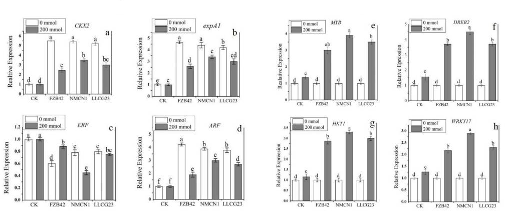
The selected Bacillus strains as PGPR, FZB42, NMCN1, and LLCG23, were able to grow on up to 10% NaCl, 18% NaCl, and 14% NaCl LB medium, respectively.
The inoculation of NMCN1 and LLCG23 significantly enhanced wheat growth parameters in terms of physiological traits, i.e., fresh weight 31.2% and 29.7%, dry weight 28.6% and 27.3%, shoot length 34.2% and 31.3% and root length 32.4% and 30.2%, respectively, as compared to control plants under high NaCl concentration (200 mmol).

It was found that salt-resistant genes in bacteria, DegU, OstB, OhrR, ComA, SodA, and OpuAC, were all up-regulated under saline conditions. And further, the plants inoculated with NMCN1 under salt stress (200 mmol NaCl) significantly overexpressed the genes in wheat related to expansin (expA1), cytokinin (CKX2) and auxin (ARF), followed LLCG23 and FZB42. The expression of ethylene encoding gene (ERF) was noticed to be highly downregulated in wheat plants treated with NMCN1 strain grown under the same stress condition.  The wheat plants treated with highly halophilic bacteria, NMCN1, were noticed to highly express the salt-resistant genes (MYB, DREB2, HKT1 and WRKY17), followed by LLCG23 and FZB42, as shown below.

Glycan binding specificity of LSEspecificity of LSECtin (CLEG4G) is different between solution NMR and Glycan

A group from Basque Research & Technology Alliance (BRTA), Chemical Glycobiology Group, CIC bioGUNE, Bizkaia, Spain, etc. has reported that solution NMR and surface-based microarray studies provide different results on the molecular recognition features of LSECtin toward bi-antennary N-glycans.
https://www.ncbi.nlm.nih.gov/pmc/articles/PMC9615123/

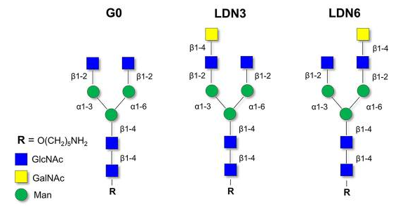
The molecular recognition features of LSECtin (CLEG4G) toward asymmetric N-glycans have been scrutinized by NMR and compared to those occurring in glycan microarrays. Strikingly, NMR studies confirmed that both asymmetric LDN3 and LDN6 N-glycans are recognized by LSECtin with similar affinities in solution, which is different in contrast to the results obtained when those glycans are presented on microarrays, where only LDN6 was efficiently recognized by the lectin.

Molecular recognition details differ from solution state to surfaces. Which one is closer to those existing in nature? Glycans are usually exposed on cell surfaces as part of glycoconjugates forming the glycocalyx. It is tempting to propose that the studies conducted using arrays are closer to those taking place on cell surfaces. However, the surface of slide glass is compretely different from actual cell surface glycocalyxes. Also, the length and chemical nature of the linkers used to attach the ligands to surfaces, and the composition of the solid support itself, could also influence the final outcome and the interpretation of the obtained results.

N-type glycosylated proteomics overview of human Alzheimer’s disease brain

A group from Department of Surgery, Beth Israel Deaconess Medical Center, Harvard Medical School, USA, etc. has reported about N-type glycosylated proteomics overview of normal, asymptomatic and symptomatic human Alzheimer’s disease brain.
https://www.mcponline.org/article/S1535-9476(22)00241-9/fulltext

The neuropathological hallmark of AD is the formation of β-amyloid (Aβ)-containing extracellular deposits and progression of intraneuronal neurofibrillary tangles from hyperphosphorylated tau.

To investigate the larger glycoprotein landscape, a qualitative N-type glycosylated proteomics analysis of brain samples was performed from 30 humans including those with normal, asymptomatic AD, and symptomatic AD. A combination of lectin affinity isolation and hydrophilic interaction chromatography (HILIC) was optimized and employed to selectively enrich N-glycoproteins prior to LC-MS/MS analysis. A combination of ConA, RCA, SNA, WGA, VVA, and AAL could target the major monosaccharide components of N-glycans in the brains. As a result, a total of 303 glycoproteins were identified from 2,035 unique glycopeptides, 580 N-linked glycosylation sites, and 124 glycans, making up a total of 1,901 glycoforms (remark: if a single glycosylation site was identified with three different glycans, they are counted as three glycoforms) that were identified from all samples.

The most abundant glycans range within the 1201-1250 MW range, which contribute to over 20% of the total glycoforms detected from the samples based on the glycan composition. The majority of glycans in this MW range are from Man5GlcNAc2 (Man5) structure. The second most frequent glycan is assigned as a biantennary glycan with a bisecting GlcNAc and core fucosylation. Other common glycans include several high-mannose, complex, and hybrid N-glycans with different degrees of fucosylation on glycopeptides were identified across the tissues. When considering the distribution based on the overall glycoforms, there is no statistical difference in the glycan molecular weight or glycan distribution among the different sample types.

To gain more information about the glycosylation pattern in different sample groups, it was analyzed at glycosylation site level, When comparing different sample types (normal, asymptomatic AD, and symptomatic AD) for the collective changes in glycosylation across different glycosylation sites, it was observed that a decrease in galactosylation, fucosylation, bisection, and the number of antennary glycans in asymptomatic and symptomatic AD samples, compared with normal brain samples. For asymptomatic compared to symptomatic AD samples, there are generally higher levels of galactosylation, fucosylation, bisection, and the number of antennary and hybrid glycans.

A definitive N-type glycan marker unique to Alzheimer’s disease has yet to be found.

Duality of the Vi Capsular Polysaccharide of Salmonella Typhi

A group from Department of Medical Microbiology and Immunology, University of California at Davis, Davis, California, USA, etc. has reported about duality of the Vi Capsular Polysaccharide of Salmonella Typhi.
https://journals.asm.org/doi/10.1128/mbio.02733-22

Bacterial pathogens overcome innate host defenses by either (i) evading opsonization and phagocytosis, which is the strategy used by extracellular pathogens, or (ii) evading killing during phagocytosis and take residence within the phagocyte, which is the approach taken by intracellular pathogens.

The enterica serovar Typhi is the causative agent of typhoid fever, a severe disseminated infection characterized by persistence of the pathogen in small granulomas, termed typhoid nodules, which are accumulations of mononuclear phagocytes and lymphocytes. S. Typhi synthesizes a virulence-associated (Vi) capsular polysaccharide, also known as the Vi-antigen, which protects bacteria from opsonization with natural IgM, thereby preventing engulfment by phagocytic host cells such as neutrophils and macrophages. However, it is paradoxical that S. Typhi has such an antiphagocytic capsule, because S. Typhi is typical intracellular pathogen.

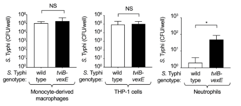
In this work, the duality of the S. Typhi Vi capsular polysaccharide was shown. The Vi capsular antigen allows S. Typhi to selectively evade phagocytosis by neutrophils, while promoting macrophage phagocytosis, by binding to DC-SIGN, C-type lectin expressed on macrophages.


where, tviB-vexE is a mutant of S. Typhi with noncapsulated

Immune-enhancing activity of polysaccharides from wild mushroom (Gymnopilus imperialis)

A group from Instituto de Ciências Ambientais, Químicas e Farmacêuticas, Universidade Federal de São Paulo, Brazil, etc. has reported about immune-enhancing activity of polysaccharides from wild mushroom (Gymnopilus imperialis).
https://www.mdpi.com/1424-8247/15/10/1179

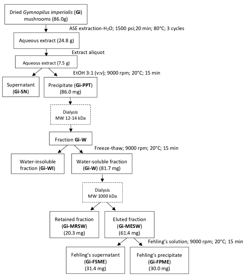
In this study, the aqueous extract was obtained from dried basidiomata of G. imperialis, which was fractioned by several steps yielding three main polysaccharide fractions (Gi-MRSW, Gi-FSME, and Gi-FPME) as shown below.

Among those polysaccharides, only the Gi-MRSW fraction displayed immune-enhancing activity on murine macrophages similar to LPS. The detailed molecular structure was not mentioned here.

Interactions between galectins and O-mannosylated core M1 glycopeptides of α-dystroglycan

A group from Frontier Research Center for Advanced Material and Life Science, Hokkaido University, Sapporo, Japan, etc. has reported about interactions between galectins and O-mannosylated glycopeptides of α-dystroglycan, especially focusing on its core M1 structure.
https://www.nature.com/articles/s41598-022-22758-0

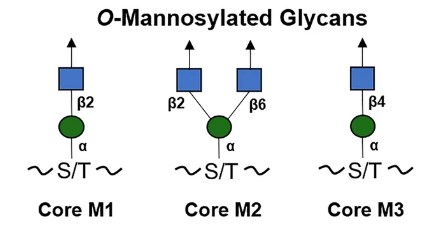
The O-linked mannose (O-Man) exists in a limited number of proteins that are required for normal development and have vital functions in muscle and neural physiology. The α-dystroglycan (α-DG) is the extracellular component of dystroglycan (DG), and is the most extensively studied mammalian O-Man glycoprotein. It is ubiquitously expressed in the skeletal muscles and the brain and is associated with cell adhesion, muscle integrity, and neurological development. α-DG possesses unique glycans, LacNac-terminated three kind of core structures (M1, M2, and M3), in its mucin (MUC)–like domain.

In this study, it was shown that Human Gal-1, -4, and -9 (except -3) can strongly recognize O-Man LacNAc-terminated glycoconjugates, and the presence of an α2,3-sialylated terminus led to a major reduction in the affinity of galectin, suggesting that this type of extension can fine-tune galectin activity towards this type of O-Man glycans. These interactions were significantly inhibited by lactose, establishing that the α-DG core M1-type glycans bind to the canonical sugar-binding site (S-face) of galectin, thus serving as a receptor for galectins.

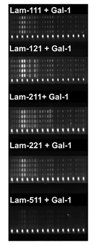
And further, it was shown in microarray experiments that Gal-1 revealed trans-bridging capabilities, linking laminin-111, -121, -211, and -221 (but little -511) and core M1 α-DG glycopeptides as shown below, providing a new insight on the therapeutic application of this galectin in muscular dystrophy.


Fluorescence images of M1 glycoconjugates microarrays with laminins plus galectins

Affects of AM fungi inoculation on soybean yield and the composition of microbial communities

A group from Engineering Research Center of Agricultural Microbiology Technology, Ministry of Education, Heilongjiang University, Harbin, China, etc. has reported about effects of inoculation of AM fungi (Rhizophagus intraradices) on soybean yield and the composition of microbial communities.
https://www.nature.com/articles/s41598-022-22473-w

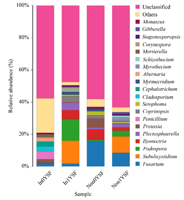
The field experiment was done in triplicate with AM fungal treatments (non-inoculated and inoculated with Rhizophagus intraradices) and continuous cropping regimes (0 and 1 year of continuous cropping for soybean) as factors, i.e., there were four conditions, In0, In1, Non0, and Non1.

The effect of AM fungal inoculation was seen greatly in the composition of fungal communities rather than the composition of bacterial communities. As shown below, the most dominant genus was Subulicystidium in In1YSF and Non1YSF. However, Fusarium was the most dominant genus in In0YSF and Non0YSF. Interestingly, the relative abundance of Fusarium decreased significantly  from 15.72% in Non0YSF to 1.58% in In0YSF.

In response to this, the disease index of soybean root rot was significantly decreased by the inoculation of AM fungi. For example, the disease index with the AM fungal inoculation decreased to 66%. The growth/yield indexes of soybean increased by the AM fungal inoculation, and it was the highest in the inoculated soybean plants under non-continuous cropping.

Modification of Glycan binding Specificity of E-selectin from sLex to 6′-sulfo-sLex with double mutations E92A/E107A

A group from Complex Carbohydrate Research Center, University of Georgia, Athens, GA 30602, etc. has reported that the specificity of E-selectin could be modified from sLex to 6′-sulfo-sialyl Lewis X with introducing double mutations.
https://www.ncbi.nlm.nih.gov/pmc/articles/PMC9564326/

Although lectins are often used to detect glycans, their application to sulfated glycans is challenging due to the paucity of sulfate-recognizing lectins as well as their broad or mixed specificities.

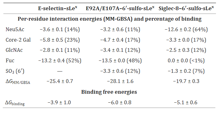
In this work, the binding specificity of E-selectin was modified by removing destabilizing steric and electrostatic interactions between the 6′-sulfate and E92 and E107 with E92A/E107A mutations, to show binding specificity to 6′-sulfo-sialyl Lewis X (6′-sulfo-sLex). As is known, E-selectin shows specific binding to non-sulfated ligand, sLex.
This new specificity mimics that of the unrelated protein Siglec-8, for which 6′-sulfo-sLex is its preferred ligand.

Powered by WordPress |Copyright © 2020 Emukk. All rights reserved