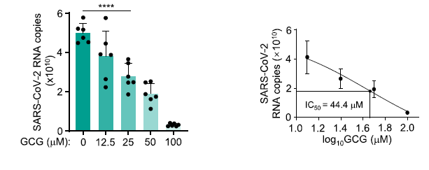
A polyphenol from green tea (GCG) could inhibit SARS-CoV-2 replication effectively

A group from Fudan University, etc. has shown that Gallocatechin gallate (GCG), a polyphenol from green tea, inhibits SARS-CoV-2 replication efficiently.
https://www.nature.com/articles/s41467-021-22297-8

SARS-CoV-2 N protein is a structural protein binding to RNA, and form a shell embracing SARS-CoV-2 RNA. This is a typical example of so called Liquid-Liquid Phase Separation (LLPS) which is a mechanism in organizing macromolecules such as proteins and RNAs into membrane-less oil droplet like organelles.
Authors has shown that GCG could could inhibit N protein LLPS in the context of SARS-CoV-2 infection effectively. Since, the amino acid sequence shares ~90% homology among corona viruses, targeting N protein by GCG could be a novel drug candidate not only for SARS-CoV-2 but also for new coronaviruses in the future.

Effects of glycosylation and disulfide bonding of SARS-CoV-2 Spike protein

A group from Harvard Medical School, etc. has reported on the effects of glycosylation and disulfide bonding of SARS-CoV-2 Spike protein onto infectivity and susceptibility to antibody inhibition.
https://pubmed.ncbi.nlm.nih.gov/33821278/

There are 22 N-glycosylation sites, 10 O-glycosylation sites and 15 disulfide bonding in the spike protein.
In this paper, it was shown experimentally how the mutations at O-glycosylation sites(676, 1170)affect binding to ACE2 and titer of sera from convalescing SARS-CoV-2-infected individuals. Let me introduce that point for this blog readers. The evaluated mutations were T676I and S1170F. As shown below, it seems that these mutations do not affect so much.

A key why children do not develop severe COVID-19 is in strong innate immune response in nasopharyngeal mucosa

In the case of influenza or respiratory syncytial virus, children are often more severe than adults. However, in the case of COVID-19, children infected with SARS-CoV-2 have a milder clinical course with significantly less morbidity and mortality than adults.
Several hypotheses have been proposed to explain why children are more protected than adults, for example, expression of angiotensin-converting enzyme 2 (ACE2) is lower in children, resulting in lower viral loads, presence of antibodies to common cold coronaviruses (229E, NL63, HKU1)that might provide partial protection is stronger in children, and a more robust innate response in children in early course of infection that mitigates against a vigorous adaptive response.

A group from Albert Einstein College of Medicine, etc. has discovered that nasopharyngeal mucosal immune response is stronger in children. It is speculated that the immune response is strengthened in nasopharyngeal mucosa because children have more frequent respiratory infections than adults. Measured cytokines in nasopharyngeal swab fluid (IFN-γ, IFN-α2, IL-1β, IL-8) was significantly upregulated in children than adults. So, the robust mucosal immune response lowers viral loads and gives protection against advancing in severity.
https://insight.jci.org/articles/view/148694

IL-1R7 antibody would be effective in COVID-19 to suppress cytokine storms

A group from University of Colorado Denver Anschutz Medical Campus has reported that IL-1R7 antibody would be effective in COVID-19 to suppress cytokine storms.
https://www.jbc.org/article/S0021-9258(21)00416-6/fulltext

Excessive inflammation observed in macrophage activation syndrome (MAS) results in severe diseases with high mortality. The cytokine storms observed in COVID-19 patients would be a typical example similar to MAS.
Interleukin-18 (IL-18), a proinflammatory cytokine belonging to the IL-1 family, is elevated in both MAS and COVID-19 patients, and its level is known to correlate with the severity of COVID-19 symptoms

IL-18 binds its specific receptor IL-1 Receptor 5 (IL-1R5, also known as IL-18 Receptor alpha chain), leading to the recruitment of the co-receptor, IL-1 Receptor 7 (IL-1R7, also known as IL-18 Receptor beta chain).

Authors found that the anti-IL-1R7 antibody significantly suppressed IL-18-mediated NFκB activation, IL-18-stimulated IFNγ production, and IL-6 production in human cell lines.

Seroprevalence of anti-SARS-CoV-2 IgG antibodies in COVID-19 patients 

A group from Juntendo University Faculty of Medicine has reported seroprevalence of IgG and IgM antibodies in COVID-19 patients, although the cohort size was small including only 34 patients.
https://www.ncbi.nlm.nih.gov/pmc/articles/PMC8023454/

Using a chemiluminescent microparticle immunoassay (CMIA)-based SARS-CoV-2 IgG test (cat. # 06R90, Abbott),
Severe/Critical cases:within a week after symptom onset=40%、1~2 weeks=88%、after two weeks=100%,
Mild/Moderate cases:within a week after symptom onset=0%、1~2 weeks=38%、after two weeks=100%.

Using an IC IgG antibody assay using the Anti-SARS-CoV-2 Rapid Test (cat. # RTA0203, AutoBio),
Severe/Critical cases:within a week after symptom onset=60%、1~2 weeks=63%、after two weeks=100%,
Mild/Moderate cases:within a week after symptom onset=17%、1~2 weeks=63%、after two weeks=100%.

In this study, IgG titers remained at significantly elevated levels for 2 months, regardless of disease severity. These results indicate that IgG serologic tests could be used as a complementary test to PCR to diagnose COVID-19 from 14 days after symptom onset. However, since this cohort is so small without including asymptomatic individuals, a larger scale cohort is needed to conclude final answer.
     

Difference in site-specific glycosylation of SARS-CoV-2 spike proteins (all recombinants) among several laboratories

A group from University of Southampton, etc. has compared site-specific glycosylation of SARS-CoV-2 spike proteins (all recombinants) among five laboratories.
The cells used for Sproten expression were as follows.
HEK293: Amsterdam, Harvard,
HEK293F: Southampton/Texas,
HEK293T: Oxford,
CHO: Swiss,
It is celarly shown that site-specific glycosylation changes considerablly with reflecting differences in cells and culture conditions.
Fundamentally speaking, it is a mixture of oligo mannose and complex type N-glycans.

Blog admin is interested in how much these differences cause difference in the infectivity, how glycosylation changes with SARS-CoV-2 mutations, and how much the glycosylation changes due to mutations affect the infectivity.
https://www.biorxiv.org/content/10.1101/2021.03.08.433764v1.full

Characteristics of  SARS-CoV-2 neutralizing antibodies targeting NTD Spike protein: Effects of Glycans and Variants

A group from Columbia University, etc. has reported about characteristics of SARS-CoV-2 neutralizing antibodies targeting NTD Spike protein.
https://www.ncbi.nlm.nih.gov/pmc/articles/PMC7953435/

Totally 17 NTD antibodies have been already published. It was found that these antibodies target a specific area (called Supersite) on NTD of the spike protein. The NTD supersite was located at the periphery of the spike protein, distal from the 3-fold axis, and facing away from the viral membrane. This surface was surrounded by four N-glycans, N17, N74, N122, and N149, and nominally “glycan free,” although there are some glycan coverage because of the dynamic motional fluctuation of glycans. And also, the supersite had strong positive electrostatic potential, while recognizing antibodies had complementary strong electronegative potential.

The SARS-CoV-2 variants, particularly B.1.1.7 and B.1.351, are concerning due to increased infectivity, and these variants escape most NTD-directed neutralizing antibodies. B.1.1.7 includes NTD deletion mutations D69-70 and D144, and strain B.1.351 includes NTD mutations D242-244 and R246I. The mutated positions including 144, 242-244, and 246 are all within the NTD supersite. While the deletion at 69-70 is outside of the supersite, it forms part of the hairpin N2 loop of NTD. So, its deletion could significantly impact the conformation of the NTD supersite.

A new pathway was discovered in activation of the complement system of the innate immunity: MASP-1 and MASP-3 have dual functions as enzymes and as PRMs 

In the lectin pathway as one of innate immunities, pattern recognition molecules (PRMs) (i.e., mannose-binding lectin (MBL), ficolins, and collectin-10/-11) bind to pathogens recognizing specific molecular structures on the membranes, make complexes with MAPSs, activate complements sequentially, and finally form Membrane Attack Complex (MAC) on the membranes to kill pathogens.

However, a group from University of Copenhagen, etc. has reported a new pathway in which MAPSs directly bind to pathogens and activate the complement system using Aspergillus fumigatus as a pathogen. This means that a new pathway in the complement system was discovered showing MASP-1 and MASP-3 have dual functions as enzymes and as PRMs
https://www.karger.com/Article/FullText/514546

A cocktail monoclonal antibody (REGN-COV2) is unaffected by B.1.1.7, B.1.351, P.1 variants, however, the vaccine Pfizer BNT162b2 shows markedly reduced inhibition against B.1.351, P.1 variants 

A group from German Primate Center, Göttingen, etc. has reported on the effectiveness of major monoclonal antibodies for COVID-19 (Casirivimab, Bamlanivimab, Imdevimab)against SARS-CoV-2 variants, that of a cocktail monoclonal antibody (REGN-COV2: consisting of Casirivimab and Imdevimab), and also that of Pfizer BNT162b2 vaccine against those variants.
https://www.ncbi.nlm.nih.gov/pmc/articles/PMC7980144/

A cocktail monoclonal antibody (REGN-COV2)efficiently inhibited infection mediated by the S proteins of all variants (B.1.1.7, B.1.351, P.1). However, infection mediated by the S proteins of the B.1.351 and P.1 variant was completely resistant to REGN10989 and Bamlanivimab.

On the other hand, the Pfizer BNT162b2 vaccine is based on an mRNA that encodes for the SARS-CoV-2 S protein and is said to be highly protective against COVID-19. All sera from 15 donors immunized twice with BNT162b2 efficiently inhibited entry driven by the WT S protein and inhibition of entry driven by the S protein of the B.1.1.7 variant was only slightly reduced. However, 12 out of 15 sera showed a markedly reduced inhibition of entry driven by the S proteins of the B.1.351 and P.1 variants


Diagnostic accuracy of COVID-19 Chest CT images with using Deep Learning methodology

A group from Sejong University, Seoul, etc. has reported on the accuracy of COVID-19 diagnosis in chest CT images with applying Deep Learning.
https://journals.plos.org/plosone/article?id=10.1371/journal.pone.0249450

The neural network using in Deep Learning was composed of 20 stages, and convolution and pooling functions were incorporated. The resolution of the input images were (224 x 224, and the sizes of convolution were (3 x 3)and(5 x 5). The obtained overall accuracy was 99.83%(sensitivity=0.9286, specificity=0.99). In the future, it will be accelerate to adopt Deep Learning in diagnostic applications.