Vaccine effectiveness of Pfizer-BioNTech or Moderna vaccine against COVID-19

CDC COVID-19 Response Team has reported about the vaccine effectiveness of Pfizer-BioNTech or Moderna vaccine.
https://www.cdc.gov/mmwr/volumes/70/wr/pdfs/mm7018e1-H.pdf

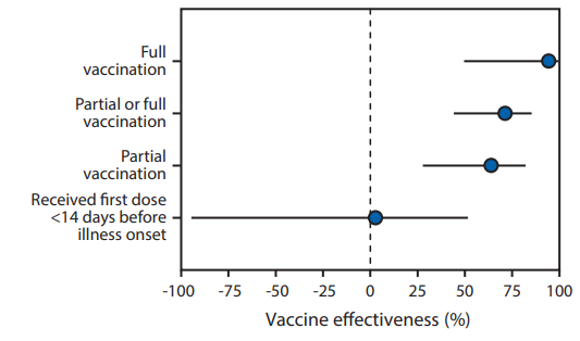
The team has reported that adjusted vaccine effectiveness for full vaccination using Pfizer-BioNTech or Moderna vaccine was 94% (95% CI = 49%–99%), and that for partial vaccination was 64% (95% CI = 28%–82%).
In this study, the vaccine effectiveness was assessed among 417 adults (including 187 case-patients and 230 controls) aged ≥65 years who admitted with COVID-19–like illness and confirmed by RT-PCR during January 1, 2021–March 26, 2021. Case-patients were those who received one or more positive test results for SARS-CoV-2. Patients meeting eligibility criteria who received negative SARS-CoV-2 RT-PCR test results served as controls. Participants in this study were considered to have received COVID-19 vaccine doses based on official CDC vaccination record card etc. and by plausible self-report.

It should be noted that there was no significant effect for receiving the first dose of a 2-dose COVID-19 vaccine series within 14 days before illness onset (adjusted vaccine effectiveness = 3%, 95% CI = −94%–51%)

Using lectin–glycan interactions to combat COVID-19 reviewing past instances against enveloped viruses

A group from Universidade CEUMA, Brazil, etc. has reviewed past instances using lectin–glycan interactions to combat enveloped viruses, and mentioned about challenge to SARS-CoV-2.
https://academic.oup.com/glycob/article/31/4/358/5934657

The presence of glycoproteins in the viral envelope opens a wide range of possibilities for application of lectins to fight COVID-19. Lectins could be leading molecules for the development of new antiviral drugs due to their ability to inhibit viral entry into the host cell by binding to glycans expressed on he virus envelop.

The overall view of glycosylation profiles of SARS-CoV-2 is as follows. There are 22 N-glycosylation sites and 6 O-glycosylation sites on the spike protein.  The oligomannose-type glycans are predominant in two sites (N234 and N709). Complex-type glycans are predominantly expressed in 14 amino acid residues (N17, N74, N149, N165, N282, N331, N343, N616, N657, N1098, N1134, N1158, N1173 and N1194), while six sites show a mixture of oligomannose- and complex-type glycans (N61, N122, N603, N717, N801 and N1074). The most common configuration of oligomannose-type glycans was Man5GlcNAc2. The short O-glycans such as Tn and core 1  structures are mainly expressed in 6 sites (T73, T76, T478, T676, T678, T1076).

Typical lectins evaluated as antiviral drugs are explained in the review paper (FRAIL, GRFT, Cyanovirin-N, BanLec, MVN, and Avaren). All of these lectins have binding specificity to oligo and high mannose, probably because envelopes of targeted viruses in the past (e.g., HIV, HCV, influenza, and Ebora) were heavily mannosylated. In that sense, we can say that glycosylation profile of SARS-CoV-2 is somewhat different from other enveloped viruses, and therefore, there might be more suitable lectins for SARS-CoV-2 besides those lectins.

Anyhow, the major limitation in using lectins as therapeutic drugs is the possible binding of the lectin in unwanted glycosylated targets. For instance, the administration of lectins could result in the agglutination and proliferation of cells. Therefore, it is so important to modify lectins with using protein engineering to suppress such side effects and to extract only the antiviral property against SARS-CoV-2. Probably, the key word would be making fusion proteins, e.g., fusion to Fab, fusion to Fc, PEGylation, and so forth. 

Characteristics of 12 monoclonal antibodies targeting to SARS-CoV-2 NTD 

A group from Israel Institute for Biological Research, etc. has reported on characteristics of 12 mAbs targeting to SARS-CoV-2 NTD.
https://pubmed.ncbi.nlm.nih.gov/33937725/

12 antibodies (from BLN1 to BLN14) targeting to SARS-CoV-2 NTD were able to efficiently neutralize SARS-CoV-2, exhibiting IC50 values ranging from 54.9 µg/ml for BLN8, to a highly potent values of 0.008 µg/ml for mAbs BLN1 and BLN12. All mAb specificity to NTD was confirmed with ELISA against NTD, and also in detail by an peptide array derived from the entire S1 protein. Then, the effects of glycan modification onto mAbs were investigate by using a glycan array and also with a competition assay adding glycans as inhibitors. While there was very low binding to most examined glycans, mAbs revealed a strong preference to LacNAc and its sialo-form suggesting that the interactions with the N-glycans are involved in the binding to NTD of BLN4 and BLN12 in particular.

It was recently reported that in addition to the “canonical” ACE2 receptor binding by RBD, SARS-CoV-2 NTD interacts with C-type lectin receptors, including L-SIGN and DC-SIGN as an alternative route for SARS-CoV-2 infection and entry into host cells. So, the ability of four representative mAbs to inhibit the interaction of the NTD with L-SIGN, was examined. In the presence of selected anti-NTD mAbs, inhibition efficiency ranged from approximately 25% binding inhibition by BLN14 to 50% inhibition by BLN4. It should be noted, that the observed binding inhibition may be the result of steric interference of L-SIGN binding to NTD, and not necessarily direct blocking of the NTD glycans, involved in binding to L-SIGN.

If L-SIGN and DC-SIGN are indeed alternative receptors for SARS-CoV-2 and mediate its infection of cells in many tissues lacking hACE2, it may be speculated that anti-NTD mAbs may also exert their therapeutic activity by limiting the spreading of the virus in the body. As a conclusion, BLN12 and BLN14 mAbs would represent excellent candidates for therapy of SARS-CoV-2, possibly in combination with anti-RBD mAbs as a cocktail mAb for SARS-CpoV-2. 

How much risk is there in SARS-CoV-2 reinfection? 

A group from Cornell University, Doha, Qatar, etc. has reported how much risk is there in reinfection of SARS-CoV-2.
https://www.thelancet.com/journals/eclinm/article/PIIS2589-5370(21)00141-3/fulltext

Among 43,044 individuals with SARS-CoV-2 antibody-positive from Apr. 16th to Dec. 31th, 2020, persons with PCR-positive swab ≥14 days after the first-positive antibody test were thought to be reinfected.

As a result of this study, the efficacy against reinfection was estimated at 92.7% (95% CI: 91.3–93.9%). It is so interesting that this efficacy estimate is similar to the efficacy reported for the two mRNA COVID-19 vaccines, and reinfections were less severe than primary infections. That is good news for all of us.

A new Biomarker for predicting therapeutic effectiveness of anti-TNF mAbs for Ulcerative colitis: Changes in Jacalin-specific IgG glycosylation

A group from University of Pisa, Italy, etc. has reported a new biomarker for ulcerative colitis.
https://www.frontiersin.org/articles/10.3389/fphar.2021.654319/full/

Ulcerative colitis (UC) is a chronic relapsing disease, characterized by an inflammation affecting the colon and the rectum with superficial mucosal ulceration, rectal bleeding, diarrhea and abdominal pain. Tumor necrosis factor (TNF) plays an important role in UC pathogenesis. Indeed, several immune cells produce high levels of TNF, and this cytokine is known to mediate several pro-inflammatory functions in the inflamed mucosa, promoting even tissue injury. On this basis, the monoclonal antibodies anti-TNF, like infliximab (IFX) and adalimumab (ADA), were developed for treatment of UC.

However, more than 30% of the UC patients receiving anti-TNF agents do not respond to treatment. Therefore, developing an early prediction method of therapeutic outcome is one of the most important challenges to optimize therapeutic management.
Authors has shown that Jacalin(JAC)-specific IgG glycosylation could be used as a biomarker of disease activity in UC, especially before starting an anti-TNF therapy. JAC is an O-glycan binder, and the putative role of JAC in predicting therapeutic effectiveness from the O-glycan point of view was not clearly demonstrated here, but it could be used as a good biomarker predicting therapeutic responses.

Many autoimmune diseases are associated with qualitative perturbations in circulating antibodies, and agalactosylated IgG is considered a marker of inflammatory condition. Agalactosylated IgG is capable to interact with FcγRIIa, induce macrophage activation and, subsequently, induce pro-inflammatory cytokines such as production of TNFα and IL6. Several studies showed that agalactosylation could be reverted in therapeutic conditions capable of ameliorating the inflammatory diseases. It has been known that in rheumatoid arthritis, steroid and anti-TNF therapy induce changes in IgG glycosylation from agalacto to sialo-complex-types.

Significant therapeutic effect of Tocilizumab was observed in patients with IL-6>100pg/mL and oxugen saturation<90%

A group from Medical University of Białystok, Poland, etc. has reported about therapeutic effects of Tocilizumab on COVID-19.
https://www.mdpi.com/2077-0383/10/8/1583/htm

In this clinical study with total 825 patients, Tocilizumab was administered to 170 patients and nothing was administered to 655 patients not only Tocilizumab but also any mAbs for cytokine receptors. The dose of Tocilizumab was 8mg/kg (the maximum dose =800mg) in a single dose.

In case of IL-6>100pg/mL, significant therapeutic effect of Tocilizumab was obtained (hazard ratio [HR]=0.21(95% confidence interval [CI]: 0.08–0.57). When IL-6>100pg/mL and either oxygen saturation <90% or requiring oxygen supplementation, HR decreased to 0.18. In addition, it was concluded that Tocilizumab has little therapeutic effect when IL-6<100pg/mL.
Anyhow, it is so great to know that Tocilizumab shows its therapeutic effect on what levels of serum IL-6 and how much dose is required for that.

Molecular structures at the binding site of the SARS-CoV-2 spike protein of N501Y variant and ACE2 receptor

A group from University of British Columbia, etc. has reported studies on  molecular structures at the binding site of the SARS-CoV-2 spike protein of N501Y variant and ACE2 receptor with using a cryo-electron microscopy.
https://journals.plos.org/plosbiology/article?id=10.1371/journal.pbio.3001237

The overall binding structure at the binding site of ACE2 and SARS-CoV-2 is almost identical to that of the unmutated version, with the exception of some local rearrangements. The aromatic ring of Y501 is sandwiched between Y41 and K353 of the ACE2 receptor, and  Y501 in the SARS-CoV-2 spike protein and Y41 in the ACE2 receptor form a perpendicular y-shaped π–π stacking interaction. This could be a reason of the increased infectivity.

The same conclusion from other researchers as this was already blogged.

Efficacy of Camostat Mesilate (TMPRSS2 inhibitor) for COVID-19 was disappointing

A group from Aarhus University Hospital, Denmark, etc. has reported on efficacy of Camostat Mesilate (which is a TMPRSS2 inhibitor) for COVID-19.
https://www.ncbi.nlm.nih.gov/pmc/articles/PMC8060682/

Authors have investigated it through a double-blind, randomized, placebo-controlled trial. The hypothesis behind it was that TMPRSS2 inhibition would block SARS-CoV-2 replication in infected patients leading to reduced viral loads, and that this in turn would lower the risk of hyper-inflammation and prevent disease progression. However, the results from the double-blind randomized placebo-controlled trial showed that camostat mesilate treatment did not significantly improve time to clinical improvement, the risk of intubation or death, time to discontinuation of supplemental oxygen, or any other efficacy outcomes among patients hospitalized with COVID-19.

Although the dose of camostat mesilate was not optimized in this trial, but Blog Admin feels that the famous infection passway through the ACE2-TMPRSS2 initiation is not major in pulmonary epithelial cells, but there might be other infection passways through C-type lectins expressed on immune cells and phagocytosis, and so forth.

The N90 glycan of ACE2 shields ACE2 from SARS-CoV-2 RBD binding, and the N322 glycan strengthens RBD binding, conversely 

A group from Max Planck Institute of Biophysics has simulated effects of ACE2 glycosylation onto binding to SARS-CoV-2 RBD.
https://www.pnas.org/content/118/19/e2100425118.long

Structurally speaking, there would be four glycosylation sites on ACE2 (N53, N90, N103, and N322)which have the possibility to interact with the RBD. The difference in glycan structures also affects binding strength to RBD, and asialo-types and high mannose show stronger interactions with the RBD at N70 and N322 positions. At the N90 position, high mannose has stronger affinity than asialo-types, and at the N322 position, vice versa.

Effects of glycans onto binding to RBD are different between N90 and N322 positions. The N90 glycan shields ACE2 from RBD binding, and the N322 glycan strengthens RBD binding, conversely. Actually, it has already reported that almost all mutations removing N322 glycosylation site are detrimental to the binding of the RBD. Since the N322 glycan interacts mainly with Y369–K378, R408, N437, N439, and V503 near the site of the N501Y mutation, it might be associated with increased ACE2 binding affinity and enhanced infectivity.

For your information, two articles related to effects of ACE2 glycosylation on to RBD binding have already introduced in this blog site.
The same thing has already demonstrated experimentally as sialic acids and high mannose weaken SARS-CoV-2 binding to ACE2.

A shore where SeviL is buried : Septifer virgatus

There is an unique lectin named “SeviL” which has binding affinity to ganglioside GM1band and its precursor form, asialo-GM1.
https://www.nature.com/articles/s41598-020-78926-7

This lectin could be extracted from Septifer virgatus.
A picture below shows its cluster.
The amount of Sevil contained in one Septifer virgatus is about 0.004mg. Taking the popuration around there into consideration, there would be 100g SeviL per 10 square meters.


That day was cloudy and windy, but here is a shore where Septifer virgus is living.