Expression status of C-Type lectin receptor 5A (CLEC5A) on Monocyte-derived Macrophages and its functions

A group from Cancer Immunology & Immune Modulation, Boehringer Ingelheim Pharma GmbH & Co. KG, Germany, etc. has reported about expression status of C-Type lectin receptor 5A (CLEC5A) on Monocyte-derived Macrophages (MdM) and its functions.
https://www.ncbi.nlm.nih.gov/pmc/articles/PMC8896916/

CLEC5A also known as myeloid DAP12-associating lectin-1 (MDL-1) is a myeloid Syk-coupled pattern recognition receptor which preferentially binds to glycans highly expressed on the surface of pathogens. CLEC5A is mainly expressed on myeloid cells (monocytes, macrophages, neutrophils, and dendritic cells) and can be further upregulated by interferon-γ. The ligand for CLEC5A was identified as terminal fucose and mannose moieties of viral glycans, expressed by dengue virus, Japanese encephalitis virus or type A influenza virus. In addition, CLEC5A binds to disaccharides (N-acetylglucosamine and N-acetylmuramic acid) of bacterial cell walls (e.g., Listeria monocytogenes and Staphylococcus aureus). Functionally, the CLEC5A receptor activation triggered by dengue virus or by other pathogens induces the production of proinflammatory cytokines (TNF-α, IL-1, IL-6, IL-8, and IL-17A) and chemokines: macrophage inflammatory protein-1 alpha (MIP-1α/CCL3), interferon-gamma induced protein (IP-10/CXCL10), and macrophage-derived chemokine (CCL22/MDC) . It was also found that CLEC5A may recognize not only pathogen-associated antigens but also some endogenous danger signals and in consequence could contribute to the pathogenesis of the aseptic inflammation.

In this report, expression status of CLEAC5A on monocyte-derived macrophages and functional consequences of the selective activation of CLEC5A by α-CLEC5A Ab were discussed in aseptic conditions as well as their impact on the activation of autologous T cells.

Expression of CLEC5A on myeloid cells
Expression of CLEAC5A was compared among several MdM: proinflammatory M1, “neutral” M0, and protumorigenic M2c, and recently described in vitro tumor-associated macrophages (TAM). The CLEC5A expression was significantly elevated in proinflammatory M1 MdM as compared to monocytes and to other MdM subsets (M0 and M2c), while monocyte differentiation towards TAM resulted in a reduction of CLEC5A expression.

Functional effects of CLEC5A activation
In order to understand the functional effects of CLEC5A agonist under noninfectious conditions, the cytokine response in M0 MdM was evaluated. M0 MdM exposed to the α-CLEC5A Ab significantly upregulated the secretion of cytokines and chemokines such as TNF-α, IL-6, IL-10, IL-1b, CCL22/MDC, CCL17/TARC, and Matrix metalloproteinase (MMP1). Interestingly, the CLEC5A activation induced up-regulation of some myeloid cell-specific surface receptors, such as CD80, PD-L1, and CD206(MRC-1), and CD209(DC-SIGN).

Finally, the selective CLEC5A-mediated reprogramming of myeloid cells in aseptic conditions appeared insufficient to promote an autologous T cell activation.

Lectin Cytotoxicity

A group from Institute of Physiology, University of Zurich, Zurich, Switzerland has reported about lectin cytotoxicity.
https://www.ncbi.nlm.nih.gov/pmc/articles/PMC8866831/

While targeting surface glycans by lectins is a promising approach for cancer therapies, lectin-based approaches (lectin alone or in a combination with other therapeutic agents) are still at an early stage of development. The development of efficient therapies based on glycan targeting requires a deep understanding of the mechanisms of cell death induced by lectins on target tumor cells. It has been known that specific lectins induce distinct modes of cell death in different type of tumor cells. Wheat germ agglutinin (WGA), for example, induces apoptosis in melanoma and leukemic cell, whereas it kills cervical carcinoma cells through paraptosis-like cell death.

In this report, using murine adenocarcinoma cells (MC-38) and young adult mouse colon cells (YAMC), the multiple cell death pathways (apoptosis, necroptosis, proptosis, paraptosis, and autophagy-dependent cell death) activated in response to treatment of cells with lectins (MAL I, MAL II, SNA, AAL, WGA, and ECL) were studied with targeting different glycan structures. In order to characterize the signaling pathways mediating cell death induced by the cytotoxic lectins, a panel of MC-38 cells with knockouts in genes involved in cell death responses was used.

Knockouted genes are as follows;
pro-apoptotic proteins BCL2 antagonist/killer 1 (BAK1) and BCL2 associated X (BAX), which mediate intrinsic apoptosis, the intrinsic apoptosis signaling cascade is activated in response to various internal cell stress factors, such as DNA damage.

Fas-associated via death domain protein (FADD), which mediates extrinsic apoptosis, the extrinsic apoptosis pathway is induced in response to activation of cell death receptors, followed by formation of the death-inducing signaling complex that includes FADD and pro-caspase-8, which cleaves the executioner caspase-3.

tumor necrosis factor receptor type 1-associated death domain protein (TRADD), receptor-interacting serine/threonine protein kinases 3 (RIPK3), mixed lineage kinase domain-like protein (MLKL) and caspase-8 (CASP8), which mediate caspase-independent necroptosis.

caspase-1 (CASP1) and gasdermin D (GSDMD), which mediate pyroptosis, CASP1 is activated by the inflammasome in response to various microbial infections and non-infectious stimuli. The pathway leads to GSDMD cleavage, which embeds in the plasma membrane and forms pores that disrupt ionic gradients and facilitate water influx, hence leading to cell swelling and osmotic lysis.

Results:
The inactivation of the BAX/BAK1 complex decreased the cytotoxic response induced by WGA, MAL I and AAL treatment. The decrease in cytotoxicity by more than 50% was similar to the effect achieved in cells treated with cisplatin, which is a classical trigger of apoptosis. By contrast, the inactivation of FADD did not impacted the cell death mediated by WGA, MAL I and AAL.

The inactivation of TRADD, another adaptor molecule required for activation of apoptosis and necroptosis downstream of tumor necrosis factor receptor 1 (TNFR1), decreased cell death in cells treated with MAL I, but not when WGA and AAL was added. The loss of MLKL decreased the cytotoxic effect of WGA, MAL I and AAL, indicating the contribution of the necroptosis pathway in cell death induced by these lectins.

Inactivation of either CASP1 or GSDMD showed only a minor decrease in MAL I-mediated cytotoxicity, supporting a partial involvement of pyroptosis in response to MAL I treatment.

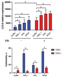
In addition to the induction of apoptosis, many lectins also up-regulate autophagy sometimes resulting in autophagy-dependent cell death. LC3-II is associated with autophagosome membranes. Treatment with MAL I, AAL and WGA up-regulated LC3-II in lysates from cells treated for 6 h with lectins. The increase in LC3-II levels indicated that cell death induced by MAL I, AAL and WGA was probably initiated through activation of the autophagic/lysosomal response rather than the classical apoptotic mitochondrial pathway. Actually by adding cycloheximide (CHX), is known to block starvation-induced autophagy, the cytotoxic response of the three lectins was significantly reduced.

Changes in rhizosphere microbiome with wheat evolution: from ancestral species to domesticated species

A group from Department of Plant Breeding and Biotechnology, Faculty of Agriculture, University of Tabriz, Tabriz, Iran, etc. has reported on changes in rhizosphere microbiome with wheat evolution (from ancestral wheat species of T. uratu and Ae speltoides (ancestral species, 1.5 to 2million years ago) to domesticated pecies of T. turgidum and T. aestivum).
https://www.ncbi.nlm.nih.gov/pmc/articles/PMC8881823/

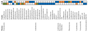
It was found that several families belonging to Actinobacteria and Proteobacteria revealed significant increases in domesticated crops (T. turgidum and T. aestivum) when compared to ancestral species (T. urartu, and Ae. speltoides) as shown below.


Differential family-level abundances in (A) domesticated and (B) ancestral wheat species during different developmental stages with color. Orange = vegetative, blue = reproductive, and green = comon in both developmental stages, respectively.

Complement activation might be strongly involved in systemic worsening of COVID-19: studies from autopsy lung and kidney samples

A group from Department of Nephropathology, University Hospital Erlangen, Friedrich-Alexander-University (FAU) Erlangen-Nürnberg, Erlangen, Germany, etc. has reported that complement activation might be involved in systemic worsening of COVID-19.
https://www.ncbi.nlm.nih.gov/pmc/articles/PMC8884149/

One pathway potentially involved in the SARS-CoV-2 driven inflammatory cytokine overproduction is activation of the complement system. This system, which belongs to the innate immune system, acts as a crucial component in the defense against infection by opsonizing pathogens or damaged cells, attracting and activating leukocytes or directly lysing bacteria or cells through the membrane attack complex.

Initiation of the complement cascade can be done by three different pathways, the classical pathway, the lectin pathway, and the alternative pathway.
The classical pathway is activated by immune complexes (antigen-IgM and antigen-IgG complexes) and many other self and non-self molecules binding to C1q, leading to a conformational change, and activating serine proteases C1s and C1r.
The lectin pathway is activated by plasma-circulating lectins (collectins like mannan-binding lectin and ficolins) recognize carbohydrate patterns on the surface of microorganisms, called pathogen-associated molecular patterns (PAMPs), and activate MAPS-1 and MAPS-2 which cleave C4 to C4a and C4b.
The alternative pathway is constantly active at a low level and is initiated by spontaneous hydrolysis of C3. Hydrolysed C3 binds to factor B (CFB) which acts as a substrate to serine protease factor D (CFD), resulting in formation of a C3 convertase.
In the end, all 3 complement pathways can lead to C3 convertase activation which cleave C3 to C3a and C3b, followed by the same pathway triggering the formation of the membrane attack complex (MAC).

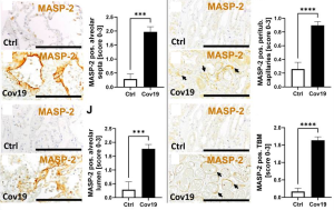
In this study, though comparative study of autopsy lung and kidney tissue samples died from COVID-19 with those of died from non-COVID-19, it was found that the lectin pathway was activated in both lungs and kidneys of patients with severe COVID-19. Activation of the lectin pathway was supported by staining for C4d in the same localization as described for MASP-2, suggesting that complement activation might be strongly involved in systemic worsening of COVID-19 inflammatory response.

< a href="">

Possible pathogenesis of worsening COVID-19 could be direct activation of platelets by SARS-CoV-2 spike protein

A group from Department of General and Laboratory Medicine, Mie Prefectural General Medical Center, Yokkaichi 510-0885, Japan, etc. has reported importance of the spike protein-induced direct platelet activation as a basis of COVID-19 pathogenesis.
https://www.ncbi.nlm.nih.gov/pmc/articles/PMC8877880/

Several mechanisms underlying the worsening of the condition of COVID-19 patients have been proposed, for instance, cytokine storm, primary pulmonary thrombosis, vascular endothelial injuries, and platelet activation. One of the clinical features of COVID-19 is the high prevalence of arterial thrombosis such as stroke and ischemic heart disease. Therefore, it was assumed that the activation in platelets would be involved in the pathogenesis. On the other hand, soluble C-type lectin-like receptor 2 (sCLEC-2) has been discovered as a new biomarker of platelet activation.

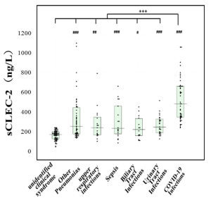
In this study, the levels of plasma biomarkers, such as sCLEC-2 and D-dimer, in 46 patients with COVID-19 were measured and compared to those in 127 patients with other infections to determine the mechanism underlying the worsening of COVID-19 infection.

Plasma sCLEC-2 levels were significantly higher in patients with COVID-19 infection than in those with bacterial infections. On the other hand, plasma D-dimer levels were significantly higher in patients with bacterial infections than COVID-19 infection. These findings suggest that COVID-19 infection tends to facilitate platelet activation, while bacterial infections tend to facilitate fibrin generation.

Possible pathogenesis of worsening COVID-19:
SARS-CoV-2 spike protein activates platelets directly via apoptosis of lymphocytes and induces thrombosis.

Glycan binding specificities of the SARS-CoV-2 spike protein

A group from Department of Medical Laboratory Science and Biotechnology, College of Medicine, National Cheng Kung University, Tainan 70101, Taiwan, etc. has reported about glycan binding specificities of the SARS-CoV-2 spike protein.
https://www.ncbi.nlm.nih.gov/pmc/articles/PMC8880561/

Assay:
Donor beads (500 ng/well) and biotin-polyacrylamide (PAA)-sugars (20 ng/well) mixed with SARS-CoV-2 spike protein S1 or S2 subunits (10–20 ng/well) were incubated at room temperature for 1 h. A mixture of acceptor beads (500 ng/well) and rabbit antisheep IgG Fc antibodies (10 ng/well) was added to the reaction to reach a final volume of 25 μL. All reactions were performed in the dark. After incubation for 2 h, the binding signals were measured and analyzed using the AlphaScreenTM detection program.

Results:
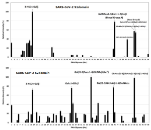
It was found that the SARS-CoV-2 spike protein S1 subunit binds specifically to blood group A antigen (strongly) and B antigen (weakly), and that the spike protein S2 subunit has a binding preference for Lewisa antigen. However, in the eyes of blog admin, it does not mean that SARS-CoV-2 Spike proteins have no affinity for glycan structures other than these ones, and the specificity is not so remarkable, especially in S2 subunit.

Pentosan polysulfate (PPS), Mucopolysaccharide polysulfate (MPS) could be potential antiviral drugs against SARS-CoV-2

A group from Department of Chemical and Biological Engineering, Center for Biotechnology and Interdisciplinary Studies, Rensselaer Polytechnic Institute, Troy, NY 12180, USA, etc. has reported that sulfated glycans and highly negatively charged compounds could show strong antiviral activities against SARS-CoV-2.
https://www.ncbi.nlm.nih.gov/pmc/articles/PMC8875565/

It was know that many sulfated glycans, such as heparin, fucoidans, and rhamnan sulfate show anti-SARS-CoV-2 activities. In this study, a small library of sulfated glycans and highly negatively charged compounds, including pentosan polysulfate (PPS), mucopolysaccharide polysulfate (MPS) were evaluated as antiviral sulfated glycans against SARS-CoV-2.

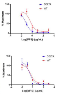
The neutralizing effect of PPS and MPS on SARS-CoV-2 pseudotyped virus in vitro were confirmed with neutralization assays. The IC50 values of PPS for the WT and Delta variant were 0.45 and 0.07 µg/mL, and the IC50 values of MPS for the WT and Delta variant were 0.42 and 0.28 µg/mL, respectively (see below). These results suggest the potential use of PPS and MPS as therapeutic and/or preventative antiviral drugs.

Microbiome in a deep hyperarid core of the Atacama Desert

A group from Departamento de Ingeniería Química, Universidad Católica del Norte, Antofagasta, Chile, etc. has reported about microbiome in a deep hyperarid core of the Atacama Desert.
https://www.ncbi.nlm.nih.gov/pmc/articles/PMC8859261/

Extensive and diverse microbial life remains to be discovered at greater soil depths within the hyperarid Atacama Desert. It was found that the moisture content changed from 2 to 11% by depth from (i) surface zone A (0–60 cm), (ii) intermediate zone B (60–220 cm), to (iii) deep zone C (220–340 cm).

The microbial composition in the soil core was dominated by Proteobacteria, Actinobacteria, Bacteroidetes and Firmicutes in all studied depth zones. However, community composition varied with soil depth.
In general,

  • Firmicutes exhibited a slightly greater detection frequency in surface zone A than in zone C, while it was not detected in zone B.
    Within Firmicutes, the occurrence of the families Lachnospiraceae and Bacillaceae was limited to zone A, while Oscillospiraceae and Salisediminibacter incerta sedis occurred only in zone C (wetter conditions).
  • Similarly, the actinobacterial families had different niches, with Sporichthyaceae being only detected in layers of zones B and C, while Illumatobacteraceae were only present in different layers within zone A.
  • The deepest part of the profile was dominated by Proteobacteria, especially by the families Comamonadaceae (330–340 cm) and Marinobacteraceae (320–330 cm).
  • The family Sphingomonadaceae of Proteobacteria showed a high abundance in several layers of zone B but a lower abundance in zone C.
  • Remarkably, Cyanobacteria of the class Oxyphotobacteria were detected with low prevalence in various samples across the soil profile.

The combined application of Bacillus spp. inoculant and sucrose can improve the growth and quality of Rheum palmatum

A group from Gansu Gaolan Field Scientific Observation and Research Station for Agricultural Ecosystem, Northwest Institute of Eco-Environment and Resources, Chinese Academy of Sciences, Lanzhou 730000, China, etc. has reported that the combined application of a bacterial inoculant and sucrose can improve the growth and quality of Rheum palmatum suppressing pathogen Fusarium spp.
https://www.ncbi.nlm.nih.gov/pmc/articles/PMC8835959/

There are many bacterial and fungal species that can function as (plant growth-promoting microbes) PGPM, of which Bacillus members are well-described in the literature for successfully promoting plant growth in diverse ways. Bacillus isolates colonize host plant roots and promote plant growth by producing bio compounds, such as the hormone indole-3-acetic acid (IAA), as well as spermidine and 2,3-butanediol, by defending against pests and pathogens by producing antibiotic substances such as hydrogen cyanide (HCN), chitinase, and siderophores.

In general, soil is usually a carbon-limited state, and less than 5% of total bacteria are in an active state under such conditions. Carbon sources, especially the most common sucrose, could impact bacteria as a direct energy source for growth. So, in this work, it was evaluated how Baillus spp. respond to the small molecular carbon addition such as sucrose and their co-effects on the rhizosphere microecology.

The combinations of three concentrations of Bacillus amyloliquefaciens EZ99 inoculant (1.0 × 10⁵, 1.0 × 10⁶, and 1.0 × 10⁷ colony-forming units (CFU)/mL, denoted as LB, MB, and HB, respectively) and with three sucrose concentrations (0.15, 1.5, and 15 g/L, denoted as LS, MS, and HS, respectively) were evaluated on R. palmatum growth and yield.

Although the HB treatment increased the growth of R. palmatum (plant length, crown weight, leaf length, and leaf weight) comparing with control (CK), it decreased the growth when coupled with sucrose, especially under the highest concentration of sucrose (HS). This indicates that a high level of bacterial inoculant (1.0 × 10⁷ CFU/mL) amended with a high level of sucrose (15 g/L) suppressed the plant growth-promoting function of PGPM. And, further, the fresh weight of R. palmatum roots were the most significantly improved under the LB + LS and LB + MS treatments (see below)

Typical differences observed in the comparisons are as follows:

  • Eight kinds of anthraquinones, the major constituents of rhubarb, were differentially identified in the comparisons. The LB + LS group accumulated the highest level of aurantio-obtusin-6-O-glucoside and torachrysone-8-O-glucoside, but reduced levels of torachrysone and laccaic acid D, while the rhubarbs under the LB treatment accumulated only 2-acetoxymethyl-anthraquinone.
  • The co-effects of PGPM and sucrose in the LB + LS treatment did not lead to a significant net difference in the total contents of soil nutrients, but they significantly increased the contents of total potassium which could mediate the bioavailability of potassium and nutrients’ cycling in the soil.
  • As for rhizobacteria community, sucrose addition had little impact on bacterial community structure in LS soil, yet it modulated the diversity induced by the PGPM addition in LB + LS soil.
  • As for fungal community, the Ascomycota and Mortierellomycota were the two most abundant phyla, together accounting for more than 92.5% of the relative abundance of the total fungal sequences. The most abundant phylum Ascomycota was enriched the most in CK (79.1%), and diminished the most in LB + LS (71.6%) treatment. At the genus level, compared with CK, the first dominant genus Fusarium was decreased, whereas the second dominant genus Mortierella was increased in all treatments, with the highest increase recorded in the LB + LS treatment.