Priority of vaccine administration for the new coronavirus (COVID-19): Should over-60s still be prioritized?

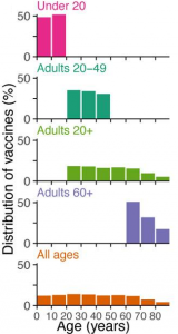
A group from The University of Colorado Boulder etc.  has simulated on the priority of vaccine administration for the new coronavirus (COVID-19).
https://www.ncbi.nlm.nih.gov/pmc/articles/PMC7743091/

Five age groups of vaccination were assumed, and simulated changing the combination of the following parameters: vaccine rollout speeds to the population (0.05% to 1%/day), infection rates (1.15, 1.5), vaccine efficacy (90%), and also the case that the effectiveness of the vaccine decreases with age (60 years old = 90% – > 80 years old = 50%).
The results vary depending on the conditions, and the best choice replaced between the case that prioritizes the over-60s and the one that prioritizes 20 ~ 59 year-olds. However, since the prerequisites are always changing in reality, overall, it would be a better choice to prioritize the over-60s .

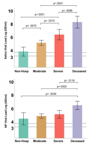
The most dynamic unifying predictor of the disease pathology of the new coronavirus (COVID-19) is the saliva viral load.

A group from Yale University School of Medicine etc. has reported that the most dynamic unifying indicator of the pathophysiology of the new coronavirus (COVID-19) is the saliva viral load.
https://www.ncbi.nlm.nih.gov/pmc/articles/PMC7805468/

Existing markers for COVID-19 include inflammatory cytokines and chemokines (CXCL10, IL-6, IL-10), inflammasome (IL-18, IL-1β), interferon (IFNα, IFNγ, IFNλ), etc.), however, they have found that the saliva virus load was very well correlated with the disease pathology. The nasopharyngeal viral load was also evaluated as a comparison. The amount of virus in saliva and nasopharyngeal (RNA copies/mL) was calculated from Ct values of RT-PCR extracting RNA from saliva. For the discrimination accuracy among non-hospitalization, moderate, severe, and deceased, strong predictive ability was obtained as follows, moderate disease (AUC = 0.96), severe disease (AUC = 0.89), and fatal (AUC = 0.91).

The upper figure of the below shows the correlation between the saliva viral load and the disease pathology, and the lower figure shows the nasopharyngeal viral load and the disease pathology.

Demonstration of ultra-high recognition method of cells combining Deep Learning and lectin microarray

A group from the National Institute of Child Health and Development demonstrated that deep learning can be combined with glycan profiling data from lectin microarrays to recognize cell differences with ultra-high accuracy.
https://www.sciencedirect.com/science/article/pii/S2352320420300742?via%3Dihub

The lectin microarray used was LecChip Ver1.0 of GlycoTechnica. The lectin microarray has 45 kinds of carefully selected natural lectins and has been widely used worldwide as a de facto standard for lectin arrays since its launch in 2007.
Deep Learning, as you know, is now used in various fields as one of methods of AI, and in this research, Google’s TensorFlow was used as the backend, and Keras was used as wrapper software. The layer configuration of this deep Leaning had an input layer of 45 (the same number of lectins) and an output layer of 5 (to discriminate five cells), and hidden layer from 1 to 5. There were five types of cells evaluated (Pluripotent stem cell, Mesenchymal stromal cell, Endometrial and ovarian cancer cell, Cervical cancer cell, Endometrial cell), and a total of 1,577 samples were used for the evaluation.
The results were astonishing as follows, and showed a high recognition accuracy of 97.4% overall.


Deep Learning software used in this paper is sold under the soft name “SA/DL Easy” from Mx. “SA/DL Easy” does not require any knowledge of programming such as Python, you can build neural networks and run deep learning just by clicking a mouse using a one-dimensional array dataset (such as glycan profiling data of lectin microarrays used in the above paper) as an input. “SA/DL Easy” is a quite user-friendly software to use Deep Learning. If you are interested in this software, please contact Mx.

U.S. Department of State releases a FACT Sheet on origin of the new coronavirus (SARS-CoV-2)

On December 15, 2020, Blog Admin uploaded an article saying that the new coronavirus (SARS-CoV-2) is likely an artificial product, citing “The 2nd Yan Report”.

On January 15, 2021, the U.S. Department of State released a FACT Sheet titled Activity at the Wuhan Institute of Virology (WIV).
https://www.state.gov/fact-sheet-activity-at-the-wuhan-institute-of-virology/

In this article, the U.S. Department of State avoids saying that SARS-CoV-2 originated in WIV, but it does show three FACTs in WIV as follows.

  1. In the fall of 2019, there were several researchers who showed similar pathology to COVID-19 were in WIV.
  2. Since 2016, WIV has been studying bat coronaviruses including “RatG13” (96.2% similar to SARS-CoV-2).
  3. Secret Chinese biological weapons researches were being conducted on WIV

Specificity of antibody responses in asymptomatic to mild new coronavirus (COVID-19) patients in Japan

A group from School of Medicine, Keio Univ. has reported a cohort study on antibody responses in asymptomatic to mild new coronavirus (COVID-19) patients who were positive for PCR testing in Japan.
https://www.ncbi.nlm.nih.gov/pmc/articles/PMC7787511/

Many overseas cohort studies have targeted patients with more symptomatic and severe cases, and antibody responses have appeared in 80% to 100% of patients three weeks after infection. Numerous studies have also shown that these antibodies correlate with age, severity, lymphopenia, and serum CRP levels.

This cohort study was conducted in asymptomatic to milder patients than in known cohort studies, and 87.5% of asymptomatic patients and 23.5% of mild patients did not have antibody responses.

Metyrene blue blocks all excess cytokines and inflammatory mediators from the new coronavirus (COVID-19)

A group of Fondazione IRCCS Estituto Neurologico Carlo Besta, Via Padova, Milan, Italy etc. believes that the low efficacy of antiviral drugs such as cytokine inhibitors is (1) probably due to delays in administration in which the virus causes an inflammatory response and is no longer the main protagonist, (2) the relatively low efficacy of cytokine inhibitors is explained because they only act on one or a few of the dozens of cytokines, and (3) other inflammatory mediators (reactive oxygen and nitrogen species) are not targeted.

When inflammatory mediators are over-generated, reactive species cause extensive cell and tissue damages. The only drug known to inhibit the over production of active species and cytokines is methylene blue, a low-cost dye with antiseptic properties that is effectively used in the treatment of malaria, urinary tract infections, septic shock, and methaemoglobinaemia. They suggest testing methylene blue to treat COVID-19 acute respiratory distress syndrome.
https://www.ncbi.nlm.nih.gov/pmc/articles/PMC7728423/

M Protein Comparison: SARS-CoV-2, SARS-CoV, MERS-CoV

The virus envelope of the new coronavirus (SARS-CoV-2) contains S proteins, E proteins, and M proteins. Because S proteins are so deeply involved in the infection, so much research has been done targeting S proteins. However, very little information is available on the structure and function of M proteins. In general, the function of the M protein is understood as the protection of the viral particle structure, and it is thought that the RNA-N protein complex and the S-protein bind to the M protein, bud out as infectious particles in the small cavity, and are released out of the cell by exocytosis.

A group of King Abdulaziz City for Science and Technology, Riyadh, Saudi Arabia focused on M proteins and its differences between SARS-CoV-2, SARS-CoV, and MERS-CoV.
https://www.sciencedirect.com/science/article/pii/S1018364720304493?via%3Dihub

The following points are pointed out:

The M protein is a transmembrane protein, and there are N-terminal domain and C-terminal domain on either side of the three transmembrane domains.
From the viewpoint of the amino acid sequence, SARS-CoV-2 has an S4 residue (221-224) inserted, and it would be an unique feature of SARS-CoV-2.
As intrinsically disordered regions, there are two regions (1-7, 205-222) in SRAS-CoV-2, three regions (1-6, 207-210, 216-221) in SARS-CoV, and two regions (1-6, 216-219) in MERS-CoV, and these differences would reflect viral particles protection capability in different environments of the virus and might be related to viral transmission mode.
Domains that could be potential B-cell epitopes would be:
SARS-CoV2    183-189 ASQRVAG, 200-217 RIGNYKLNTDHSSSSDNI
SARS-CoV     183-188 SQRVGT, 199-215 RIGNYKLNTDHAGSNDN
MERS-CoV     180-188 MVKRQSYGT, 200-211 AGNYRSPPITAD

Evaluation of saponins and tannins targeting SARS-CoV-2 precursor protein main protease (Mpro) as an inhibitor of the new coronavirus (SARS-CoV-2)

Similar to the last blog, a group of Ladake Akintola Unyv. of Technology, Ogbomoso, Oyo State Nigeria has evaluated the inhibitory effects of saponins and tannins by molecular docking and molecular dynamics simulations, targeting SARS-CoV-2 precursor protein main protease (Mpro).
https://link.springer.com/article/10.1007/s40203-020-00071-w

Saponins are found in plant roots, leaves, stems, etc., but are especially found in beans and are known to have antioxidant properties. On the other hand, tannins are astringent components contained in many seeds, and astringency of persimmons is a good example of tannin. Tannins strongly bind to proteins and have the effect of causing denaturation, and the effect of tannins on their properties is called “Astringent.”

The results are as follows, but may show potential antiviral effects that are no less so than remdesivir. Evaluation in vivo is expected.

Ligands Binding affinity (ΔG) kcal/mol Inhibition constant (Ki), µM
Saponins
Priverogenin A − 8.3 0.83
Arjunic acid − 8.1 1.16
Theasapogenol B − 8.1 1.16
Euscaphic Acid − 8.0 1.37
Tannins
Punicalagin − 9.0 0.25
Punicalin − 8.6 0.5
Ellagic acid − 8.4 0.7
Corilagin  − 8.2  0.98
Gallagic acid − 8.1 1.16
Reference
Remdesivir − 7.6 2.7


Screening plant-derived inhibitors targeting SARS-CoV-2 precursor protein main protease ( Mpro)

When the new coronavirus (SARS-CoV-2) infects host cells, a huge precursor protein is synthesized from RNA derived from the virus. The precursor protein is cleaved by various proteases and becomes functional proteins necessary for the growth of viruses. The main protease (Mpro) is one of the such precursor proteases and is an essential one for the growth of viruses.

A group of Univ. of Medical Sciences and Technology, Khartoum, Sudan has used this main protease to evaluate the inhibitory effects of plant-derived components used as traditional Sudanese medicines by molecular docking and molecular dynamic simulations.
https://link.springer.com/article/10.1007/s40203-020-00073-8

As a result, the following plant-derived ingredients are expected as inhibitors of the novel coronavirus. Expect in vivo ratings.

Name Energy (kcal/mol) Predicted LD50 mg/kg
Gallic acid − 17.45 2260
Quercetin − 15.81 159
Naringin − 14.50 2300
Capsaicin − 13.90  47
Psychotrine − 13.50 480


Presence of neutral antibodies against all β-coronaviruses

Four types of coronavirus (OC43, HKU1, NL63 and 229E) are known as coronaviruses that cause common human colds. On the other hand, there are three types of coronaviruses (SARS-CoV, SARS-CoV-2, MERS-CoV) that are highly pathogenic. As you know, SARS-CoV-2 is the new coronavirus that caused the pandemic (COVID-19) from the end of 2019.

A group of Univ. of Washington etc. has report on neutralize antibodies that respond broadly to this type of β-coronaviruses.
https://www.biorxiv.org/content/10.1101/2020.12.29.424482v2.full

The cross-reaction of antibodies is a well-known phenomenon, but for example, antigen testing using antibodies does not accurately identify antigen viruses when there is such a cross-reactivity. However, a strong cross-reactivity can help inhibit the infection of new viruses if the antibodies of past-affected viruses are still active.
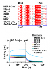
A monoclonal antibody against coronavirus, which was named as B6, has a wide range of cross-reactivity and is attracting attention as a neutralize antibody. As shown in the figure below, this B6 antibody has an amino acid sequence common to these β-coronaviruses in the S2 sub unit as an epitope, and the relevant part is involved in membrane fusion in the event of infection. The MHV in binding curve in the figure below is a mouse hepatitis virus belonging to the coronavirus family.