A group from Research Team for Geriatric Medicine (Vascular Medicine), Tokyo Metropolitan Institute of Gerontology, Tokyo, Japan. etc. has reported about spatiotemporal changes of tissue glycans depending on localization in cardiac aging.
https://www.ncbi.nlm.nih.gov/pmc/articles/PMC9841240/
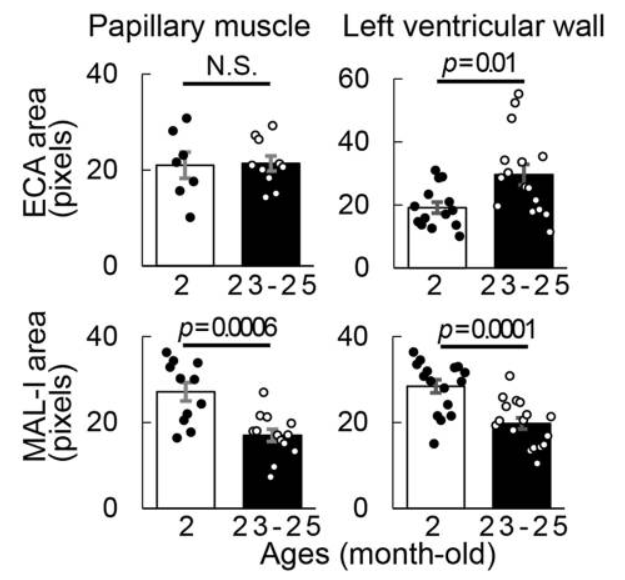
To investigate the characteristics of glycans in mouse cardiac tissues, eight parts of the short-axis slices from normal female mouse hearts, including the left ventricular wall (ventral sides and dorsal sides), the papillary muscle, and the ventricular septum were analyzed for three age groups (2 months, 12–14 months, and 23–25 months) using the lectin microarray.
The glycan profiles of the eight areas of the hearts of 2-month-old mice were analyzed in detail using PCA. Interestingly, it was fond that glycan profiles differ in each region of the cardiac tissue. For instance, papillary muscle and ventricular septum were characterized by sialic acid residues, and ventral left ventricular wall were characterized by O-glycan residues and large-branched and asialo N-glycan residues.
It was also found that sialic acid resides decreases with aging, although the rate of change varied from area to area.