Neutralization of SARS-CoV-2 using polyvalent Nano-Lectin

A group from School of Chemistry and Astbury Centre for Structural Molecular Biology, University of Leeds, United Kingdom, etc. has reported about neutralization of SARS-CoV-2 using polyvalent Nano-Lectin.
https://www.ncbi.nlm.nih.gov/pmc/articles/PMC10302749/

The SARS-CoV-2 S protein trimer is heavily glycosylated with 22 N-linked glycans on each monomer subunit, consisting of oligomannose, hybrid, and complex glycans. Mutations in spike (S) protein epitopes allow SARS-CoV-2 variants to evade antibody responses induced by infection and/or vaccination. In contrast, mutations in glycosylation sites across SARS-CoV-2 variants are very rare, making glycans a potential robust target for developing antivirals. So, it was thought that lectins could be potentially potent antiviral activity against SARS-CoV-2 variants.

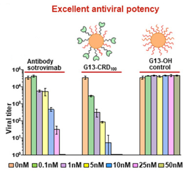
It is know that DC-SIGN carbohydrate recognition domain (CRD) binds specifically to mannose- and fucose-containing glycans found on virus surfaces, including SARS-CoV-2, with low to moderate monovalent affinities (Kd’s: 0.1–3 mM). In order to increase the affinity of the lectin DC-SIGN, the DC-SIGN CRDs were conjugated with Gold nanoparticles (GNP). 13 nm Gold nanoparticles (G13) were synthesized by citrate reduction of H[AuCl4], and first partially PEGylated. Then, these G13s were incubated with linker-labeled DC-SIGN CRDs.

As a result, it was shown that G13-DC-SIGN CRD, as the 1st polyvalent nano-lectin, has broad activity against SARS-CoV-2 variants.
Mannose- and Mannan-coated fucoidan/chitosan nanoparticles can activate macrophages

A group from LAQV, REQUIMTE, Departamento de Ciências Químicas, Faculdade de Farmácia, Universidade do Porto, Rua de Jorge Viterbo Ferreira, Porto, Portugal, etc. has reported that mannose- and mannan-coated fucoidan/chitosan nanoparticles can activate macrophages.
https://www.ncbi.nlm.nih.gov/pmc/articles/PMC10298651/

In order to activate macrophages, mannose receptors on the macrophage surface were stimulated by drug-free fucoidan/chitosan (F/C) nanoparticles functionalized using mannose (M) and mannan (Mn). The polyelectrolyte complexed nanoparticles were electrostatically self-assembled through Coulombic interactions between cationic chitosan and anionic fucoidan.

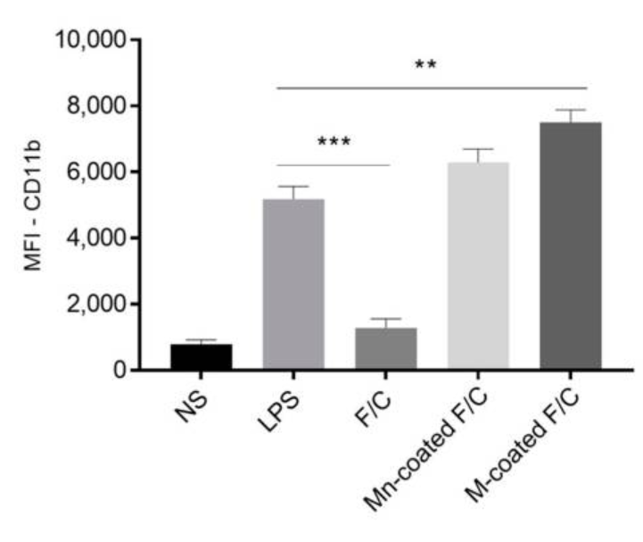
A significant increase in CD11b expression on macrophages occurs in the presence of mannan and mannose-coated F/C nanoparticles, similar to exposure to LPS, and has a statistically significant difference compared to the non-stimulated (NS) and uncoated-F/C nanoparticles cases. Thus, it was demonstrated that the carbohydrate-coated nanoparticles elicit the activation of macrophages as observed for the LPS stimulation.

As a result, it was shown that targeting macrophage receptors via the functionalization of drug-free polymeric nanoparticles represents a promising approach to modulate the immune system.

prediction of glycan structures from lectin binding using a Boltzmann model prediction

A group from Department of Physics, University of California, San Diego, etc. has reported about prediction of glycan structures from lectin binding yusing a Boltzmann model prediction.
https://www.ncbi.nlm.nih.gov/pmc/articles/PMC10274649/

In this study, it was tested if glycan structures can be predicted from lectin binding patterns. Each lectin has a small set of motifs to which it binds. Since lectins are not expected to have logically complex binding rules, it was thought that shallow neural network topologies to be adequate. A reasonable minimal model for this interaction would be hought to be a fully visible Boltzmann machine, which can be conceptualized as a two layer network.

To be honest, I think that it’s not so accurate than expected. This will be partly because the glycans are a very heterogeneous group, and partly because the binding specificity of lectins is ambiguous. There are many people who are against glycan structure estimation using this kind of lectin panels, and there are a large number of MS/MS adherents.

However, in the context of this paper’s discussion, their thought is the same as mine.
“Information got from lectins is accurate enough in a biological context!”

Human milk is very effective in protecting preterm infants from necrotizing enterocolitis

A group from USDA Children’s Nutrition Research Center, Department of Pediatrics, Baylor College of Medicine, Houston, TX 77030, USA, etc. has reported that pre- and probiotics are not sufficient for protection from Necrotizing enterocolitis (NEC) in an exclusively formula-based diet.
https://www.ncbi.nlm.nih.gov/pmc/articles/PMC10255242/

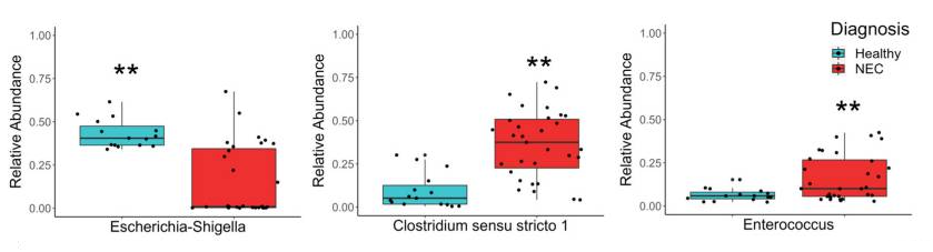
NEC is the leading cause of death resulting from gastrointestinal disease in premature infants, with a mortality rate of 15–40%. It was found about intestinal bacteria that (1)Escherichia-Shigella is Significantly more abundant in healthy piglets and negatively correlates with disease severity, (2) Clostridium sensu stricto 1 and Enterococcus are significantly more abundant in the colon of diseased piglets and correlates with disease severity (see below).

Although dietary probiotic Bifidobacterium longum subsp. infantis (BL. infantis) and sialylactose (3′SL) supplementation had no effect, donor human milk (DHM) significantly reduced the incidence of NEC.

Changes in complement component C3 glycosylation in type 1 diabetes complications

A group from Faculty of Pharmacy and Biochemistry, University of Zagreb, Zagreb, Croatia, etc. has reported about a new biomarker (c3.Asn939-N2H10) for type 1 diabetes (T1D) complications.
https://www.frontiersin.org/articles/10.3389/fendo.2023.1101154/full

N-glycan profiles of complement component C3 have been profiled in 189 T1D subjects with different status of complication severity. The analysis was conducted by high-mannose glycoprotein enrichment from human blood serum using Con A lectin matrix, Glu-C digestion, glycopeptide purification and subsequent nano-LC-ESI-MS technique.

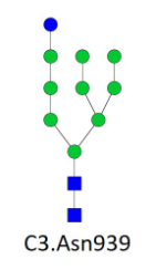
It was found that only one glycoform, C3.Asn939-N2H10, significantly changed in albuminuria and retinopathy, and also there was the biggest correlation between C3.Asn939-N2H10 and HbA1c.

A serum biomarker for non-small cell lung cancer (NSCLC): chondroitin sulfate proteoglycan

A group from Department of Laboratory Medicine, Shanghai Tongji Hospital, School of Medicine, Tongji University, Shanghai, China,etc. has reported about a serum biomarker for non-small cell lung cancer (NSCLC) .
https://respiratory-research.biomedcentral.com/articles/10.1186/s12931-023-02423-4

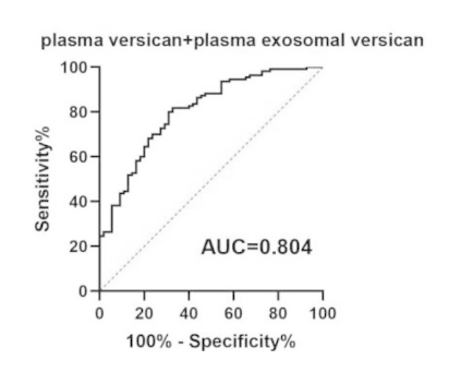
Plasma versican and plasma exosomal versican expression in NSCLC patients was significantly upregulated and was significantly higher in T3 + T4 patients compared with T1 + T2 patients (P < 0.05) as shown below (ROC curve). Versican is a large chondroitin sulfate proteoglycan. The plasma versican and plasma exosomal versican were detected using an ELISA kit (made in China).

n-hexadecanoic acid secreted by tomato roots most strongly promotes biofilm formation of Pseudomonas stutzeri

A group from School of Biotechnology and Pharmaceutical Engineering, Nanjing Tech University, Nanjing, China, etc. has reported that n-hexadecanoic acid secreted by tomato roots most strongly promotes biofilm formation of Pseudomonas stutzeri.
https://www.ncbi.nlm.nih.gov/pmc/articles/PMC10220591/

Plant growth-promoting rhizobacteria (PGPR) have been widely used in agricultural applications because of their safety, biological control of diseases and insect pests, and ability to induce c abiotic tolerance. It is know that the colonization, chemotaxis, and biofilm formation by PGPR are induced by the root exudates and certain metabolites.

In this study, it was shown that inoculation with a certain concentration of Pseudomonas stutzeri NRCB010 significantly promoted tomato growth and induced significant changes in tomato root exudates. Among those root exudates, n-hexadecanoic acid induced the most remarkable growth, chemotactic response, biofilm formation, and rhizosphere colonization of Pseudomonas stutzeri.

Effects of Tricodelma coating on Maize and Watermelon

A group from Jiangsu Collaborative Innovation Center for Solid Organic Wastes, Educational Ministry Engineering Center of Resource-Saving Fertilizers, Nanjing Agricultural University, Nanjing, China has reported about Trichoderma Coating on maize and watermelon.
https://pubmed.ncbi.nlm.nih.gov/37195176/

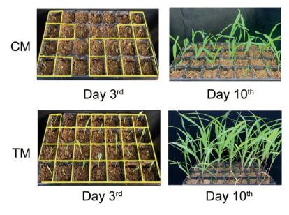
In this study, the fungus Trichoderma guizhouense NJAU4742 was introduced to both maize and watermelon seed microbiomes by seed coating, and it was shown that seed coating on watermelon and maize with Trichoderma sp. improves plant growth and rhizosphere soil enzyme activities significantly.
The germination rate of coated seeds were significantly increased by 25% for maize after 3 days postplanting and 35% for watermelon after 8 days postplanting, compared with the control.
effects of tricodelma coating on maize

Compared with the control, the enzyme activities of urease, sucrase, acid phosphatase, alkaline phosphatase, α-glucosidase, peroxidase, and cellulase increased by 97.9%, 51.7%, 38.3%, 51.6%, 61.2%, 82.3%, and 27.0%, respectively, in the coated maize, and the enzyme activities of sucrase, acid phosphatase, neutral phosphatase, α-glucosidase, peroxidase, polyphenol oxidase, and cellulase increased by 51.7%, 25.7%, 21.1%, 62.6%, 39.1%, 34.3%, and 32.8%, respectively, in the coated watermelon.
The incidence of maize stalk rot in the coated treatment was significantly reduced by 28% compared with that in the control, and the incidence of watermelon with coating was reduced by 37% at 60 days after pathogen application.

PET imaging targeting cell surface glycans for pancreatic cancer using a 18F-labeled rBC2LCN lectin

A group from Department of Gastrointestinal and Hepato- Biliary- Pancreatic Surgery, Faculty of Medicine, University of Tsukuba, Tsukuba, Japan, etc. has reported about PET imaging targeting cell surface glycans for pancreatic cancer using a 18F-labeled rBC2LCN lectin.
https://onlinelibrary.wiley.com/doi/epdf/10.1111/cas.15846

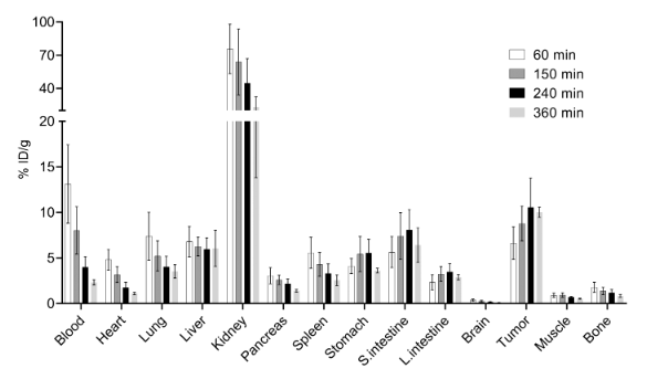
In this study, the potential application of an rBC2LCN–fluorine-18 (18F) conjugate was explored as a novel positron emission tomography (PET) probe. Capan-1, an H-type-3-positive human pancreatic cancer cell line, was was selected and Capan-1 cells (2×10⁶) were injected subcutaneously into the right dorsa of nude mice. It is known that rBC2LCN lectin has a binding specificity to H-type-3 glycan. Importantly, rBC2LCN does not induce hemagglutination (induced when exogenous lectins are introduced into the blood), allowing it to be harmlessly admin-istered intravenously.

As aresult, it was shown that 18F-labeled rBC2LCN lectin offers a novel class of tumor-specific probes for PET that are based on targeting cell surface glycans. However, I feel that this probe is actually specific to cancer cells, but it might be a problem to know that this binds to other normal tissues as well as shown below.

Human serum albumin nanoparticles conjugated with WGA for targeted delivery of antibiotics to combat urinary tract infections

A group from University of Vienna, Faculty of Life Sciences, Division of Pharmaceutical Technology and Biopharmaceutics, Vienna, Austria, etc.has reported about human serum albumin (HSA) nanoparticles conjugated with WGA for targeted delivery of antibiotics to combat urinary tract infections.
https://www.sciencedirect.com/science/article/pii/S1549963423000369?via%3Dihub

It is so interesting to learn how NPs were prepared. For the production of NPs, 20 mg HSA was dissolved in 10 ml 100 mM phosphate buffer pH 8. Afterwards, 1 ml olive oil was layered over the aqueous protein solution. The probe micro tip of the sonifier was positioned at the interface of the two phases. The sample was sonicated with an acoustic power of ≈253 W cm−2 at 40 % amplitude for 2 min. Subsequently the NPs were washed four times by centrifugation (5204 ×g, 40 min, 4 °C). For production of active pharmaceutical ingredients (API) containing NPs, 2.5 mg, 5 mg, 10 mg or 20 mg rifampicin (RIF) was dissolved in 1 ml isopropyl alcohol, mixed with olive oil and treated as described above. For the preparation of trimethoprim (TMP) loaded particles, 2.5 mg, 5 mg, 10 mg or 20 mg API was dissolved in 10 ml 100 mM phosphate buffer pH 8.

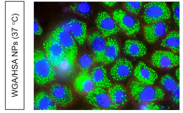
When human urothelial cells were incubated with targeted NPs, NPs (stained with green dyes) were observed around the blue stained nucleus and within the red stained membrane, which indicates an effective internalization of the NPs into the cells. Comparing the cases between with WGA and without WGA, 60 % higher cell binding potential was observed with WGA.

Powered by WordPress |Copyright © 2020 Emukk. All rights reserved