Glycan profiling analysis of serum NRG1 in papillary thyroid cancer with using Lectin Microarrays

A group from Department of Head and Neck Surgery, Fudan University Shanghai Cancer Center, Shanghai, China, etc. has reported on glycan profiling analysis of serum NRG1 in papillary thyroid cancer with using Lectin Microarrays.
https://www.ncbi.nlm.nih.gov/pmc/articles/PMC8798758/

This study is evaluating if BRAF V600E mutant status in papillary thyroid cancer (PTC) patients could be detected by analyzing multiple glycan profiles of serum NRG1 through lectin microarray assays.

It has been reported that NRG1 overexpression was related in various cancers. NRG1 protein can bind to ERBB3 or ERBB4 protein, activating ERBB receptor tyrosine kinases, followed by the initiation of signal cascades, including PI3K/AKT pathways. NRG1 can be secreted to serum as reported.

lectin microarrays were used to analyze the different glycan profiles of NRG1 from sera of BRAF(+) PTC and BRAF wild type(-) PTC patients. Lectin microarray is a rapid and sensitive high-throughput technique to get glycan profiles, allowing researchers to directly analyze glycoproteins without liberation of glycans from the core substrate.

It was found that LEL, BPL and NML tended to have bound to NRG1 in BRAF(+) PTC patients compared to BRAF(-) controls.

Rhizosphere Biofilm: Characteristics of Mucigel exuded from plant roots and EPS exuded from Rhizobacteria

A group from Division of Biogeochemistry of Agroecosystems, Georg-August University of Göttingen, Göttingen, Germany, etc. has reported on Biofilm Matrix in the Plant Rhizosphere.
https://www.ncbi.nlm.nih.gov/pmc/articles/PMC8792611/

Roots exude a diverse set of compounds into the rhizosphere, including mucigel, amino acids, and secondary metabolites, which regulate rhizosphere functions. Mucigel is a gelatinous high-molecular-weight substance produced by almost all plants. The mucigel backbone is know to be built of polysaccharides, but proteins, minerals, and lipids are also part of the biogel. Rhizobacteria also exude extracellular polymeric substances (EPS). EPS are mainly composed of polysaccharides, but also contain proteins, nucleic acids, lipids, and minerals, and forms biofilms on root surfaces functioning as comfortable place to live. Thus, the polysaccharides were the major chemical constituent of both biogels (77.4% and 74.6% for mucigel and EPS, respectively) and did not significantly differ between them.

The polysaccharide backbone did not significantly differ in proportion between mucigel and EPS, namely galactose (mucilage = 23.8%; EPS = 22.8%), fucose (13.9%; 9.9%), glucose (16.7%; 28.7%), rhamnose (12.4%; 15%), xylose (13.4%; 8.1%), and glucuronic acid (8%; 12.8%). In contrast, mannose (3.9%; 18.6%) was significantly higher in EPS than in Mucigel (nearly fivefold higher), whereas arabinose (16.3%; 4.8%) and galacturonic acid (27.3%; 7.8%) had higher proportions (3.4-fold and 3.5-fold higher, respectively) in Mucigel than in EPS.

Alginate is an anionic polysaccharide found in EPS, consisting of only uronic acids such as glucuronic acid, galacturonic acid, and mannuronic acid. Alginate participates in the formation of microcolonies at the beginning of the biofilm formation process, increases EPS hydration, and assists in trapping cations such as Ca2+, Zn2+, Cd2+, and Ni2+. The high proportion of galacturonic acid in mucigel is likely one of the major reasons for the higher water absorption capacity of mucigel than EPS.

Mucigel can also be decomposed and consumed by microorganisms. Enzymatic release of highly abundant sugars in mucigel such as galactose, fucose, and arabinose can feed microorganisms residing in the mucigel. The presence of endogenous glycosyl hydrolase enzymes in mucilage augments this claim. It seems that microorganisms utilize mucigel as an energy source, with average times of 7–15 days for the consumption of 50% of the mucigel carbon added to the soil. The high protein content of mucigel leads to a C:N ratio of approximately 16:1, which is approximately double the C:N ratio of microorganisms. Thus, considering that 50% of the C is utilized via catabolism and oxidized to gain energy, mucigel has the ideal composition to function as a sole energy, C and N source for microorganisms. Consequently, microorganisms solely need to be supplied with mineral nutrients (P, K, Ca, Mg, etc.)—a common nutrients shared with their mucigel-providing plants.

H84T BanLec shows broad-spectrum antviral activities against Herpesvirus

A group from Department of Microbiology and Immunology, SUNY Upstate Medical University, Syracuse, NY USA, etc. has reported that H84T BanLec shows broad-spectrum antviral activities against Herpesvirus.
https://www.ncbi.nlm.nih.gov/pmc/articles/PMC8803833/

As antivirals, one emerging strategy is the use of lectins to bind glycoproteins on the viral envelope. Naturally occurring lectins have been explored as broad-spectrum antimicrobials to inhibit viruses, bacteria, and other microbes. One promising lectin, BanLec, was derived from a banana lectin and shows high affinity to mannose N-glycans, and proved an effective strategy to combat HIV. Unfortunately, wild-type BanLec was a T-cell mitogen and activated basophils and mast cells. To address these issues, we developed H84T BanLec, replacing histidine at position 84 with threonine, which markedly reduced mitogenicity while maintaining the antiviral properties of the lectin.

High-mannose N-linked glycans are present on the envelope proteins of many viruses, including human herpesviruses. Human herpesviruses (HHVs) are enveloped DNA viruses that cause a variety of diseases, ranging from cold sores to skin rashes and infectious mononucleosis. HHVs cause lifelong infection, and after the initial infection, remain latent until reactivation. HHVs are prevalent worldwide, with up to 95% of the population infected with multiple types. Three of the most common HHVs are herpes simplex virus 1 (HSV-1), varicella-zoster virus (VZV), and human cytomegalovirus (HCMV).

The mechanism of action for H84T BanLec is virus dependent, and authors have found the following differences.
For VZV, H84T BanLec does not block attachment or entry. Instead, H84T BanLec interferes with post-entry steps in the viral replication process to prevent VZV spread, suggesting that H84T BanLec likely interacts with VZV-infected cells by inhibiting glycoprotein maturation, trafficking, virion assembly, or cell fusion.
For HCMV, H84T BanLec does not interfere with virion synthesis, but that the mechanism of action is through prevention of virus attachment and entry.
For HSV-1, H84T BanLec is also acting post-entry.

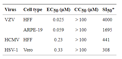
SI (selectivity index) calculated as CC50/EC50

Wheat rhizosphere: The contribution of seed- and soil-originated microbiomes

A group from Institute of Applied Microbiology, Justus-Liebig-University, Giessen, Germany, etc. has reported comprehensive studies about the contribution of seed and soil-originated microbiomes on the formation of the wheat rhizosphere and the effects of differences in wheat strain and soil.
https://www.ncbi.nlm.nih.gov/pmc/articles/PMC8789879/

Seed-transmitted microbes specifically enriched under specific plants in a particular location. Overall, seed-derived bacterial and fungal microbiome were higher in the endosphere compared with the rhizosphere. The strong effect of plant genotype (A. tauschii, T. aestivum, T. dicoccoides, and T. durum) on the bacterial and fungal microbiome composition was observed. The rhizosphere microbiome composition of wheat species, specially between cultivated T. durum and its ancestor T. dicoccoides, were similar in all three locations. However, the enriched genera were different from location to location. The analyses of environmental variables on the rhizosphere microbiome showed that the bacterial and fungal species were significantly affected by the ammonium and nitrate content in soil.

Roughly speaking, the assembly process of the rhizosphere microbiome composition starts immediately after the seed is placed in the soil, and the seed microbiome, the plant genotype, and the soil microbiome cooperatively shape the rhizosphere microbiome composition as the result of release of specific secondary metabolites and signaling molecules by the plant roots.

Mannose-binding Lectins exist in cell walls of Marchantia polymorpha

A group from Laboratoire de Recherche en Sciences Végétales, Université de Toulouse, CNRS, UPS, Auzeville-Tolosane, France, etc. has reported on cell wall proteome (CWP) of Marchantia polymorpha.
https://www.ncbi.nlm.nih.gov/pmc/articles/PMC8792609/

Plant cell walls are composite structures made of polysaccharidic polymers like pectins, hemicelluloses and cellulose as well as of lignins in lignified secondary walls. In this study, 410 CWPs of M. polymorpha were identified.

Typical examples are as follows,

Several families of CWPs involved in the remodeling of the cell wall polysaccharides networks have been identified.
Fifteen GH17 (b-1,3-glucosidases) could be involved in the hydrolysis of callose which has been found in the cell plates of all land plants.
Three GH5 (endo-b-1,4-glucanase) and five GH16 (endoxyloglucan transferases) were identified. Their substrates in cell walls are assumed to be hemicelluloses like xyloglucans or mannans and they are known to play roles in polysaccharides rearrangements during cell growth.
The identification of two GH28 (polygalacturonases), six pectin methylesterases (PMEs) and two pectin acylesterases (PAEs) is consistent with the presence of galacturonic acid in the acidic hydrolysate of M. polymorpha cell wall polysaccharides.
Finally, five GH18 (chitinases) and seven GH19 (chitinases/lyzozymes) have been identified. They have been shown to exhibit chitinase and chitinase/lysozyme activities, respectively. They have anti-fungal and anti-bacterial activities and are involved in defense reactions against biotic and abiotic stresses.

Several protein families are known to be involved in oxido-reduction reactions involving aromatic compounds in M. polymorpha cell walls. The antagonistic enzymatic activities of CIII Prxs allow them to participate in cell wall remodeling events in two opposite ways: (i) they play a role in cell wall loosening by generating reactive oxygen species able to cut non-enzymatically the cell wall polysaccharides; or (ii) they can participate in the cell walls stiffening by oxidizing aromatic compounds such as aromatic amino acids, monolignols or cinnamic acid in the presence of H2O2, or in the cross-linking of structural proteins like extensins, thus forming covalent networks.

The acquisition of a cuticle has been a major event in land plant colonization. Twenty-three GDSL lipases/acylhydrolases have been identified. A GDSL lipase/acylhydrolase has been shown to be involved in cuticle formation.

Eight proteins predicted to be D-mannose binding lectins were identified. The abundance of such proteins in the CWP could be linked to the presence of high amounts of mannans as suggested by the presence of significant amount of mannose residues in pectins- and hemicelluloses-enriched cell walls extracts.

Neutralizing antibody responses against SARS-CoV-2 Omicron

A group from Wellcome Centre for Human Genetics, Nuffield Department of Medicine, University of Oxford, Oxford, UK, etc. has reported on neutralizing antibody responses against SARS-CoV-2 Omicron.
https://www.ncbi.nlm.nih.gov/pmc/articles/PMC8723827/

Here is some presented data showing that the huge number of mutational changes present in Omicron lead to a substantial knockdown of neutralizing capacity of commercially available mAb.
“Commercial neutralizing antibody responses against SARS-CoV-2”

Degradation of neutralization titers of Omicron by sera from vaccines might be ineludible, but it is unlikely that vaccines will completely fail and it is hoped that although vaccine breakthroughs will occur, protection from severe disease will be maintained, perhaps by T cells. It is likely that the vaccine-induced T cell response to SARS-CoV-2 will be less affected than the antibody response.

Wheat Rhizosphere: elevated atmosphric CO2 changes rhizosphere microbe and accelerate mineralization of organic phosphorous

A group from Department of Animal, Plant and Soil Sciences, Centre for AgriBioscience, La Trobe University, Melbourne Campus, Bundoora, Victoria, Australia, etc. has reported on changes in the rhizosphere of wheat due to elevated atmosphric CO2 and mineralization of organic phosphorus.
https://www.ncbi.nlm.nih.gov/pmc/articles/PMC8785599/

Phosphorus (P) is fundamentally important to soil biota as a major building block of life. Since organic P comprises up to 80% of total P in soils, the mineralization of organic P by soil microorganisms could have the potential to be a prominent process in P transformation, especially the mineralization of a dominant component of organic P such as phytate.
While plant roots release extracellular phosphatases, much of this enzyme activity is restricted to the root surface. Thus, active mineralization of soil organic P mainly takes place in rhizosphere where soil microorganisms interact with plant-derived C.

Climate change has the potential to impact organic P transformation. One of most important climate change factors, elevated atmospheric CO2 concentration (eCO2), would considerably accelerate the mineralization of soil organic P. The acceleration of organic P mineralization is attributed to modified plant-soil-microbe interactions due to changes in the plant carbon (C) flow belowground.

Authors quantified the microbial contribution to mineralization of organic P under eCO2 by exploring the community-wide genetic profiling of soil microorganisms, including bacterial and fungal communities and their functions in relation to mineralization of a major organic P compound, phytate, in the rhizosphere of wheat.

Microbial diversity was more clearly affected by eCO2 in the Chromosol than in the Vertosol. Elevated CO2 significantly increased bacterial species richness by 33% across the within 3 mm rhizosphere compartments in the Chromosol. In the Vertosol, however, rhizobacteria were not significantly affected by eCO2. Abundances of two major bacterial phyla, Bacteroidetes and Gemmatimonadetes were relatively enriched in the rhizosphere under eCO2 and positively associated with the mineralization of phytate. The most prevalent Bacteroidetes families were Chitinophagaceae and Microscillaceae, followed by Sphingobacteriaceae and Hymenobacteraceae.

The fungal community had also greater species richness in the within 3 mm rhizosphere compartments when wheat plants were grown under eCO2 in the Chromosol. However, in the Vertosol, CO2 treatment did not significantly affect any indicators of α-diversity of fungal community. eCO2 significantly increased the abundances of Basidiomycota genus Agaricus, and the genera Claroideoglomus and Funneliformis of Glomeromycota in the Chromosol.

In SARS-CoV-2 unvaccinated cases, Omicron infection shows limited cross-reactive neutralization against other variants

A group from Gladstone Institutes, San Francisco, CA, USA, etc. has reported that infection with Omicron variant in unvaccinated individuals may not elicit effective cross-neutralizing antibodies against other variants. In vaccinated individuals, however, Omicron infection effectively induces immunity against itself and enhances protection against other variants.
https://www.medrxiv.org/content/10.1101/2022.01.13.22269243v1.full-text

To determine humoral immune responses induced by infection with the three different strains, sera from infected mice and tested their neutralization efficiency against SARS-CoV-2 strains were collected: WA1, Alpha, Beta, Delta, and Omicron. Efficient virus neutralization was defined by more than 50% reduction in plaque forming units at the lowest serum dilutions. Sera from Delta-infected mice showed the broadest cross-variant neutralization, effectively neutralizing all the strains except Beta. By contrast, while Omicron infection effectively neutralized Omicron itself, it exhibited limited (<50%) cross-neutralization of other strains. Sera from WA1-infected mice conferred effective protection against WA1, Alpha, and Delta, but not against Beta and Omicron. These results indicate limited immunity induced by Omicron relative to other strains, which may be due to its highly mutated spike protein or its lower replicative capacity. In human cases, interestingly, sera from vaccinated individuals with confirmed Omicron breakthrough infection showed the highest level of protection (>80%) against all strains, including Omicron as shown below.

Specific glycan structures observed in Cisplatin resistant Testicular Cancer

A group from Institute of Chemistry, Slovak Academy of Sciences, Bratislava, Slovakia, has reported about difference in glycan structures between cisplatin sensitive and resistant testicular cancer (TC) cell lines.
https://pubmed.ncbi.nlm.nih.gov/35044085/

Testicular cancer is the most frequent type of cancer among young men aged between 15 and 34 years and represents 1.5% of all cancer types in men and 5% of urological tumours overall.
Cisplatin, as one of the very first metal-based chemotherapy drugs, is still widely used to treat patients with various types of cancers including TC. This drug is also effective in curing TC patients having metastases with cure rates of up to 90% and with a general survival rate of up to 95%. The reason why cisplatin works so effectively in treatment of GCTs is that such cells produce the embryonal stem cells. In turn, damaged embryonal stem cells need to be eliminated through apoptosis in order not to pass on mutations to the next generation. Loss of this embryonic feature might lie behind the development of cisplatin resistance.

Cisplatin binds to DNA and creates lesions (i.e. protein–DNA complexes and inter/intra-strand DNA adducts) which cannot be repaired by the DNA repair mechanisms, resulting in a disruption of synthesis of DNA, mRNA and proteins, promoting the accumulation of reactive oxygen species, activating signalling pathways and finally resulting in cell death.

Authors have found that DBA lectin could be the best probe for evaluating the resistance of TC lines, and also that HHL lectin could be used to prospect for cisplatin sensitive TC lines.

Correlation between lectin binding to human chorionic gonadotropin (hCG) of sensitive/resistant testicular cancer cell lines with cisplatin ID50

SARS-CoV-2 omicron variant shows attenuated lung disease in mice and hamsters comparing to other variants

A group from the collaborative network of the SARS-CoV-2 Assessment of Viral Evolution (SAVE) program of the National Institute of Allergy and Infectious Diseases (NIAID) has reported that SARS-CoV-2 omicron variant shows attenuated lung disease in mice and hamsters, which parallels preliminary human clinical data.
https://www.ncbi.nlm.nih.gov/pmc/articles/PMC8722607/

Some recent studies (as also introduced in this blog) speculated that because mutations in the omicron spike protein overlap with ones known to promote adaptation to mouse hosts, the progenitor of omicron jumped from humans to mice, and then back into humans. In support of this, omicron but not Wuhan-1 RBD binds to murine ACE2. Hamsters also are highly susceptible to SARS-CoV-2 infection with similar pathological changes seen in lung tissues from COVID-19 patients. Using experimental data from multiple laboratories of the SAVE/NIAID consortium, authors report on the infectivity and pathogenesis of multiple omicron isolates in mice and hamsters.

Here is the data of comparison between omicron in BALB/c mice and beta variant. At 2 days-post-infection (dpi), infectious virus levels in the nasal turbinates and lungs were significantly lower (~1,000-fold, P < 0.001) in BALB/c mice infected with omicron compared to beta virus.

A comparison of infectious viral burden in hamster tissues at 3 dpi between delta and omicron viruses showed virtually no difference in nasal turbinates but substantially less infection of omicron in the lungs of hamsters.

Here is the comparison of lung pathology in Syrian hamsters after infection with delta or omicron viruses. Macroscopically, the lungs obtained from the delta-infected hamsters showed congestion and/or hemorrhage, but this was absent in omicron-infected animals. Overall, the Omicron variant replicates less efficiently in the lungs of Syrian hamsters, which results in less severe pneumonia compared to the delta variant.

Our experiments suggest that compared to other SARS-CoV-2 isolates (e.g., beta or delta), the Omicron variant infection is attenuated in laboratory mice and hamsters for causing infection and/or disease. While these results are consistent with the very preliminary clinical data in humans suggesting that omicron causes a more transmissible yet possibly milder respiratory infection, the basis for the attenuation in rodents remains unknown.

One pre-print study (also introduced in this blog) suggests that omicron9 replicates faster in the human bronchus and less in lung cells, which may explain its greater transmissibility and putative lower disease severity; although it remains unclear if these observations extend to rodents, we observed less infection of hamster bronchial cells in vivo with omicron than delta virus. Lower viral burden in nasal washes and turbinates in 129 mice were also measured comparing to other SARS-CoV-2 strains. The attenuation in mice was unexpected given that omicron has multiple mutations in the RBD that are sites associated with adaptation for mice.
Whereas the more than 30 substitutions in the omicron could impact receptor engagement and cell entry, sequence changes in other domains could affects replication, and so forth. Thus, detailed genetic and functional studies are required to define the basis of virological and clinical attenuation of omicron in mice and hamsters.

Powered by WordPress |Copyright © 2020 Emukk. All rights reserved