Effectiveness of Pfizer and Moderna vaccines for various SARS-CoV-2 variants occuring in the world: B.1.351 and P.1 variants are critical

A group from Massachusetts General Hospital, etc. has reported on effectiveness of two typical vaccines, BNT162b2 (Pfizer) and mRNA-1273 (Moderna), for various SARS-CoV-2 variants occuring in the world.
https://www.ncbi.nlm.nih.gov/pmc/articles/PMC7899476/

Distribution of typical SARS-CoV-2 variants in the world.

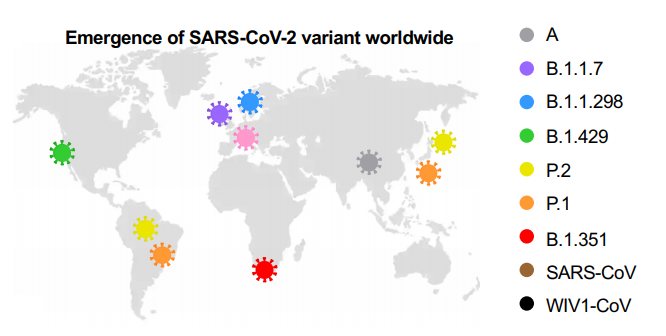
Effectiveness of Pfizer and Moderna vaccines is shown by the neutralization response. There seems to be almost no change for B.1.1.7 variant, but the neutralization activity of these vaccines are significantly reduced in B.1.351 v2 and P.1 variants. Two common mutations, K417N and E484K, exist in B.1.351 and P.1 variants, and these mutations do not exist in B.1.1.7. 

Anti-complement Therapy would be effective for Membranous Nephropathy

A group from Ohio State University Wexner Medical Center, etc. has reported that excess compliments are deposited in Membranous Nephropathy (MN).
https://www.ncbi.nlm.nih.gov/pmc/articles/PMC7879111/pdf/main.pdf

Glomeruli tissue samples were captured by laser microdissection, and IgGs and complements in the samples were analyzed by mass spectrometry comprehensively. As a result, elevated glomerular levels of all complements (except for CR1) and IgG1~IgG4 were observed in MN patients comparing with health controls. On the other hand, mannose-binding lectin (MBL), Ficolin, and Collectin were not detected. These results suggest that Glomeruli tissue could be damaged by excessively activated complements not through the lectin pathway. Loss of CR1 at the site of the damaged filtration barrier might accelerate excess complement activation resulting in significant damages. In addition, it is thought that M-type phospholipase A2 receptor (PLA2R) could be an antigen in membranous nephropathy.

Cross-reactivity of  hCoV-specific IgGs against SARS-CoV-2 uninfected individuals

A group from NMI Natural and Medical Sciences Institute at the University of Tübingen, etc. has reported on the cross-reactivity of hCoV-specific IgGs against SARS-CoV-2 infected and uninfected individuals using 1,173 serum/plasma samples.
https://www.nature.com/articles/s41467-021-20973-3.pdf

Up to now, it has been known that there are 4 kinds of epidemic human coronaviruses (hCoVs: NL63, 229E, OC43, HKU1) which cause seasonal colds.
As shown below, there is certainly correlation between SARS-CoV-2 and hCoVs specific IgGs to a certain degree. 10% of SARS-CoV-2 infected individuals do not develop detectable SARS-CoV-2-specific IgG, but it has not been identified if this result could come from cross-reactivity of hCoVs-specific IgGs or just from innate immunity against SARS-CoV-2.


Once again: C-type Lectins, CD209/DC-SIGN, CD209L/L-SIGN, could be infection receptors of SARS-CoV-2 as well as ACE2

There have been some papers reporting that C-type Lectins could be SARS-CoV-2 infection receptors as well as well-know ACE2.
Harvard Medical School, July 30, 2020
https://www.biorxiv.org/content/10.1101/2020.07.29.227462v1
Boston University, Dec. 9, 2020
https://www.biorxiv.org/content/10.1101/2020.06.22.165803v2
There was a review paper from Boston University, (Dec. 22, 2020) in addition to the above paper, let me introduce two kinds of interesting data for your reference.
https://www.mdpi.com/2079-7737/10/1/1

Although ACE2 is a really important virus receptor, the expression level of ACE in lung is very low, however, CD209 and CD209L are more broadly expressed in human tissues rather than the ACE2.
A result of infection experiments using SAR-CoV-2 pseudotyped lentivirus and HEK293 cells overly expressing CD209L, CD209, and ACE2 is also shown below.
Taking these things into consideration, it would be clear that CD209 and CD209L could mediate SARS-CoV-2 entry as infection receptors even for tissues lacking ACE2 expression.
The CRDs of C-type Lectins (DC-SIGN, L-SIGN) recognize strongly high mannose type glycans.

In order not to develop COVID-19 even if infected with the new coronavirus (SARS-CoV-2): A way to coexist with SARS-CoV-2, survive virus wars

What I am always thinking about COVID-19 was written in the following paper published from a group of Central University of Tamil Nadu etc.
https://academic.oup.com/femspd/article/79/1/ftaa076/6027506

In the case of COVID-19, there are so many asymptomatic individuals (some studies have estimated that up to 80%).
Generally speaking, the incubation period for COVID-19 is reported to be between 5 to 6 days, although this can be up to around 14 days, but, asymptomatic individuals show an extended median duration time of 19 days.

SARS-CoV-2 activates the STING pathway and NLRP3 inflammasome assembling, and induces cytokine storm as a result of excess production of IL-1β, and IL-18, TNF-α, IFN-γ, IL-6, etc. However, bats do not develop any disease, although they are reservoir of various viruses. In the case of bats, they are coexisting with virus preparing the following features evolutionally.
(1)limiting the assembly of the NLR family pyrin domain containing three (NLRP3) in monocytes、
(2)loss of the ability to effectively sense exogenic cytoplasmic DNA via loss of PYHIN genes
(3)excessively high levels of the anti-inflammatory cytokine IL-10,
(4)lack of functional killer cell Ig-like (KIR) and killer cell lectin-like (KLR) receptors.

It is know that the levels of inflammatory cytokines are relatively low, and the SARS-CoV-2-specific IgG and neutralizing antibody levels are significantly lower in asymptomatic individuals. This might mean that asymptomatic is a result of weak immune response. And on the contrary, it might be a result of existing antibody cross-reactivity of classical seasonal cold-causing coronaviruses 229E, HKU1, OC43 and NL63.

In the context of coexistence with virus, mechanisms uncovered in bats likely hint the development of therapeutic strategies against COVID-19 in humans. That could be a wisdom, to survive virus wars.
 

Agalacto (i.e. GlcNAc) binding lectins: BLL, PVL, GSL-II, and BGL 

Agalacto type complex N-glycans without terminal Gal are often seen in autoimmune disease. Well known Lectins with glycan binding specificity to agalacto are BLL, PVL, GSL-II as shown below. BLL and PVL are mushroom derived, and GSL-II is bean derived.

Boletopsis leucomelaena: BLL lectin


Psathyrella velutina: PVL lectin


Griffonia simplicifolia: GSL-II lectin


A group from New England Biolabs, Inc. has identified gene sequence of Boletopsis grisea, a close North American relative of Boletopsis leucomelaena, and investigated glycan binding specificities of recombinant lectin (named rBGL) with using glycan arrays. Curiously, rBGL has bi-specific glycan binding characteristics, not only to agalacto type N-glycans but also to O-glycans with Thomsen–Friedenreich antigen (TF-antigen; Galβ1,3GalNAc-α-). Depending on applications, the binding specificity to O-glycan would be interesting.
https://www.nature.com/articles/s41598-020-80488-7

Chitinase 3-like-1 could be a good Therapeutic Target for the new coronavirus (COVID-19)

A group from Brown University, etc. has reported that Chitinase 3-like-1 (CHI3L1/YKL-40) could be a good therapeutic target for the new coronavirus (COVID-19).
https://pubmed.ncbi.nlm.nih.gov/33442679/

In the case of COVID-19, majority is asymptomatic or mild, but 10-20% of patients require hospitalization. Especially, it is so interesting that the severity of COVID-19 is deeply related to aging and comorbid disorders (such as diabetes, hypertension, obesity and metabolic syndrome, cardiovascular disease and chronic lung diseases like COPD and asthma). It is know that CHI3L1 is secreted from a spectrum of cells in response to a variety of injury and inflammation, and regulates innate and adaptive immunity and also protects apoptosis. What’s even more interesting is that CHI3L1 increases with aging and also with risk factors of COVID-19 (i.e., presence of comorbid disorders). Taking these things into consideration, authors focused on the relationship between CHI3L1 and angiotensin-converting enzyme 2 (ACE2), transmembrane protease serine 2 (TMPRSS2), cathepsin L (CTSL).

In an in vivo experiment using mice, it was found that CHI3L1 stimulates expression of pulmonary ACE2, TMPRSS2, and CTSL. In other experiments using Calu-3 lung epithelial cells, the expression of ACE2, TMPRSS2, CTSL increased monotonically with CHI3L1 dose, and also it was found that a monoclonal antibody for CHI3L1 (FRG) strongly inhibits the expression of ACE2, TMPRSS2, and CTSL. In actual COVID-19 patients hospitalized, it was also indicated that there is a significant correlation between severity and CHI3L1.

These evidences suggest strongly that CHI3L1 could be a good therapeutic target for COVID-19. Let’s anticipate the future research. 

IgM plays a major role unexpectedly in preventing the new coronavirus (SARS-CoV-2) infection

A group from University de Monterey, etc. has reported an evaluation result of SARS-CoV-2 neutralization activities of IgM, IgA, and IgG using plasma samples from 25 convalescent patients with the new coronavirus (COVID-19).
https://pubmed.ncbi.nlm.nih.gov/33596407/

IgM, IgA, and IgG were selectively depleted using isotype-specific ligands immobilized on beads, respectively. A comparison of the SARS-CoV-2 pseudoviral inhibitory dilution (ID50) for each plasma is shown below. In the IgM-depleted plasma, neutralization activity was greatly reduced by 5.5 times, the secondary one was the IgG-depleted plasma with the reduction of 4.5 times, and the reduction of IgA-depleted plasma was 2.4 times. Considering that IgM is only 5% of total immunoglobulin, the neutralization activity of IgM is very curious.

Q677 mutations in clade 20G (lineage B.1.2) of the new coronavirus (SARS-CoV-2) are increasing rapidly since late 2020

A group from University of Bern, Switzerland, etc. has reported a sharp increase of Q677 mutations (Q677H and Q677P) in clade 20G (lineage B.1.2) of the new coronavirus (SARS-CoV-2) in the USA. Since this mutation is present in the proximity of the furin cleavage site, it may have an effect on infectivity, but detailed research will be a future issue.
https://pubmed.ncbi.nlm.nih.gov/33594385/

Excessive complement activation occurs in kidneys of patients with the new coronavirus (COVID-19)

A group from Friedrich-Alexander-University (FAU) Erlangen-Nürnberg, etc. has reported excessive complement activation in kidneys of patients with the new coronavirus (COVID-19).
https://www.ncbi.nlm.nih.gov/pmc/articles/PMC7878379/

COVID-19 causes severe acute respiratory syndrome (ARDS), and the tissue damage is not restricted to lungs but also develop cardiac and kidney injury. In the case of kidneys, the glomerules and tubules seem to be damaged. Excessive activation of complements develops excessive membrane attack complex (MAC) resulting in damage of the glomerular and tubular tissues.
The table below shows a comparison of the level of complement expression among COVID-19 and typical kidney diseases comparing with a control.


ATI: acute tubular injury
HUS: hemolytic uremic syndrome
DIC: disseminated intravascular coagulation