blog

Prediction of a SARS-CoV-2 animal host using random forests model (one of machine learning methods)

A group from University of Liverpool has reported results of random forests model (one of machine learning methods) to predict the animal host of SARS-CoV-2.
https://journals.plos.org/plospathogens/article?id=10.1371/journal.ppat.1009149

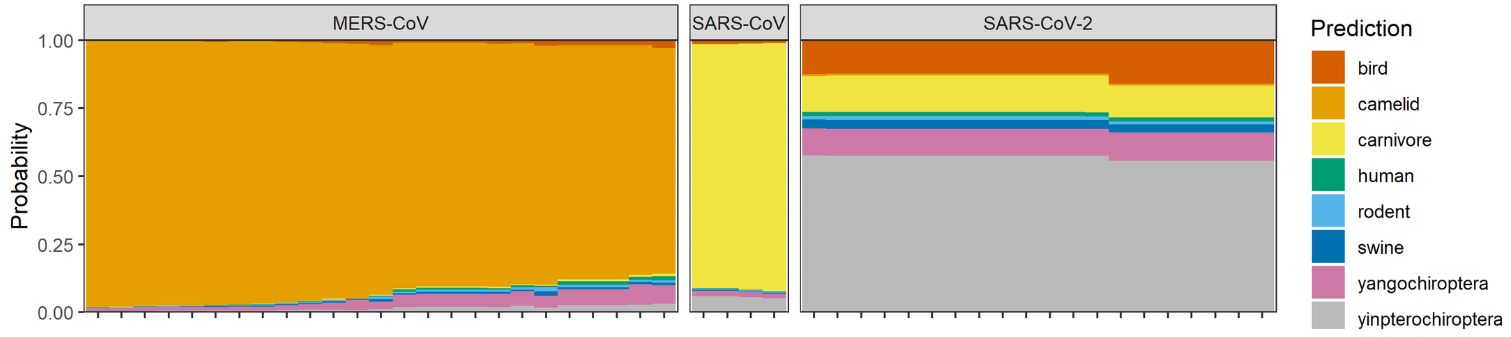
A figure below shows stacked bar plots of predicted probabilities of each host category for coronavirus RNA sequences, assuming bird, camelid, carnivore, human, rodent, swine, yangochiroptera, and yinpterochiroptera as potential hosts. It is clearly shown that MARS-CoV has camelid host, SARS-CoV has carnivore host, and SARS-CoV2 seems to have a bat host (suborder Yinpterochiroptera). While the random forests model supports bats as the ultimate origin of SARS-CoV-2, the involvement of intermediate hosts remains unclear.
Maackia amurensis lectin reduces the expression levels of ACE2, ADAM17, Furin, etc. : A new finding in suppressing SARS-CoV-2 infection

A group from Rowan University, Stratford, USA, etc. has suggested that Maackia amurensis lectin (MAL, MAA, MASL as abbreviated names) could be effctive in inhibiting SARS-CoV-2 infection.
https://www.ncbi.nlm.nih.gov/pmc/articles/PMC8019238/

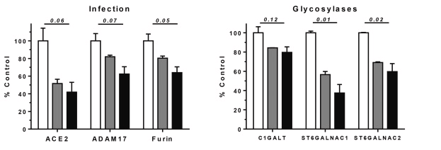
Oral squamous cells were used as a model cell in this study. Transcriptome analysis was done to investigate effects of MAL onto ACE2, ADAM17, Furin, and Glycosyltransferases (GalNAc-T, ST6GalNAc-1, and ST6GalNAc-2).
Interestingly, it was shown that those decreased in a MAL dose dependent manner. For instance, at a dose of 1925nM of MAL, ACE2 mRNA level decreased by 60%, ADAM17 by 40%, and ST6GalNAc-1 by 60%. As a result of these events, MAL decreases inflammatory signaling events that would otherwise lead to activation of the IL6 amplifier implicated in COVID-19 induced ARDS

MAL is known to have binding specificity to α2-3Sia.

Functional Roles of Dexamethason as a therapeutic drug for COVID-19 

As a special case, Japanese Ministry of Health, Labor and Welfare approved “Remdesivir” as a therapeutic drug for COVID-19 in May, 2020. In Sept., 2020, Dexamathason as a corticosteroid was also approved as a therapeutic drug for COVID-19. Let me introduce a paper explaining what kinds of functional roles Dexamethason plays.

It is a paper from a group of University of Huddersfield, UK, etc.
https://link.springer.com/article/10.1007/s10753-021-01464-5

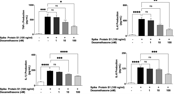
Stimulation of human PBMCs with a recombinant spike glycoprotein S1 resulted in significant release of pro-inflammatory cytokines TNFα, IL-6, IL-1β and IL-8. Pre-treatment with dexamethasone (100 nM) caused significant reduction in the release of these cytokines. SARS-CoV-2 spike glycoprotein S1 induced exaggerated inflammation in PBMCs through mechanisms involving activation of NF-κB transcription factor, p38 MAPK and the NLRP3 inflammasome, and it was found that the pre-treating PBMCs with dexamethasone inhibited NF-κB DNA binding by ~46%.

Effects of SARS-CoV-2 P.1 variant to therapeutic antibodies and SARS-CoV-2 vaccines 

A group from University of Oxford, etc. has reported the effects of P.1 variant to therapeutic antibodies and SARS-CoV-2 vaccines.
https://www.ncbi.nlm.nih.gov/pmc/articles/PMC8008340/

P.1 contains the following mutations:
L18F, T20N, P26S, D138Y, and R190S in the NTD;
K417T, E484K, and N501Y in the RBD;
D614G and H655Y at the C terminus of S1;
and T1027I and V1176F in S2

Neutralization of both Lilly antibodies (LY-CoV16 and LY-CoV555) was severely impacted. There was also escape from neutralization of P.1 by Regeneron antibody (REGN10933) and a modest reduction in neutralization of P.1 by AstraZeneca antibody (AZD8895), while AstraZeneca antibodies (AZD1061 and AZD 7442) showed equal neutralization of all SARS-CoV-2 variants. The three Adagio antibodies (ADG10, ADG20, and ADG30) neutralized all variants, with all reaching a plateau at 100% neutralization; interestingly, ADG30 showed a slight increase of neutralization of P.1.

Geometric mean neutralization titers against P.1 were reduced 2.6-fold (p < 0.0001) relative to the Victoria virus for the Pfizer-BioNTech vaccine serum and 2.9-fold (p < 0.0001) for the Oxford-AstraZeneca vaccine. 

Pathological changes of bronchoalveolar lavage fluid (BALF) collected from COVID-19 patients

A group from San Martino Policlinico Hospital, Genoa, Italy, etc. has reported on characteristics of bronchoalveolar lavage fluid (BALF) collected from COVID-19 patients.
https://www.ncbi.nlm.nih.gov/pmc/articles/PMC8049078/

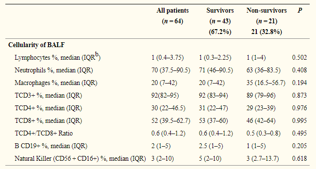
BALF cellularity was mainly composed of neutrophils and macrophages (neutrophils were more abundant than macrophages). The median value of macrophage percentages was higher in non-survivors compared to survivors (35% vs 20%). However, we must be cautious that all the differences shown in a table below are not statistically significant.

Esophageal squamous cell carcinoma (ESCC) -specific glycan biomarkers 

A group from Northwest University, Xi’an, China, etc. has reported a potential glycan biomarker for Esophageal squamous cell carcinoma (ESCC)
.
https://www.frontiersin.org/articles/10.3389/fchem.2021.637730/full

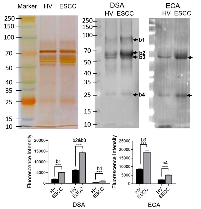
Saliva was used as a sample in this study, and saliva was centrifuged to remove insoluble components and a protease inhibitor was added. Lectin microarrays using 37 lectins were used for investigating differences in glycosylation patterns between healthy volunteers (HV) and ESCC patients. As a result, it was found that DSA and ECA lectins are specific to ESCC, and Galβ1-4GlcNAc-containing N-glycans could be potential biomarkers for ESCC.

Farmed minks were infected with SARS-CoV-2 from humans: A future threat with new virus variants  

A group from University Göttingen, etc. has alarmed a possibility in reinfection of mutated SARS-CoV-2 in farmed mink to human.
https://pubmed.ncbi.nlm.nih.gov/33857422/

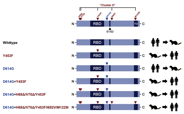
Transmission of SARS-CoV-2 from humans to farmed mink has been observed in Europe and USA. Substitution D614G, which is dominant in SARS-CoV-2 from humans, was also found in viruses from mink, and a mink-specific mutation Y453F (mutant D614G+Y453F) or Y453F in conjunction with H69Δ, H70Δ (mutant D614G+H69Δ/H70Δ/Y453F) were also found in mink. The Y453F mutation locates in RBD, and reduced inhibition by human serum samples tested. The neutralizing titer IC50 increased by 1.62 times with this mutation, and the neutralizing titer of REGN10933, which is one component of a cocktail therapy (REGN-COV2), also increased with this mutation.

New emerging variants generated through transmission from humans to wild animals could represent a future threat to human health.

Differences in SARS-CoV-2 infectivity between M1 alveoli macrophages and M2 macrophages 

A group from Peking Union Medical College, etc. has reported on differences in SARS-CoV-2 infectivity between M1 macrophages and M2 macrophages including alveolar epithelial type II (AT2) cells.
https://www.nature.com/articles/s41421-021-00258-1

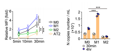
Generally speaking, M1 macrophages are inflammatory and M2 macrophages are anti-inflammatory. Comparing AT2, M1 macrophages, and M2 macrophages, SARS-CoV-2 infection and its replication was higher in macrophages than AT2, M1 macrophages took up SARS-CoV-w2 with higher efficiency than M2 macrophages, and the viral loads increased in an exponential fashion in M1 macrophages but in a flat fashion in M2 macrophages. It is considered that ACE2 could be the receptor that mediates the infection with SARS-CoV-2, and ACE2 has been reported to be expressed also by macrophages. However, there were no difference in the infectivity between ACE2-overexpressing macrophages and ACE2-knockeddown macrophages. So, virus takeup through phagocytosis could initiate SARS-CoBV-2 infection.
Authors related the difference in the infectivity between M1 and M2 to difference in cell softness, endosomal, and lysosomal pH.

Dectin-2 is related to Infection of Influenza virus and the induction of inflammatory responses

A group from Niigata University, etc. has reported that Dectin-2 is related to Infection of Influenza virus and the induction of inflammatory responses.
https://www.jstage.jst.go.jp/article/biomedres/42/2/42_53/_pdf/-char/en

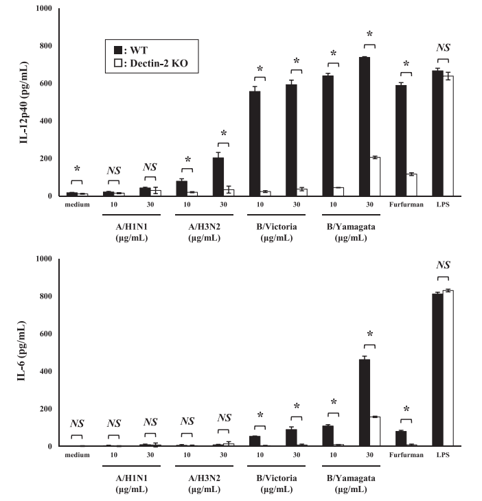
Although the importance of relationship between hemagglutinin (HA) and sialic acids has been emphasized, agents targeting HA and sialic acids are not effective in suppressing severe influenza unless they are administered within 48 h after symptoms begin. Antigen-presenting cells (APCs), such as dendritic cells (DCs) and macrophages, recognize pathogen-associated molecular patterns (PAMPs) and damage-associated molecular patterns (DAMPs) of dead cells or damaged tissues, and activate inflammatory immune responses. Toll-like receptors (TLRs), Nod-like receptors (NLRs), and C-type lectin receptors (CLRs) are known as such pattern recognition receptors.

Since HA is strongly high mannosylated, there must be a signal path activating inflammatory immune responses through the interaction between C-type Lectins and glycosylated. C-type Lectin family includes DC-SIGN, Dectin-1, Dectin-2, Mincle, etc. Authors have found that Dectin-2 expressed on BM-DCs recognizes glycosylated HA from the type A and type B strains, and induce production of inflammatory cytokines. Dectin-2 recognized high mannose polysaccharides.
A figure below shows that production of inflammatory cytokines is greatly reduce by knocking out Dectin-2.

Powered by WordPress |Copyright © 2020 Emukk. All rights reserved