Cortical-bone-derived stem cells (mCBSCs) treatments effectively improve myocardial structure and functions after myocardial infarction compared to MSC: Difference in glycosylation

A group from Research Team for Geriatric Medicine (Vascular Medicine), Tokyo Metropolitan Institute of Gerontology, Japan, etc. has reported that cortical-bone-derived stem cells (mCBSCs) treatments effectively improve myocardial structure and functions after myocardial infarction compared to MSC.
https://www.ncbi.nlm.nih.gov/pmc/articles/PMC8584423/

Recently, it have been shown that mouse cortical-bone-derived stem cells (mCBSCs) improve cardiac remodeling and functions. The mCBSC-treated hearts showed increased neovascularization, and newly formed cardiac myocytes were also observed. mCBSCs produce a unique combination of immunomodulatory and angiogenic and proangiogenic factors, which may be the reason why mCBSCs were more effective in improving the post-myocardial infarction hearts compared to cardiac-derived stem cells and MSCs.

The self-renewing ability in mCBSCs was higher than that in mMSCs, but the mCBSCs exhibited only chondrogenic differentiation, while the mMSCs exhibited adipogenic, osteogenic, and chondrogenic differentiation. It was also found that mCBSCs secrete a greater amount of TGF-β1 compared to mMSCs, and that the TGF-β1 contributed to the self-migration of mCBSCs and activation of fibroblasts. Migrated CBSCs expressing TGF-β1 may contribute to converting cardiac fibroblasts into myofibroblasts at an infarcted site.

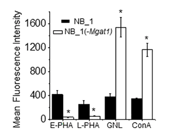
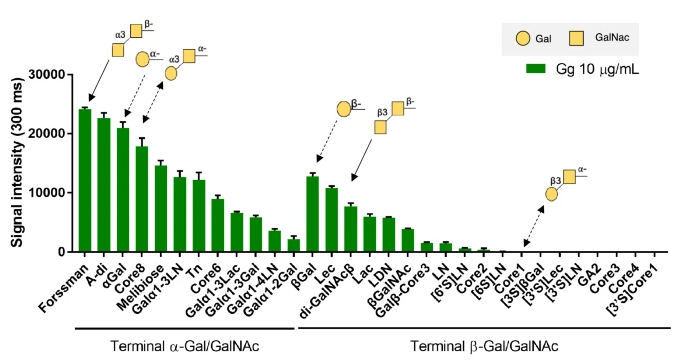
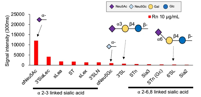
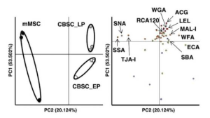
As for cell surface glycans, the relative intensities of three lectins WFA (lacdiNAc), ECA (lactosamine-binding lectin), and MAL-I (α2-3 sialic acid binding lectin) in mCBSCs were higher than those in mMSCs. Moreover, the relative intensities of the three lectins, SNA, SSA, and TJA-I (α2-6 sialic acid binding lectins), in mCBSCs were significantly lower than those in mMSCs. It was considered that this lower expression of α2-6sialic acid may be an indication of specific differentiation towards chondrogenic lineage and not towards adipogenic or osteogenic differentiation.

Previous research has shown that glycans contribute to regulation of the signaling mediated by leukemia inhibitory factor (LIF), Wnt, FGF, bone morphogenetic protein BMP, and Notch, which are required for the maintenance of stem cells. It has been also known that WFA-binding glycans on LIF receptorβ and gp130 regulate LIF/STAT3 signaling, which is required for self-renewal of mouse embryonic stem cells.

In this study, it was found that WFA-binding glycans are more specific to CBSCs than MSCs, and this suggested that WFA-binding glycans may contribute to the self-renewal of CBSCs by regulating LIF/STAT3 signaling, although further studies is required.

Lactobacillus agilis isolated from rhizosphere shows potential as a novel biotherapeutic agent

A group from Department of Biosciences, COMSATS University Islamabad (CUI), Islamabad, Pakistan, etc. has reported that Lactobacillus agilis isolated from rhizosphere of the medicinal plants Ocimum tenuiflorum, Azadirachta indica, Ficus carica shows potential as a novel biotherapeutic agent.
https://www.ncbi.nlm.nih.gov/pmc/articles/PMC8568817/

Potential probiotic bacteria can be used as a biotherapeutic agent and a sustainable alternative to antibiotics, as an anti-oxidative, anti-inflammatory, and anti-diabetic agent without causing any serious side effects. In this experiment, the following assays were performed to select potentials.

Safe status assays
First of all, to exclude potentially harmful bacterial strains, testing of safe status was done before selection of bacterial strains for human as probiotics.
(1) should be negative for blood hemolytic activities and (2) did not show a breakdown of gelatin.

Antibiotic susceptibility
The strains which show less or no resistance to antibiotics were selected, and L. agilis was the best.

In-vitro antibacterial assay
The antibacterial results showed the significant marked antagonistic activity of L. agilis NMCC-15 to kill or inhibit the growth of all the tested bacterial pathogens (Escherichia coli, Pseudomonas aeruginosa, Staphylococcus aureus, Listeria monocytogenes and Bacillus cereus).

Antioxidant, anti-diabetic, and anti-inflammatory assays
Percentage inhibition of the free radical from the mean values showed the comparable effects of L. agilis supernatant to the standard drug with values 68% and 73%, respectively.
L. agilis inhibited the activities of the α-amylase (anti-diabetic potential) by 51.3% than control.
The supernatant of L. agilis showed 61.6% for denaturation albumin protein while aspirin showed 69% activity.

The most abundant bacteria and fungi in seedlings rhizobacteria came from their seeds, not the soil

A group from MaxPlanck Tandem Group in Plant Microbial Ecology, Universidad del Valle, Cali, Colombia, etc. has reported that the most abundant bacteria and fungi in seedlings rhizobacteria came from their seeds, not the soil.
https://www.frontiersin.org/articles/10.3389/fmicb.2021.737616/full

Until recently, scientists have traditionally believed that all the rhizosphere microbiome “is recruited from the main reservoir of microorganisms present in soil. However, Is it really true?

In this experiment, plants were grown in hermetically sealed jars on sterile sand as a way to observe microbiome development in the absence of any other source of inoculum except seeds. In this experiment, 18 kinds of plant were evaluated.

Amazingly enough, in this experiment it was confirmed that soil contributes to microbial diversity in the rhizosphere, however the most abundant bacteria and fungi in rhizospheres derive from seeds. Soil significantly increased bacterial diversity in rhizospheres, however, the highest read abundance was of seed-transmitted bacteria. Only an average of 26% of bacterial taxonomic units (OTUs) came from seeds, but these were responsible for an average of 72% of the reads. These OTUs were mostly Proteobacteria of the genera Pantoea, Enterobacter, Pseudomonas, and Massilia, which were observed in seeds or spermospheres and have also been observed associated with a variety of plant seeds.

Compared to bacteria, there was less seed-transmitted fungal diversity in rhizospheres, with only an average of 12%; however, these OTUs tended to become abundant, representing an average of 42% of the reads. Of these seed-transmitted rhizospheric fungi, Fusarium was the most abundant, occurring in all soil-grown rhizospheres, as it did in all spermospheres.


In this experiment, unsterilized seeds were planted in either sterile sand or farm soil inside hermetically sealed jars. The sand was sterilized by autoclaving twice for 20 min at 121°C, and after transfer to glass jars, it was autoclaved a third time for 20 min at 121°C, and the soil was excavated from a fallow cassava field at a CIAT property near Palmira, Colombia.

Changes in N-glycosylation on neuroblastoma affect on cell proliferation and invasion

A group from Department of Biochemistry and Molecular Biology, East Carolina University, Greenville, North Carolina, USA, etc. has reported that changes in N-glycosylation on neuroblastoma affect on cell proliferation and invasion.
https://journals.plos.org/plosone/article?id=10.1371/journal.pone.0259743

Two types of neuroblastoma cells were used. One is an ​engineered N-glycosylation mutant cell line with Mgat1 knockdown, NB_1(-Mgat1), expressed solely oligomannose N-glycans, and the other was its parental cell line, NB_1 expressed significant levels of higher order N-glycans, complex and hybrid N-glycans as shown below (GNL and ConA have higher affinity to oligomannose type N-glycans, and E-PHA and L-PHA have higher affinity to complex type N-glycans).

Cell growth was faster in NB_1 cells with complex type N-glycans than in NB_1(-Mgat1) cells without complex type N-glycans. Quantitatively speaking, there was about a 54% decrease in cell proliferation between NB_1(-Mgat1) cells and NB_1 cells.

NB_1(-Mgat1) cells had more and longer protrusions than NB_1 cells, indicating that the glycosylation mutant cells solely expressing oligomannose type N-glycans were more invasive than those expressing complex types of N-glycans. To quantify cell invasiveness, the ratio of measured invasive area to the measured sphere area was measured. The cell invasiveness of the NB_1(-Mgat1) cell line was 2.3-fold greater than the NB_1 cell line.

Two types of multivalent lectins produced from hemagglutinins of clostridium botulinum: Gg binds to Gal/GalNAc and Rn binds to Neu5Ac

A group from Tokyo University of Agriculture and Technology, Tokyo, 183-8509, Japan, etc. has reported two types of multivalent lectins produced from hemagglutinins of clostridium botulinum.
https://www.nature.com/articles/s41598-021-01501-1

Clostridium botulinum is a gram-positive anaerobic bacterium that produces a neurotoxin. Botulinum bacteria are classified into serotypes A to G depending on the antigenicity of the toxin produced. The type C 16S progenitor toxin is composed of a neurotoxin, non-toxic non-hemagglutinin component and several hemagglutinin proteins (HA) designated as HA1, HA2, HA3a and HA3b.

It has been know that type C HA1 has two binding sites: site I and site II. Site I binds to N-acetylneuraminic acid (Neu5Ac), N-acetylgalactosamine (GalNAc) and galactose (Gal) while site II binds to galactose only.

Authors have developed two new multivalent lectins from the above mentioned hemagglutinins of clostridium botulinum designed to have different binding specificities were produced: namely Gg and Rn:
Gg is an Alexa 488 labeled complex with HA1 WADF -HA2 WT-HA3 WT, which binds to Gal/GalNAc,

and, Rn is an Alexa 594 labeled complex with HA1 NQAA-HA2 WT-HA3 WT, which binds to Neu5Ac only.

Auto-antibodies against AngII were developed with SARS-CoV-2 infection

A group from Pritzker School for Molecular Engineering, University of Chicago, Chicago, Illinois, USA, etc. has reported that auto-antibodies against AngII were developed with SARS-CoV-2 infection.
https://www.ncbi.nlm.nih.gov/pmc/articles/PMC8575143/

Surprisingly, it was found that a substantial proportion, 63% (73/115), of the COVID-19 patients had positive levels of anti-AngII autoantibodies, where 115 were hospitalized COVID-19 patients convalescent from a SARS-CoV-2 infection.

First, it was pointed out that the levels of anti-AngII were significantly higher in patients with dysregulated blood pressure as shown below.

Further, the effect of anti-AngII autoantibodies on oximetric saturation (SF ratio) was measured. The lowest daily mean SF ratio was compared between the COVID-19 patients who developed auto-antibodies against AngII (73 patients) and those who did not (42 patients) as shown below.

It was considered that SARS-CoV-2 and AngII to ACE-2 might lead to their co-phagocytosis by antigenpresenting cells, thus providing a strong immune adjuvant (the virus molecules) to the self-peptide AngII, leading to an anti-AngII autoimmune response.

Antibody response induced by a 3rd boost SARS-CoV-2 vaccination: after 8 months from the 2nd vaccination

A group from The Institute of Medical Biology, Chinese Academy of Medical Sciences and Peking Union Medical College, Kunming, Yunnan, China, etc. has reported on antibody response induced by a third boost dose of inactivated SARS-CoV-2 vaccine.
https://pubmed.ncbi.nlm.nih.gov/34666622/

In this study, 53 volunteers, who joined in the development and production of inactivated COVID-19 vaccines (it is not sure what company developed the relevant ones, though), received two doses (at 0 and 28 days) of the vaccines in 2020, and they received a 3rd dose 8 months after the 2nd dose recently.

At 0 days, 5 days, 7 days, and 14 days after the 3rd dose, blood was collected from 6 volunteers for the evaluation. It was found that both the anti-S antibody and neutralizing antibody against the original Wuhan strain gradually increased after 5 days, and the positive conversion rate of antibodies reached 100% at 14 days. Interestingly, the memory of IFN-γ-T cells against S, N, M, O antigens of SARS-CoV-2 can be quickly awakened after the 3rd dose.

These results indicate that although the neutralizing antibodies gradually decrease after two doses of inactivated vaccines, the antibody response could be awakened quickly by the 3rd vaccination and the T cell immune memory is still active even after 8 months from the 2nd vaccination.

SARS-CoV-2 specific antibody transfer to breastmilk is dominated by IgA and IgM

A group from Department of Biological Engineering, Massachusetts Institute of Technology, Cambridge, MA, USA, etc. has reported that SARS-CoV-2 specific antibody transfer to breastmilk is dominated by IgA and IgM.
https://www.ncbi.nlm.nih.gov/pmc/articles/PMC8531199/

Previous studies clearly illustrated the evolution and transfer of Fc-effector function in pregnant women via placenta to their infants, but less is known about the transfer of Fc-effector function across breastmilk.

As expected, mothers infected with SARS-CoV-2 possessed SARS-CoV-2-specific antibodies in serum and breastmilk that were not present in SARS-CoV-2-uninfected specimens. But, interestingly, robust transfer of IgA and IgM was observed in breastmilk, but the transfer of IgG1 was more limited as shown below.

Importance of surfactin-type lipopeptide: Bacillus velezensis recognizes plant root cell pectin and thereby improve symbiotic relation

A group from University of Liège‐Gembloux Agro‐Bio Tech, Gembloux, Belgium, etc. has reported Bacillus velezensis recognizes plant root cell pectin and thereby improve symbiotic relation.
https://pubmed.ncbi.nlm.nih.gov/34724831/

Molecular interactions driving the early steps of partnership establishment between plant roots and rhizobacteria are not yet fully understood. Authors have found that Bacillus velezensis, one of beneficial bacteria, recognizes plant root cell wall pectin in synergy with soluble root exudates, and thereby the bacterium stimulates the production of a specific surfactin-type lipopeptide as key components of its secretome to improve the partnership in the course of early colonization.

Actually, an 8-fold increase of the surfactin production was detected at the early exponential growth phase with homogalacturonan low methylated (HGLM) as shown below, however, the bacteria seem not to recognize oligomers with a lower polymerization degree (DP) than HGLM, as is clear from the fact that oligogalacturonides (OGs) did not stimulate surfactin biosynthesis. This might mean that longer polymers could indicate a healthy host suitable for bacterial colonization, while the monomers or low DP oligomers may reflect a dead or infected plant that is unable to adequately provide resources to the bacteria.


homogalacturonan low methylated (HGLM), DP of >150; oligogalacturonides (OG), DP of 15; galacturonic acid (GA), DP of 1; where REM means a root exudates mimicking medium.

But, there is no information about a lectin-like protein on the bacteria which can recognize the pectin backbone as a plant molecular pattern was not mentioned here anything.

Development of mannosylated macromolecules specific to each human mannose-binding lectins such as DC-SIGN, Dectin-2, and DEC-205

A group from Department of Chemistry, Massachusetts Institute of Technology, MS, USA, etc. has developed mannosylated macromolecules specific to each human mannose-binding lectins.
https://www.ncbi.nlm.nih.gov/pmc/articles/PMC8549053/

Carbohydrate-binding proteins (lectins) play vital roles in cell recognition and signaling, including pathogen binding and innate immunity. Thus, targeting lectins, especially those on the surface of immune cells, could advance immunology and drug discovery. Lectins are typically oligomeric; therefore, many of the most potent ligands are multivalent. An effective strategy for lectin targeting is to display multiple copies of a single glycan epitope on a polymer backbone; however, a drawback to such multivalent ligands is they cannot distinguish between lectins that share monosaccharide binding selectivity (e.g., mannose-binding lectins such as DC-SIGN, DC-SIGNR, MBL, SP-D, langerin, dectin-2, mincle, and DEC-205) as they often lack molecular precision.

Authors developed β-mannosylated glyco-IEGmers specific to each lectins with mannose binding specificity.

To generate the target mannosylated glycomacromolecules, iterative exponential growth (IEG) cycles beginning from (R)- or (S)-glycidyl propargyl ether (GPE, > 99% ee) were conducted to yield oligo/polytriazoles with precisely 8, 16, or 32 allyl side chains. Macromolecules were produced with three different overall absolute configurations: all (R) (“isotactic”), all (S) (“isotactic”), and alternating (R-alt-S) (“syndiotactic”). To append the mannose residues, these IEGmers were exposed to pure β-thiomannose sodium salt under UV light (λ = 365 nm).

The binding affinity changed from sample to sample over several orders of magnitudes. For example, KA values for DC-SIGN ranged from approximately 1 × 10⁴ for (S)-8mer to about 1 × 10⁸ for (R)-16mer. For dectin-2, it also ranged from ∼1 × 10⁴ to ∼1 × 10⁸. DEC-205 bound all of the glyco-IEGmers with high KA values in the range of ∼1 × 10⁶ to 1 × 10⁹, with the highest KA of all pairwise interactions tested (2.2 × 10⁹).

This wide range in affinity is impressive as the mannose binding epitopes presented was identical among these lectins.