A group from Institute for Advanced Biosciences, Université Grenoble Alpes, Grenoble, France, etc. has reported changes in glycosylation of melanoma tumor cells and its impacts on functionality of dendritic cells.
https://www.ncbi.nlm.nih.gov/pmc/articles/PMC9986448/
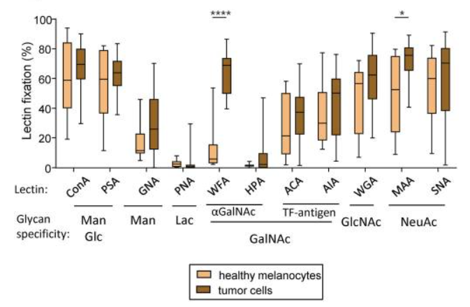
It was found that higher levels of GalNAc and NeuAc residues (revealed by the interaction of WFA and MAA respectively) were observed in melanoma tumor cell lines when compared to healthy melanocytes.
The global view of tumor glyco-code profiles upon separation of patients based on better or worse overall survival (median OS) or progression-free survival (median PFS) revealed a pattern of higher expression of Thomsen-Friedenreich antigen (TF-antigen), GlcNAc, Fuc and NeuAc residues (detected by ACA, WGA, RPL-αMan, UEA-I, MAA and SNA) in tumor cell lines from patients with worse clinical outcome. Strikingly, tumor cells with higher levels of f TF-antigen and GlcNAc residuesi (detected by ACA and WGA respectively) were found in patients with worse OS, whereas tumor cells with higher levels of terminal αGalNAc (seen by HPA) were found in patients with a better PFS.
It should be highlighted that higher levels of Man/Glc residues on tumor cell lines (detected by ConA) correlate with better PFS, whereas higher levels of NeuAc and Fuc residues (detected by SNA, MAA or UEA-I respectively) predict a worse clinical outcome in melanoma patients
Interestingly, there was positive correlations between levels of Man/Glc and GlcNAc residues on tumor cells and proportions of tumor-infiltrating cDC1s. Man/Glc were linked with a good clinical outcome, and GlcNAc was a candidate to boost cDC1s’ functionality. Strikingly, levels of Fuc residues on tumors negatively correlated with infiltration by T cells, and were associated with a poor outcome. In addition, levels of TF-antigen residues on tumor cells from melanoma patients negatively correlated with tumors’ infiltration by CD8+ T cells, and were linked to a shorter survival.