Spatial and abundance mapping of plant N-glycosylation cellular heterogeneity inside Soybean Root Nodules

A group from Environmental Molecular Sciences Laboratory, Earth and Biological Sciences Directorate, Pacific Northwest National Laboratory, Richland, WA, USA, etc. has reported about spatial and abundance mapping of plant N-glycosylation cellular heterogeneity inside Soybean Root Nodules.
https://www.ncbi.nlm.nih.gov/pmc/articles/PMC9150855/

Although glycans are significant mediators of the nodulation process and plant-microbe interactions, there are no studies on whether protein N-glycosylation is affected by nitrogen fixation. Therefore, we compared N-glycome spatial and abundance profiles in soybean nodules infected with wild-type rhizobia and infected with nifH-mutant rhizobia incapable of efficiently fixing atmospheric nitrogen. The nifH-mutant still infects the root and forms the nodules, but cannot fix the nitrogen.

It was found that the majority of N-glycans showed an overall higher abundance in the nifH-mutant nodules, only a few N-glycans showed non-significant changes between WT and nifH-nodules, while no N-glycan was found significantly more abundant in WT. Notably, all glycans with LewisaN-がた-epitope showed significantly higher abundance in the nifH-mutant nodule tissue. In contrast, the level of truncated N-glycans was conserved between the two types of nodules.

Proteomic results identified nine glycoproteins as potentially major carriers of Lewisa glycans as possible candidates for further characterization.
The nine proteins identified were as follows; phytocyanin domain-containing protein (I1LWP0), amidohydro-rel domain-containing protein (I1L921), dirigent protein (I1JL51), peroxidase (I1MP39), germin-like protein (C7S8D5), an uncharacterized copper ion binding protein with oxidoreductase activity (I1MUX7), an uncharacterized enzyme with mannosyl-oligosaccharide glucosidase activity (I1K3K7), and two uncharacterized proteins with unknown molecular function (I1K380, and C5HU39).

Glycoproteins with those specific glycans may be involved in mediating efficient biological nitrogen fixation or downstream effects during bacteroid infection. Indeed, all these nine proteins have known or predicted functions related to biological nitrogen fixation or root development, and it is plausible that their Lewisa-type glycosylation may be involved in their function.

Serum O-glycosylated hepatitis B surface antigen (HBsAg) level could be used to evaluate serum HBV virion levels

A group from Department of Gastroenterology and Hepatology, Juntendo University Shizuoka Hospital, 1129 Nagaoka, Izunokuni-shi, Shizuoka, 410-2295, Japan, etc. has reported that the serum O-glycosylated hepatitis B surface antigen (HBsAg) level can be used to evaluate serum HBV virion levels through conventional immunoassay and may be a novel potential biomarker of viral kinetics, especially in patients receiving NA therapy.
https://pubmed.ncbi.nlm.nih.gov/35641912/

Currently, oral administration of nucleos(t)ide analogs (NAs) is the most popular treatment strategy for patients with CHB because of the excellent virologic efficacy and safety profile of NAs. Long-term administration of NAs suppresses HBV replication in most patients, resulting in biochemical remission and histological improvement, including the regression of fibrosis and cirrhosis. However, HBV infection cannot be completely eliminated because of the persistence of intrahepatic covalently closed circular DNA (cccDNA). Measuring the intrahepatic cccDNA concentration would be the most direct way to assess the replication-competent viral reservoir. However, there are limitations, including the need for liver biopsy and the lack of a standardized method to quantify cccDNA.

HBsAg has long served as a qualitative serological marker for the diagnosis of HBV infection. Quantitative HBsAg assay has demonstrated that serum HBsAg levels are correlated with serum HBV DNA levels and intrahepatic cccDNA levels, and show prognostic significance. However, currently available HBsAg assays cannot distinguish between HBV virions and non-infectious subviral particles (SVPs). Recently, O-glycosylation of the PreS2 domain of M-HBsAg was identified as a distinct characteristic of genotype C HBV virions via a glycan-based immunoassay, and a recombinant antibody that specifically recognizes O-glycosylated M-HBsAg (anti-Glyco-PreS2 antibody) was developed.

Authors have found that the serum O-glycosylated HBsAg level can be used to evaluate serum HBV virion levels through conventional immunoassay and may be a novel potential biomarker of viral kinetics, especially in patients receiving NA therapy.

Palmitoylethanolamide (PEA) would be a promising adjuvant in the therapy of COVID-19

A group from Retrovirus Center, Department of Translational Research and New Technologies in Medicine and Surgery, University of Pisa, 56100 Pisa, Italy, etc. has reported that palmitoylethanolamide (PEA) would be a promising adjuvant in the current therapy of COVID-19 or against emerging RNA viruses that share the same route of replication as coronaviruses.
https://pubmed.ncbi.nlm.nih.gov/35632821/

One of the candidate inhibiting molecules against SARS-CoV-2 was PEA, one of the members of the N-acyl-ethanolamine family. PEA has been shown to inhibit peripheral inflammation and mast cell degranulation as well as to exert neuroprotective and antinociceptive effects in rats and mice. Recently, it was discovered that the anti-inflammatory activity of PEA does not follow the same route as anandamide. PEA-induced analgesic and anti-inflammatory activities are mediated by the activation of peroxisome proliferator receptor alpha (PPAR-α). PPAR-α generates a signaling cascade that leads to the disruption of lipid droplets by the activation of β-oxidation within mitochondria and peroxisomes, and the concomitant stimulation of omega-oxidation in microsomes. The efficacy of PEA in the prevention or treatment of bacterial and viral infections has also been reported. This encouraging evidence in the literature has stimulated research as to whether PEA can be used to inhibit the pathogenesis of SARS-CoV-2.

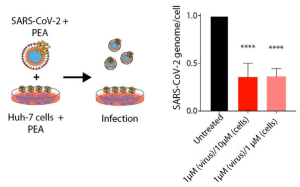
No PEA toxicity was detected, even when PEA was present as high as 100 μM in Huh-7 and 293T cells. When Huh7 cells were infected with SARS-CoV-2 virions incubated or not with PEA at 1 μM, the amount of SARS-CoV-2 genomes were reduced by ~64%, as detected by qRT-PCR 72 h after infection.

The effect of PEA was evaluated against SARS-CoV-2 two variants (Delta and Omicron). It was shown that PEA-pretreated Huh-7 cells at 10 and 1 μM reduced the numbers of viral genomes by 62.4% and 51.2% for SARS-CoV-2 Delta and by 43.4% and 77.3% for the Omicron variant, respectively. Interestingly, it was found that SARS-CoV-2 Delta and Omicron virions exposed to PEA showed a reduction in viral genomes/cell up to nearly 65% for both variants with 10 μM PEA. Also, when PEA was applied to both cells and virions simultaneously. PEA administered at 1 μM led to significant reductions in the number of viral genomes in cell lysate that were quantified as 75.3% and 72.5% for the Delta and Omicron VOCs, respectively.

A novel method to detect O-GlcNAcylated proteins with a disaccharide-tag and WJA lectin

A group from Faculty of Integrated Biosciences, Graduate School of Frontier Sciences, The University of Tokyo, Kashiwa, Chiba, Japan, etc. has reported a novel method to detect O-GlcNAcylated proteins specifically and with high sensitivity.
https://www.ncbi.nlm.nih.gov/pmc/articles/PMC9126400/

O-GlcNAc modification of serine or threonine occurs only on nuclear or cytoplasmic proteins. This modification is significantly different from N-glycosylation or O-glycosylation from several perspectives.
(1) O-GlcNAcylation occurs in the nucleus or cytoplasm of the cell, whereas general glycosylation occurs in the luminal regions of the endoplasmic reticulum and Golgi apparatus.
(2) O-GlcNAcylation is a reversible reaction via the actions of O-GlcNAc transferase (OGT) and O-GlcNAcase (OGA), although N- and O-glycosylation are irreversible.
(3) general N- and O-glycans on proteins are sequentially processed and elongated with many types of glycosyltransferases. However, O-GlcNAc residues cannot be modified by other monosaccharides.

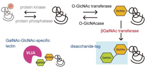
In this study, to detect O-GlcNAc modification of proteins, especially in the nucleus of the cell, expression vectors encoding soluble β4GalNAc-TA were constructed with a nuclear localization signal (NLS) from the SV40 large T antigen at the N- or C-terminus, respectively, and let β4GalNAc transferase transfer GalNAc to the GlcNAc residue of the O-GlcNAcylated proteins forming GalNAcβ1-4GlcNAc disaccharide. Then, Wisteria japonica agglutinin (WJA) was used as a probe to detect GalNAcβ1-4GlcNAc disaccharide, because WJA is highly specific for GalNAcβ1-4GlcNAc.
This method was named as disaccharide-tag method.

The binding between GalNAc-GlcNAc used in this method and WJA lectin has a stronger Ka of 1.4 x 10⁵ M-1 which enables higher sensitivity than any other methods. In addition, it should be emphasized that sugar chains with the GalNAc-GlcNAc sequence were hardly expressed in animal cells. The GalNAc-GlcNAc structure is found at the non-reducing end of the N-type sugar chain of pituitary glycoprotein hormones (leutropin, thyrotropin, follitropin), which serves as a clearance signal from the blood stream. These glycoprotein hormones are present only in the pituitary gland and are not expressed in other tissues and cells.

Roles of PSGL-1 on HIV infection

A group from Department of Biological Sciences, University of Toronto Scarborough, 1265 Military Trail, Toronto, ON Canada, etc. has reported that P-selectin glycoprotein ligand-1 (PSGL-1/CD162) is expressed on HIV-1 envelops and can mediate virus capture and subsequent transfer to permissive cells (CD4+ T-cells etc).
https://www.ncbi.nlm.nih.gov/pmc/articles/PMC9123692/

P-selectin glycoprotein ligand-1 (PSGL-1/CD162) has been studied extensively for its role in mediating leukocyte rolling through interactions with its cognate receptor, P-selectin. Structurally, PSGL-1 is a highly glycosylated homodimeric transmembrane protein, with an extracellular domain (ECD) of 50–60 nm in length that extends far out from the cellular surface.

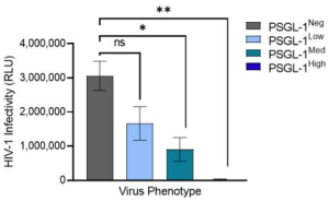
In reality, PSGL-1 plays diverse roles in the physiology of HIV-1 infection. It has been shown that the viruses with high levels of PSGL-1 are not infectious as show below.

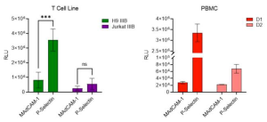
Since the primary target of HIV-1 infection, CD4+ T cells, are often found on activated endothelial tissues which display P-selectin in inflammatory conditions, it was interested in testing whether virions captured by P-selectin could be transferred to nearby permissive cells to elicit infection.
It was decided to test this model with T cell line and PBMC viruses, which contained lower levels of PSGL-1 and higher levels of gp120. We observed that viruses produced in T cell lines and primary PBMC were captured by P-selectin at levels that were markedly higher than the control. Most importantly, both viruses were effectively captured by P-selectin and transferred to HIV-1-permissive cells, suggesting that this mechanism of PSGL-1-mediated virus capture and transfer might also occur when HIV viruses encounter P-selectin on cell surfaces in vivo.

Burkholderia vietnamiensis B418 could be an effective biological nematicide for nematode management

A group from School of Bioengineering, Qilu University of Technology (Shandong Academy of Sciences), Jinan, 250353, China, etc. has reported that Burkholderia vietnamiensis B418 is an effective biological nematicide for nematode management.
https://www.nature.com/articles/s41598-022-12472-2

Root-knot nematodes (RKN), Meloidogyne spp., are highly polyphagous sedentary parasites capable of infesting a wide range of crops especially in greenhouse vegetable cultivation. During infestation, RKN can incite obvious knots or galls on plant roots, destroy the normal structure of the roots, compete with the host for water and nutrition, and make the host susceptible to secondary pathogens. Globally RKN damage is estimated to cause an annual economic loss of over $100 billion, accounting for about 12.6% of total crop losses.

The effects of different treatments on the reduction rate of nematode density and control efficacy against RKN are as follows.
All treatments (Burkholderia vietnamiensis B418 inoculation, fosthiszate, and avermectin) reduced nematode density to some extent compared with the negative control.
The highest reduction rate and control efficacy was observed with B. vietnamiensis B418 inoculation alone by 74.84% and 71.15%, respectively.
The introduction of B418 enhanced the inhibitory effects of chemical nematicide fosthiazate (from 38.92% to 62.71%) and biological nematicide avermectin (from 59.24% to 67.87%), which were still lower than that of B418 inoculated alone, indicating there was slight incompatibility within the combinations of B418 with fosthiazate and avermectin.

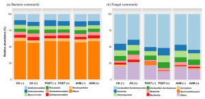
The application of B418 increased bacterial 16S rRNA sequences from 9.1% to 34.6%, and fungal ITS-2 rRNA sequences from 7.1% to 30.3%, which both exhibited more variation than the disease control treatments (9.1%).

The beneficial effects of PGPR on plant growth involve either direct mechanism such as biofertilization (facilitation of nutrient uptake including nitrogen and phosphorus primarily) and phytostimulation (production of plant growth promoting hormones), or indirect mechanism as biocontrol agents that antagonize the deleterious effects of phytopathogens by producing inhibitory substances or by inducing plant systemic resistance. The reason why inoculation of B. vietnamiensis B418 was effective in inhibiting RKN. However, it was found that the increment of Mortierella with B418 application was in accordance with the control effect against RKN. These variations in fungal community indicated that B418 inoculation resulted in the inhibition of pathogen-related species and the enhancement of plant-beneficial ones.

CK: control; FOST: fosthiazate; AVM: avermectin. (+ /−) stands for with and without B418 inoculation.

Novel serum glycan monosaccharide composite-based biomarkers for colorectal cancer (CRC) and colorectal adenoma (CRA)

A group from Department of Hepatobiliary and Pancreatic Surgery, The Affiliated Hospital of Qingdao University, Qingdao, China, etc. has developed novel serum glycan monosaccharide composite-based biomarkers for colorectal cancer (CRC) and colorectal adenoma (CRA).
https://www.ncbi.nlm.nih.gov/pmc/articles/PMC9099097/

HPAEC-PAD method was used to quantify the concentrations of two free monosaccharides (Glc-F and Man-F) and the six hydrolyzed monosaccharides (Fuc-H, GalN-H, GlcN-H, Gal-H, Glc-H, and Man-H) in the sera of healthy individuals, CRA, and CRC patients.
The concentrations of monosaccharides in serum were significantly higher in CRA and CRC patients than in healthy individuals. In concreat, concentrations of Fuc-H, GalN-H, GlcN-H, Gal-H, Glc-H, and Man-H in serum were significantly higher in CRC patients compared with healthy individuals. In addition, concentrations of Fuc-H, Gal-H, Glc-H, and Man-H in serum were significantly higher in CRA patients than healthy individuals

The logistic regression analysis was used to develop diagnostic models based on the above results. Tow types of the models were developed, one is MC1 and the other is MC2.
The logistic regression equation for MC1 was as follows: MC1 = -8.18 – 0.021 Fuc-H – 0.004 GlcN-H + 0.011 Man-H, where the Fuc-H, GlcN-H and Man-H were in μmol/L, and
the logistic regression equation for MC2 was as follows: MC2= -6.639 – 0.022 Fuc-H + 0.003 Gal-H + 0.003 Man-H, where the Fuc-H, Gal-H and Man-H were in μmol/L

MC1 was used to distinguish between healthy individuals and CRC, and MC2 was used to distinguish between healthy individuals and CRA. Area under receptor operating characteristic curve (AUC) of MC2 and MC1 was 0.8025 and 0.9403 respectively.
As a reference, the AUC of the existence marker CEA between healthy individuals and CRC was 0.7384.

Cocaprin (a β-trefoil fold lectin) from the mushroom Coprinopsis cinerea inhibits both cysteine and aspartic proteases

A group from Department of Biochemistry and Molecular and Structural Biology, Jožef Stefan Institute, 1000 Ljubljana, Slovenia, etc. has reported that a lectin with β-trefoil fold from the mushroom Coprinopsis cinerea, named cocaprins, inhibits both cysteine and aspartic proteases.
https://www.ncbi.nlm.nih.gov/pmc/articles/PMC9104457/

Sequence-based structural analysis about Coprinopsis cinerea predicted that the two encoded proteins, CCP1 and CCP2, respectively, and their paralog CCP3, containing a Ricin-type β-trefoil lectin-like domain. All three proteins lack a signal peptide for classical secretion and are, thus, predicted to be cytoplasmic.

It was found that cocaprins inhibit plant cysteine proteases belonging to the C1 family with Ki in the low micromolar range, and also inhibit aspartic protease pepsin with Ki in the low micromolar range.

It has shown that cocaprins have lectin activity in addition to protease inhibition. Glycan microarrays were used to analyze carbohydrate binding specificity of cocaprins. For CCP1, very weak binding was observed on a mammalian glycan array to structures including LacNAc or polyLacNAc and for CCP2 the binding was even weaker. This shows a potential for glycan-binding activity in cocaprins.

However, the biological function of cocaprins is unknown. Regarding a potential role in defense, it is noteworthy that CCP2 expression was induced upon challenge with a fungivorous nematode . However, no toxicity of the protein was detected against nematodes or dipteran insect larvae, although all of which have been shown to be targeted by other β-trefoil protease inhibitors and lectins. Strange isn’t it.

Diagnosis of IgA nephropathy (IgAN): A sandwich assay focusing on aberrant glycosylation of serum IgA1 with WFA lectin

A group from Reagent Engineering, Protein Technology Group, Sysmex Corporation, Kobe 651-2271, Hyogo, Japan, etc. has reported about evaluation of serum WFA+-IgA1 as IgA nephropathy (IgAN) diagnostic marker.
https://www.ncbi.nlm.nih.gov/pmc/articles/PMC9104065/

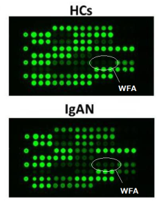
Authors have reported results of an automated sandwich immunoassay system for diagnosing IgA nephropathy (IgAN), using Wisteria floribunda agglutinin (WFA) lectin and anti-IgA1 monoclonal antibody focusing on aberrant glycosylation of IgA1.

The usefulness of WFA for the immunoassays was investigated by lectin microarray. A figure below shows the typical images of microarray results in HC and IgAN patients, and it was confirmed that the WFA signal increased significantly in IgA1 derived from IgAN patients compared with that in HCs.

Unfortunately, the results seemed to be disappointing from a view point of diagnostic assay, i.e., Sensitivity = 66% and Specificity = 62%.

The importance of booster vaccine doses for protection against all Omicron variants

A group from Center for Retrovirus Research, The Ohio State University, Columbus, OH 43210, USA, etc. has reported about the importance of booster vaccine doses for protection against all Omicron variants.
https://www.sciencedirect.com/science/article/pii/S1931312822002207?via%3Dihub

There are several Omicron sub-lineages with significant variations in their S proteins, including BA.1, BA.1.1, and BA.2. While the constellation of mutations varies between isolates, the BA.1.1 lineage is defined by the presence of a single R346K mutation that is absent from the BA.1 lineage, whereas the BA.2 lineage is defined by key S mutations T19I, L24S, Δ25/27, V213G, T376A, and R408S. Although BA.1 was the major variant during the Omicron wave of the pandemic, the BA.2 variant, and to a lesser extent BA.1.1, has begun to account for an increasing proportion of cases. In particular, the BA.2 variant exhibits enhanced transmissibility relative to BA.1 and can reinfect previously BA.1-infected individuals.

In this study, authors have demonstrated the followings:
Omicron BA.1.1 is effectively neutralized by Omicron patient sera, and Omicron sub-lineages are comparably neutralized by Omicron patient sera.
Omicron BA.1 and BA.2 are resistant to neutralization by two-dose mRNA vaccination but sensitive to neutralization after a booster dose.