A counter-receptor of Siglec-7 was identified as  leukosialin (CD43)

A group from Nagoya Univ. has identified a counter-receptor of Siglec-7.
https://www.jbc.org/article/S0021-9258(21)00251-9/fulltext

Siglec-7 binds to sialic acids and are expressed on natural killer (NK) cells and monocytes. Siglec-7 suppress cytotoxicity of NK cells through interaction with a counter-receptor on it. However, the counter-receptor of Siglec-7 has not been identified yet completely. Authors purified the receptor with using K562 cells, FC-Siglec-7 fusion protein, diSia-dextran polymer, and identified it as leukosialin (CD43) with MS analysis. Additionally, they demonstrated that the cytotoxicity of NK cells toward K562 cells was suppressed by overexpression of leukosialin in a Siglec-7-dependent manner.
 

SARS-CoV-2 NTD could bind to A blood type I antigen expressed on respiratory epithelium cells

A group from Harvard Univ. and Emory Univ. has reported on the interaction between ABO(H) blood group and SARS-CoV-2 RBD.
https://ashpublications.org/bloodadvances/article/5/5/1305/475250/The-SARS-CoV-2-receptor-binding-domain

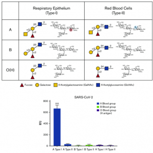
It has been suggested that SARS-CoV-2 NTD might be bid to ABO(H) blood antigens, because SARS-CoV-2 RBD shares sequence similarity with a galectin family. Authors have confirmed that SARS-CoV-2 RBD could bind to A blood type I antigen expressed on respiratory epithelium cells with using glycan arrays.

The difference in Biomarkers between ICU patients with COVID-19-related ARDS and classical ARDS: Vascular Endothelial cells are heavily damaged

A group from University of Ferrara, etc. has reported on the difference in biomarkers between ICU patients with COVID-19-related ARDS and classical ARDS.
https://ccforum.biomedcentral.com/articles/10.1186/s13054-021-03499-4

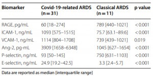
Evaluated marker were as follow, Receptor for Advanced Glycation End Products (RAGE), Angiopoietin-2 (Ang-2)、soluble intercellular adhesion molecule-1 (ICAM-1), soluble vascular cell adhesion molecule-1 (VCAM-1), E-Selectin, and P-Selectin.

Ang-2 is a growth factor (glycoprotein) promoting vascularization,
ICAM-1 is a member of immunoglobulin superfamily and a ligand for activated leukocytes,
VCAM-1 functions as a cell adhesion molecule like ICAM-1, but is usually not expressed on endothelial cells, and needs IL-1, TNF-α stimulations,
E-Selectin is a leukocyte-endothelial cell adhesion molecule expressed on vascular endothelial cells,
P-Selectin is a leukocyte adhesion molecule, and expressed in platelets and vascular endothelial cells.

RAGE has almost no relationship with COVDI-19 suggesting that the ARDS is caused by virus infection. Ang-2, ICAM-1, BVCAM-1, and E-Selectin are higher in COVID-19 (P-Selectin is lower on the contrary) suggesting that vascular endothelial cells are heavily damaged in ICU patients of COVID-19. 

The effect of Q677 variants (Q677P and Q677H)on SARS-CoV-2 infectivity: Conformational changes around RRAR motif

On a blog article posted on Feb. 19th, 2021, it was reported that new variants (Q677H, Q677P) are increasing from the end of the last year in North America.

A group from Univ. of Bern, etc. has reported on the effect of Q677 variants on SARS-CoV-2 infectivity with using a SWISS MODEL.
https://www.ncbi.nlm.nih.gov/pmc/articles/PMC7885944/

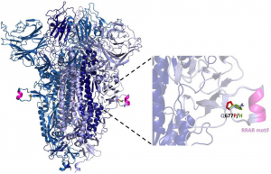
The S1/S2 cleavage site contains the multibasic cleavage site (RRAR Motif). Although the position 677 is outside the polybasic site (furin binding pocket), the presence of a proline at this site may introduce a favorable kink that promotes the dynamic conformational changes necessary for cleavage at the S1/S2 junction, which is governed not only by furin-like activities, but also by trypsin-like proteases (e.g., TMPRSS2) and cathepsins. Moreover, the introduction of a proline in this model appears to be 3.7 angstroms away from the carbon backbone of S689 (relative to 4.9 angstroms for the native glutamine), which may promote atomic interactions that encourage conformations favorable for proteolytic cleavage. In the case of the S:Q677H substitution, histidine protonation could similarly act as a conformational switch affecting accessibility to proteases. These conformational changes may accelerate virus infection. 

Disease severity of the new coronavirus (COVID-19)and its relationship with balance of humoral immunity and cellular immunity

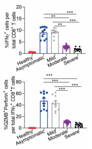
In the new coronavirus (COVID-19), there is a broad spectrum of disease severity from asymptomatic to severe. How the immune reaction is different in the disease spectrum? There are so many studies are ongoing in the world to uncover this problem. A group from Third Military Medical University, Chongqing, China, etc. has reported there is a clear difference in the balance of humoral immunity and cellular immunity between asymptomatic/mild and moderate/severe.
https://www.nature.com/articles/s41392-021-00525-3

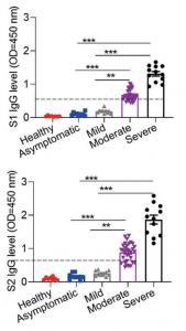
Let’s classify disease severity into two categories, asymptomatic/mild and moderate/severe. In the case of asymptomatic/mild, the expression of SARS-CoV-2 specific IgG is weak and transiently terminated without developing germinal center. In contrast, in the case of moderate/sever, the expression of SARS-CoV-2 specific IgG increases greatly and circulating cTFH cells and chemokine CXCL13, which indicate the magnitude of ongoing germinal center, are upregulated, suggesting that the humoral immunity is strongly activated.
On the other hand, in the case of asymptomatic/mild, IFN-γ producing CD8+ T cells、SARS-CoV-2 specific TH1 cells, Granzyme B(GZMB)are greatly upregulated, suggesting that cellular immunity is strongly activated. (Granzyme B(GZMB)is a Serine Protease expressed on cytoplasmic granules of activated cytotoxic T cells and NK cells, and induce apoptosis of target cells)

That is, asymptomatic/mild is a result of SARS-CoV-2 infection suppression with activate cytotoxic T-cells and NK cells (not mentioned here), and if the viruses are not cleared with this process, the humoral immunity comes to the front to combat viruses, or we can say that it is a result of the balance between humoral immunity and cellular immunity.
Further, recent studies found there exists cross-reactive T cell recognition between circulating “common cold” coronaviruses and SARS-CoV-2I. So, it is of great interest to examine whether the history of “common cold” coronavirus infection with pre-existing SARS-CoV-2 cross-reactive T cells could account for the clinically asymptomatic state in COVID-19 patients.

Monochronal Antibody Cocktails would be effective in rapidly mutating RNA viruses such as SARS-CoV-2 

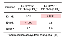
A group from Fred Hutchinson Cancer Research Center, etc. has investigated the effect of genomic mutations onto two types of monochronal antibodies (LY-CoV555 and LY-CoV016) against RBD of SARS-CoV-2.
https://www.biorxiv.org/content/10.1101/2021.02.17.431683v1

It was found that LY-CoV016 is very weak for K417N mutation, and LY-CoV555 is so weak for E484K mutation. In both cases, IC50 increased more than 1,000 times with these mutations. Therefore, a strategy using antibody cocktails with different monochronal antibodies with different target epitopes would be quite effective in developing therapeutic drugs for rapidly mutating RNA viruses such as SARS-CoV-2.

SARS-CoV-2 UK variant (B.1.1.7) and South African variant (B.1.351) have stronger affinity to ACE2

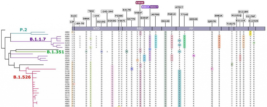
Two types of recently appeared SARS-CoV-2 variants named UK variant (B.1.1.7) and South African variant (B.1.351) are very famous. And, there have been several reports on the efficacy of Pfizer’s and Moderna’s vaccines for those variants. On Feb. 6th, 2021, a short report was introduced in this blog that the current vaccines are not so affected by UK variant, but are greatly affected by South African variant.

A group from School of Medicine, Stanford University has reported that these variants have higher affinity to ACE2, in concrete, about two times with B.1.1.7 and about five times with B.1.351.
https://www.ncbi.nlm.nih.gov/pmc/articles/PMC7924271/ 

Aberrant glycosylation in Breast Cancer and its relationship with Macrophages: Functions of Lectins such as SIGLEC-9, SIGLEC-10, SIGLEC-15, and MGL 

There was a review on aberrant glycosylation in breast cancer and its relationship with macrophages. Important points from this revie was summarized here.
https://www.mdpi.com/1422-0067/22/4/1972/htm

The following things have been reported about aberrant glycosylation observed in breast cancer,
increase in Lewisa Lewisx,
increase in Core Fucose,
increase in branched N-glycans,
truncation of O-glycans,
and, increase in sialo-glycans for both N-glycans and O-glycans.

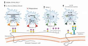
The breast cancer microenvironment is composed of mainly by stromal cells and immune cells infiltrated. It would be so important to understand interactions between breast cancer cells and immune cells (typically macrophages) from a view point of cancer therapy.
From this point, SIGLEC-9, SIGLEC-10, SIGLEC-15, and MGL would be hot spots. SIGLEC-9 binds to Sialyl-T enriched MUC1, and activate MAPK-ERK signal path leading to increase in immune checkpoint PD-L1 expression and and IL-10 secretion. SIGLEC-10 binds to Sialyl-T and Sialyl-Tn antigens, and suppress macrophage’s phagocytosis. SIGLEC-15 binds to Sialyl-Tn antigens, and seems to activate SYK/MAPK signal path, rather than stimulating TGF-ベータecretion, although the functions are not clearly identified. On the other hand, MGL bids to GalNAc, and is related to activation of ERK signal path and secretion of IL-10. These points would be drug development targets for the breast cancer.

The role of neuclear chromatin protein HMBG1 released from cells in Sepsis and a relationship with blood pH and serum Zn concentration

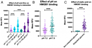
A group from UC San Diego has reported that blood pH, serum Zn concentration, existence of plasma sialoglycoproteins are deeply related to binding formation of High mobility group box 1(HMGB1), which is released from cells into blood with developing sepsis, to leukocyte receptors.
https://www.pnas.org/content/118/10/e2018090118

It was know that HMBG1 works as a chaperone protein controlling gene expression through the interaction with chromatin in healthy cells, and further is passively or actively released from cells into blood in conditions like sepsis, leading to activation of innate immunity, migrating neutrophils to necrotic tissues, and removing such tissues.
http://www.med.osaka-u.ac.jp/introduction/research/endowed/therapy

Authors has shown that the binding of HMBG1 to neutrophils is greatly inhibited by the decrease in blood pH and serum Zn concentration, and also the binding is inhibited by preferential binding of HMBG1 to plasma sialoglycoproteins like a lectin binding to sialic acids.
Blood pH is tightly maintained between 7.35 and 7.45 in healthy people. However, it goes down below 7.3 with developing sepsis, and also serum Zn concentration decreases to a level of few µM. In a healthy condition, even if HMGB1 was released from cells into blood, it’s binding to neutrophil receptors could be inhibited by preferential binding to sialoglycoproteins, and thereby no extra inflammation would not be induced. However, with decreasing blood pH and Zn concentration, sialoglycoproteins lose its function as inhibitors, and HMGB1 is able to bind to neutrophil receptors inducing inflammation. That means, a treatment targeting inhibition of HMGB1 with controlling blood pH and the Zn concentration might be also effective in sepsis-related Sequential Organ Failure happening in COVID-19.