The universal tendency of age to be involved in the severity of the new coronavirus (COVID-19) and its reasons

The fact that the new coronavirus (COVID-19) is more likely to develop severe respiratory illness with the age is well known. This behavior is not specific to COVID-19 and shows a similar trend also in SARS and MARS that have occurred in the past.
Below is the result of the latest COVID-19 fatality rates by ten-year age-groups in seven countries.
https://journals.plos.org/plosone/article?id=10.1371/journal.pone.0241031


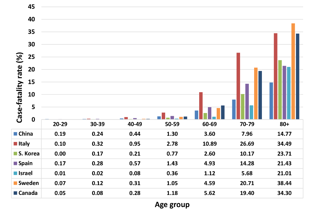
The figure above proves splendidly that the elderly persons are really dangerous. But why does the fatality rate increase as the age increases? Unfortunately, we still don’t know exactly what the mechanism is for this.

However, generally speaking, when the age increases, (1) dendritic cells and alveolar macrophages decrease, (2) the expression level of TLR7 and MDA5 is reduced, which are virus RNA receptors to activate innate immunity, and (3) although neutrophils and alveolar macrophages are on the increase, it can be said that the immune function of these cells such as phagocytosis etc. tends to decrease with the age. In other words, it seems to be a rough picture that the impaired innate and adaptive immune responses develops severe disease.
https://www.jci.org/articles/view/144115

Relationship between the new coronavirus (COVID-19) and ABO/Rh± blood types

A group of University of Bologna has evaluated the effects of ABO blood types on the risk of infection with the new coronavirus (COVID-19) using 7,503 cases and 296,216 controls.
https://www.ncbi.nlm.nih.gov/pmc/articles/PMC7500631/

The reported results are as follows,

Type A: OR: 1.23 (95% CI: 1.09-1.4)
Type B: OR: 1,05 (95% CI: 0.96-1.15)
AB Type: OR: 1.09 (95% CI: 0.94-1.26)
Type O: OR: 0.77 (95% CI: 0.67-0.88)

Further, a group of Sakarya University has reported that the Rh+ blood group was found in a significantly high number of patients who were admitted to ICU.
https://pubmed.ncbi.nlm.nih.gov/32965363/

Using CRISPR/Cas12a to detect new coronavirus (COVID-19:SARS-CoV-2): Sensitization effect adding Mn2+

CRISPR/Cas9 won the 2020 Nobel Prize in Chemistry about a week ago.
We have found several papers that used this technique as a high sensitive detection method for the new coronavirus. The following paper reports that detection using CRISPR/Cas12a could be further 13 times more sensitive by adding Mn2+.
https://www.ncbi.nlm.nih.gov/pmc/articles/PMC7536916/#advs2020-bib-0005

A brief description of the protocol is as follows:

  • Extract an RNA sample from a sample.
  • Make cDNA using reverse transcription, and amplify by PCR (dsDNA is made).
  • Pre-design crisper RNA (crRNA) using RNA sequences specific to the E protein of the new coronavirus.
  • Add Cas12a enzyme and crRNA to the dsDNA sample, the new coronavirus-specific sequence is cut out when it is in the sample dsDNA.
  • Thus, since the inhibitory interaction between the fluorescent probe and the quencher that have been incorporated in the PCR process is lost, the fluorescence comes out by applying the excitation light.

Of course, this fluorescence is read by a detector, but it can be confirmed by the eyes.
The addition of Mn2+ to this system increased the sensitivity by 13 times.

Why is the humoral response (antibody-centric adaptive immune response) in the new coronavirus (COVID-19) non-persistent?

A group of Harvard medical school reported about why the humoral response (antibody-centric adaptive immune response) is non-persistent in the new coronavirus (COVID-19)?
https://pubmed.ncbi.nlm.nih.gov/32877699/

In addition to innate immunity, adaptive immunity centered on antibodies plays an important role in the elimination of foreign substances such as viruses that invade the living body. In the early stages of infection, low affinity IgG antibodies are produced, but IgG antibodies produced over time mature and increase affinity for antigens. This is because B cells that produce high affinity antibodies differentiate in germinal center (a small structure formed in the immune system, such as the spleen and lymph nodes, during the immune response). In the early stages of infection, antigen-specific B cells quickly differentiate into plasma cells to produce low-affinity antibodies, but some B cells express transcription factor Bcl6 (Bcl-6 Tfh cells) to form the germinal center.

In fact, in COVID-19, the fact that the formation of this germinal center is suppressed seems to be the cause of the title. Why is the formation of the cup center suppressed? The detailed mechanism is not known, however,  it is said that excessive production of cytokines such as TNF-α suppresses the differentiation of Bcl6-Tfh cells and results in loss of germinal center.

This behavior seems to occur not only in COPID-19, but also in Ebora, Marburg disease, and H5N1 influenza.

How glycan targeted drug discovery for cancer should be

Glycans, as is said to be the face of cells, greatly change the structure depending on the state of the cell. It is known that specific changes in the glycan structure occur in canceration, and various diagnostic and therapeutic drugs are under development using glycans as the drug discovery targets. Changes in glycan structure caused by canceration depend on the type of cancer, but common characteristics include followings:

(1) Increase in multi-branching of N-type sugar chains
(2) Shortening of O-type glycan chains
(3) Increase in terminal-sialic acid modification
https://jitc.bmj.com/content/8/2/e001222.long

However, in addition to these, there seems to be a feature that the amount of glycan modification increases when it becomes cancerous, and the high mannose structure increases, too.
https://pubmed.ncbi.nlm.nih.gov/32143591/

Based on the cited papers above, I will summarize the strategy of glycan targeted drug discovery as follows.

  • Antobody-drug conjugates (ADC): target on cancerous cell surface expressing targeted glycans using glycan binding agents (i.e., lectins etc.). In this case, it might work better to adopt bispecific antibodies to glycans on target cancerous cell surface more precisely  (e.g., bispecific to GD2 ganglioside and MUC1)
  • Siglec inhibitors: Siglec is expressed on most of the immune cells, the immune response is suppressed when the signal is triggered by binding to sialoglycan.
  • Galectin inhibitors: tumor cells highly express galectins, and bind to immune checkpoint CTLA-4 to suppress the immune response. Galectin-1 induces apoptosis of T- cells.
  • Immune checkpoint inhibitors: PD-1/PD-L1 are also strongly glycosylated, and molecular-targeted drugs with those excellent inhibitory effects are promising.
  • C-type lectins: DC-SIGN, Dectin-1 and others promote anti-tumor activity, while NK62DG and Mincle work immuno-suppressively

Characteristics of IgG Responses in The New Coronavirus (COVID-19)

A group of Chulalongkorn Univ. reported the IgG response of patients with the new coronavirus (COVID-19) in Thailand as follows:
The study is based on data from 384 patients analyzed between March 10 and May 31, 2020.
https://journals.plos.org/plosone/article?id=10.1371/journal.pone.0240502

IgG in serum comes up two weeks after infection.
In patients with mild infection, IgG was not detected in 20% of patients 2 weeks after infection.
The expression level of IgG increases as mild, moderate, severe and symptoms become heavier.
Men have higher expression of IgG than women.


The reported tendency on IgG correlates with other reports published.

Mutations in S-proteins of the new coronavirus (SARS-CoV-2): 25 mutations, including A930V, D614G, A706S, and A879S

From the RNA sequence of the new coronavirus (SARS-CoV-2) registered from India in NCBI-Virus-database as of June 6, 2020, a research result has been reported on what mutations occurred compared to the original strain that began to spread in Wuhan, China.
https://www.ncbi.nlm.nih.gov/pmc/articles/PMC7521409/

There are 25 mutations, and they are divided into four clusters (1-100, 148-255, 570-680, 820-930).

Mutations in T572I, A879S, A892V, and A930V have caused significant changes in the secondary structure of proteins, T572I causes a structural change to coilded→helix and the other three to helix→beta sheet. Mutations in the A930V, D614G, A706S, and A879S seem to make a relatively large difference in protein structure. Of particular interest is that the mutation of D614G occurs in 88% of RNA sequences, which is a mainstream among new coronaviruses that are spreading in India. For the D614G, it is reported that it promotes the open conformation of RBD, and as a result, the infectivity of the new coronavirus is increased.

R408I, E471Q is said to be a mutation that is seen only in India.

Diagnosis of serum SARS-CoV-2 nucleocapsid protein (N-protein) is highly effective in early diagnosis of new coronavirus (COVID-19)

It is well known that the diagnosis for the new coronavirus is twofold; RT-PCR to check for the infection and antibody testing to check for the infection history.
Antibody testing can not be used for early diagnosis of SARS-CoV-2, because it take more than a week before the antibody rises up. However, by focusing on serum nucleocapsid protein of SARS-CoV-2, it seems that the infection of the new coronavirus can be detected even early before the antibody (IgG) rises up.
https://www.ncbi.nlm.nih.gov/pmc/articles/PMC7498565/

The ROC curve showed that AUC was 0.9756 (95% CI), sensitivity was 92%, and specificity was 96.8%. The cut-off value of the capsid protein was 1.85 pg/mL.

Wouldn’t it be a great achievement?

Fusion protein of lectins derived from Ricinus communis and Phytolacca americana expected as a novel therapeutic agent for the new coronavirus (COVID-19)

As a treatment for the new coronavirus (COVID-19), the usefulness of the fusion protein (RTAM-PAP1) between a mutant of the lysine A chain extracted from Ricinus communis (RTAM) and a lectin extracted from the leaves of Phytolacca americana (PAP1) is reported from the viewpoint of affinity evaluation between various proteins of SARS-CoV-2 and RTAM-PAP1 and also from toxicity testing using mice.
https://www.mdpi.com/2072-6651/12/9/602/htm

The binding affinity between a patient-derived antibody B38 of SARS-CoV-2 (as a control), ACE2, RTAM-PAP1 and various proteins of SARS-CoV-2 were evaluated using three-dimensional molecular structure analysis software named CoDockPP, HASSOCK2.2, and ZDOCK. The binding of RTAM-PAP1 was stronger than ACE2, and showed the same binding affinity as B38 comprehensively.
In addition, the toxicity tests using mice showed no side effects even at 1 mg/kg of dose.

This paper shows the usefulness of the fusion protein of lectins derived from Ricinus communis and Phytolacca americana as a novel drug for the new coronavirus.


Ricinus communis


Phytolacca americana

How to reduce cross-reactivity to other coronaviruses in the detection of new coronaviruses (SARS-CoV-2) using RT-PCR

RT-PCR is used as you know to test for suspected infection of the new coronavirus (SARS-CoV-2). Sensitivity and specificity are very important in testing, but there are some doubts as to how high the generally implemented RT-PCR could detect SARS-CoV-2 without cross-reactivity to other coronaviruses including influenza viruses. Therefore, the following groups have reported a novel methodology for improving the specificity of SARS-CoV-2 detection without sacrificing sensitivity.
https://www.mdpi.com/2075-4418/10/10/775

As the methodology, they adopted the followings, (1) in the domain of structural protein of SRAS-CoV-2, design dual-target PCR primers targeting on coding region of the accessory and envelope proteins (ORF3ab-E primers) and that of the capsid protein (N primers), and (2) use a peptide nucleic acid (PAN) designed to target on the N region as a blocker of PCR reaction.

Peptide nucleic acids are artificial compounds in which the deoxyribose phosphate backbone is replaced by a pseudo-peptide polymer to which the nucleobases are linked, and the binding affinity for target DNA and RNA is remarkably increased by nearly 1000 times, and therefore, it does not act as a primer but act as an inhibitor of PCR.

As a result, cross-reactivity to other coronaviruses and influenza viruses disappeared completely, and the detection rate of SARS-CoV-2 was 100% for ORF3ab-E and 82.6% for PNA-N.