Archive 20/11/8

RT-PCR testing for the new coronavirus (SARS-CoV-2) is critical to the number of days since the first signs of infection and how samples are obtained.

RT-PCR is Gold Standard for testing for the new Coronavirus (SARS-CoV-2).

However, you need to be careful about RT-PCR results. Rt-PCR tests from the nasopharyngeal are highly reliable until the fourth day after signs of infection, but after 10 days, the detection accuracy falls and the probability of false negative gets very high.
On the other hand, the test from the feces is not reliable at the beginning of infection, but it is possible to detect the virus even after a number of days of infection, and it is better to use feces than to obtain samples from the nasopharyngeal to determine whether the virus has been eliminated in the end.
https://bmcmedicine.biomedcentral.com/articles/10.1186/s12916-020-01810-8


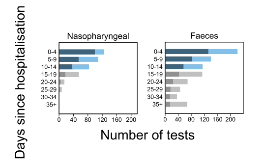
In this figure, dark blue and dark gray indicate virus detection two weeks before and after infection, respectively, and light blue and light gray indicate false negatives.

The presence of cross-antibodies between the new coronavirus (SARS-CoV-2) and HCoVs

The following group has reported the results of a study of cross-antibodies between the new coronavirus (SARS-CoV-2) and coronavirus (HCoVs) that contribute to the seasonal cold.
https://science.sciencemag.org/content/early/2020/11/05/science.abe1107

The HCoVs antibodies seem to have the effect of suppressing SARS-CoV-2 infection with the cross-reactivity for people who have never infected with the new coronavirus.
This cross-antibody is not targeting RBD of SARS-CoV-2 as the epitope, but common amino acid sequences in the S2 region seem to be the ones as shown below.


In addition, since the retention rate of the cross-antibody is high in young people, it may be related to the phenomenon that young people are less likely to develop the severe illness of COVID-19.

The synergetic effects of TNF-α and IFN-γ may be at the heart of the severity of the new coronavirus (COVID-19).

When COVID-19 develops and becomes severe with the new coronavirus, cytokines such as IL-6, IL-18, IFN-γ, IL-15, TNF-α, IL-1α, IL-1β are upregulated, and fall into the so-called cytokine storm state. It is widely known that acute respiratory distress syndrome (ARDS) in COVID-19 is directly correlated with this cytokine storm.

In the expression of such various cytokines, it has been pointed out by the following group that the synergetic effects of TNF-α and IFN-γ in particular may be causing severe disease.
https://www.ncbi.nlm.nih.gov/pmc/articles/PMC7605562/

Here is an example of in vivo experiment using a mouse that the synergetic effect of TNF-α and IFN-γ mimics the symptoms of COVID-19.


It has also verified in vivo that applying TNF-α and IFN-γ neutralizing antibodies to hACE2 transgenic mice infected with SARS-CoV-2 increases survival rates significantly.


So, inhibitors for accurate points in the downstream signal path of including TNF-α and IFN-γ will lead to development of effective drugs for ADRS of COVID-19.

Diabetes boosts severity and mortality of new coronavirus (COVID-19)

Clinical studies in South Korea have reported statistical analysis of the effects of diabetes on the new coronavirus (COVID-19).
https://e-dmj.org/journal/view.php?doi=10.4093/dmj.2020.0141

The OR (odds ratio: 95% CI) of diabetes statistically calculated by taking into account the effects of age, sex, hypertension, dyslipidemia, chronic kidney disease, chronic obstructive lung disease, cardiovascular disease, atrial fibrillation, end-stage renal disease, cancer on COVID-19 was as follows:

Hospitalization: 1.071 (0.722 – 1.588)
Oxygen inhalation: 1.349 (1.099 – 1.656)
Ventilators: 1.930 (1.276 – 2.915)
Death: 2.659 (1.896 – 3.729)

In the new coronavirus (COVID-19), the effect of hydroxychloroquine on viral proliferation was not observed.

Hydroxychloroquine is one of the candidates for the treatment of the new coronavirus (covid-19). This drug is said to prevent the proliferation of the virus by inhibiting RNA polymerase.
As for the mechanism of this medicine, by opening a gate on the cell membrane so that zinc ions can enter into the cell, and the zinc inhibits the function of RNA polymerase.

For the new coronavirus, the effect of this hydroxychloroquine was investigated by comparing the administrated group with the non-administered group. As an evaluation index in this study, the amount of the new coronavirus was evaluated with the number of cycles (Ct) to reach the PCR reaction threshold in RT-PCR.
Unfortunately, it turned out that there was not a significant difference between the two groups.
https://www.ncbi.nlm.nih.gov/pmc/articles/PMC7592126/

Over the past days, the mechanism is different from Hydroxychloroquine, but the WHO has announced that Remdesivir, which also inhibits the proliferation of virus, was not effetive.
More and more, the battle against coronavirus is not in sight.

ACE2, a receptor for the new coronavirus, is fairly locally expressed in motile cilia on epithelial cells in the Upper respiratory tract.

In the new coronavirus, it is well known that ACE2 is the main receptor. The results of a detailed investigation of the expression status of ACE2 in human have been reported.
https://www.nature.com/articles/s41467-020-19145-6

Ace2 is said to be expressed in the nasopharynx, lungs, small intestine, kidneys, and testes by gene expression analysis, but immunostaining using ACE2 antibodies shows that ACE2 is relatively less expressed in the lungs than in other organs. In addition, ACE2 seems to be expressed in a fairly locally expressed in motile cilia on epithelial cells of the Upper respiratory tract.

On the other hand, it was also found that the expression level of ACE2 was little correlated with age, gender, and smoking. The administration of drugs such as Angiotensin-converting enzyme inhibitors (ACEI) and angiotensin II receptor blockers (ARBs) used in the treatment of hypertension, which is a concern for SARS-CoV-2 infections, was also found to have no effect on ACE2 expression.

Therefore, as for the prevention and severity of the new coronavirus (COVID-19), the followings are pointed out as a conclusion.
(1) SARS-CoV-2 inhibitors in the oral nasal cavity are effective in preventing infection,
(2) The expression level of ACE2 itself has nothing to do with the severity of COVID-19.

The severity of influenza correlates with Host Cell’s high Mannose modification

For influenza viruses, it is well known that the binding of α2-6Sia in human respiratory tract cells and HA (hemaggulutinin) with influenza virus causes infection. However, the relationship between the severity of influenza and the glycosylation is not well known.
The following groups use lectin microarrays to study the severity of influenza and the glycosylation of Host Cells using ferret model.
https://www.pnas.org/content/117/43/26926.long


The result is that the glycosylation of Host cells changes when infected with the influenza virus, and the increase in the high mannose structure is correlated with the severity of the disease.
This is an interesting phenomenon. The high Mannose-binding C-type lectin is expressed on immune cells, so its association resulting in self-tissue damages might be suspected.

S-protein of SARS-CoV-2 has a staphylococcal enterotoxin-like sequence in the shadow of the severity of the new coronavirus (COVID-19)

The fact that the S-protein sequence of the new coronavirus (SARS-CoV-2) contains an unique sequence “PRRA” not found in other coronaviruses is inserted at the boundary between the S1 and S2 domains. It has also been frequently pointed out that the presence of this sequence has increased the infectivity of this virus to humans.

There is a paper discussing about the presence of a staphylococcal enterotoxin-like sequence around the inserted “PRRA” sequence (661-685) of SARS-CoV-2, and its potential effect on hyperinflammation of COVID-19.
https://www.pnas.org/content/117/41/25254.long

By binding the T cell receptors (TCR) to this bacterial toxin-like sequence, the resulting toxic shock syndrome might be behind of the severe hyperinflammation of COVID-19? It says. This inference was made from molecular structural analysis of proteins, and is expected to be tested in practice.


SEB represents staphylococcal enterotoxin and has a quite structure similar to the 661-685 sequence of SARS-CoV-2.

Relationship between the incidence rate of the new coronavirus (COVID-19), temperature, and BCG

Using data on the new coronavirus (COVID-19) in the United States, Canada, Italy, Spain, Germany, France, Australia, Japan, South Korea, Malaysia, and Thailand, the results of analyzing the effects of temperature and BCG vaccination on the incident rate of COVID-19 have been reported.
https://journals.plos.org/plosone/article?id=10.1371/journal.pone.0240710

Effects of Temperature (Average Temperature Model):
The temperature of -21 to 5.5 degrees Celsius is used as a reference.
In the region from 6 to 10.5 degrees C, the incident rate ratio is 1.18 (95% CI: 0.87 – 1.59)
In the region from 11 to 18.5 degrees C, the incident rate ratio is 0.97 (95% CI 0.65 – 1.45)
In the region from 19 to 36.5 degrees C, the incident rate ratio is 0.87 (95% CI: 0.47 – 1.62)

About BCG Vaccination:
If vaccinated with BCG vaccine, the incident rate ratio is 0.37 (95% CI: 0.17 -0.79)

In conclusion, although the average temperature of 6 to 10.5 degrees Celsius tends to increase the incident rate slightly, there is little change in the incident rate even if the average temperature rises. As for the inoculation of BCG vaccine, it is clearly effective in suppressing the incident rate.

Serum-IgG response in mild and severe COVID-19 patients infected with the new coronavirus (SARS-CoV-2)

In COVID-19 patients infected with the new coronavirus (SARS-CoV-2), IgG antibody tests of (Architect and iFlash) were used to measure serum-IgG in mild and severe cases. In some mild cases, IgG was not detected. However, when a method of testing for a neutralizing antibody was used, it was really detected from such mild cases in which IgG was not detected. So, this means that we must be careful in choosing IgG assay for COVID-19.

IgG rises up on the average 11th day (range of 7 to 20 days) after symptoms appear in severe patients, while on average the 22nd day (range of 14 to 79 days) for mild patients, and the total amount of IgG produced was higher in severe patients than in the mild.
https://journals.plos.org/plosone/article?id=10.1371/journal.pone.0241104

Powered by WordPress |Copyright © 2020 Emukk. All rights reserved