Chest X-rays and CT are used as a diagnosis of the new coronavirus (COVID-19). The Univ. of Oklahoma group has succeeded in improved diagnostic accuracy by using Deep Learning techniques to determine whether or not the breast X-ray images are from COVID-19-derived pneumonia.
https://www.sciencedirect.com/science/article/pii/S138650562030959X?via%3Dihub
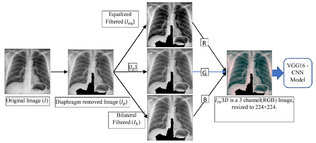
Deep Learning uses a six-layer Convolutional Neural Network (CNN), with a chest X-ray image resized into 224 x 224 x 3, and a Convolution of 3 x 3. The x3 in the input image indicates that it is three colored (R, G, and B). Since the X-ray image is black and white gray, the three colors of R, G, and B are pre-processed for the image as shown in the figure below. In the figure below, (Ip) is an image with the diaphragm removed, (Ieq) is an image processing method that adjusts the contrast using the intensity histogram of the image, and (Ib) is an image obtained by bilateral filtering on (Ieq). R, G, and B images are simulated by using these three (Ip), (Ib), and (Ieq) images.
The results of Deep Learning are,
with the simple model using the X-ray image as it is, 88% of the accuracy was obtained, and the accuracy was improved to 94.5% by adding the pre-processing of the above mentioned image editing method.
Wouldn’t it be time for diagnosis using Deep Learning to be used more and more in the medical field?