Niclosamide inhibits SARS-CoV-2 infection: Inhibition of SARS-CoV-2 infection mediated by endocytosis pathway 

A group from National Centre for Biological Sciences (TIFR), Bengaluru, India, etc. has reported on SARS-CoV-2 infection mechanisms based on endocytosis.
https://journals.plos.org/plospathogens/article?id=10.1371/journal.ppat.1009706

A key step in successful virus infection is the release of viral genomic content into the host cell cytoplasm. To achieve this, viruses bind to specific cell surface receptors and subsequently undergo membrane fusion either directly at the plasma membrane or following endocytic uptake. Both alternatives of entry are feasible for SARS-CoV-2 infections depending on the availability of receptors and proteases at the host cell surface. Although angiotensin converting enzyme 2 (ACE2) is a well-studied receptor for SARS-CoV-2, other receptors and co-receptors have been discovered from a number of groups. Additionally, SARS-CoV-2 requires proteolytic processing of the viral envelope Spike protein by host cell proteases to gain entry. Therefore, these viruses can directly fuse at the cell surface if the Spike protein is cleaved by a cell surface serine protease like TMPRSS2, or utilize an endo-lysosomal route for fusion, where the Spike protein is primed by cysteine protease cathepsins. So, viral entry and infection in different host cells is dependent on the expression of these key host factors (receptors such as ACE2 and protease like furin, TMPRSS2, and cathepsin.

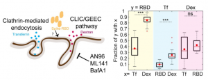
The CLIC/GEEC (CG) pathway is a clathrin-independent endocytic pathway mediated by uncoated tubulovesicular primary carriers called clathrin-independent carriers (CLICs) which arise directly from the plasma membrane and later mature into tubular early endocytic compartments called Glycosylphosphotidylinositol- anchored protein (GPI-AP) enriched compartments (GEECs).

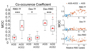
Authors studied the endocytosis of receptor binding domain (RBD) of SARS-CoV-2 Spike protein in gastric epithelial cells (AGS) in the presence and absence of ACE2. AGS can be considered as a cell line with undetectable levels of endogenous ACE2. It was shown that RBD is endocytosed via the CG endocytic pathway (rather than clathrin-mediated endocytosis (CME) pathway) and its uptake is sensitive to pharmacological perturbations of this pathway in AGS cells.

To determine the effect of ACE2 on uptake of RBD in AGS cells, a stable AGS cell line ectopically expressing ACE2 (AGS-ACE2) was generated; the expression of ACE2 was confirmed using qPCR and western blot analysis. RBD uptake in AGS-ACE2 was about 3-fold higher than AGS cells. On characterizing the RBD endocytic itinerary in AGS-ACE2 cells, an increase in the co-occurrence of RBD with transferrin was observed, and slightly reduced co-occurrence of RBD with dextran compared to AGS cells was also observed. This indicates that in addition to trafficking via the CG pathway, RBD is now trafficked via the CME in AGS-ACE2 cells.

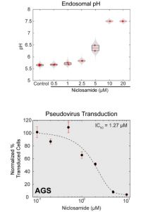
Finally, it was shown that niclosamide neutralizes endosomal pH and inhibits SARS-CoV-2 infection as follows.

Protection from kappa variant (B.1.617.1) and delta variant (B.1.617.2) by Pfizer-BioNTech or Oxford-AstraZeneca vaccine serum  

A group from University of Oxford, Oxford, UK, etc. has reported on protection from B.1.617.1 and B.1.617.2 by vaccine serum.
https://www.ncbi.nlm.nih.gov/pmc/articles/PMC8218332/

Kappa variant = B.1.617.1 is characterized by two mutations L452R and E484Q in the RBD, and delta variant = B.1.617.2 is characterized by two mutations L452R and T478K in the RBD). The L452R mutation is the common in those variants.

Authors tested neutralization of B.1.617.1 and B.1.617.2 using serum from individuals who had received 2 doses of the BNT162b2 Pfizer-BioNTech or ChAdOx1 nCoV-19 Oxford-AstraZeneca vaccine.

Geometric mean neutralization titers against B.1.617.1 were reduced 2.7-fold (p < 0.0001) relative to the Victoria virus for the Pfizer-BioNTech vaccine serum, and 2.6-fold (p < 0.0001) for the Oxford-AstraZeneca vaccine. The Victoria virus is a Wuhan-related strain isolated early in the pandemic from Australia. For B.1.617.2, titers were reduced 2.5-fold (p < 0.0001) relative to the Victoria virus for the Pfizer-BioNTech vaccine serum and 4.3-fold (p < 0.0001) for the Oxford-AstraZeneca vaccine. Although significant reductions in neutralization titers of sera collected from recipients of the Oxford-AstraZeneca and Pfizer-BioNTech vaccines were observed as such, but make sure that there is no evidence of complete escape from neutralization.  

Serum sphingosine could be a GOOD serologic biomarker for the early identification of asymptomatic versus symptomatic COVID-19 patients 

A group from Medical University of South Carolina, Charleston, USA, etc. has reported that reduced sphingosine levels provide a sensitive and selective serologic biomarker for the early identification of asymptomatic versus symptomatic COVID-19 patients.
https://www.nature.com/articles/s41598-021-93857-7

There was a slight but significant increase in the levels of sphingosine (p < 0.05) in individuals who are antibody positive (n = 134) compared to negative (n = 130), with sphingosine levels 28.96 versus 23.25 pmol/5 × 10−5 L serum, respectively. And further, COVID-19 patients’ serum sphingosine levels were around 15-fold decreased compared to that of asymptomatic donors from 28.96 to 1.88 pmol/5 × 10−5 L serum, respectively (Sphingosine (Sph), dihydro-sphingosine (dhSph), sphingosine 1-phosphate (Sph-1p)).

From the ROC analysis, a sphingosine threshold (or cut-off) value of 8.2 pmol/5 × 10−5 L resulted in 98.47% (95% CI 94.60–99.73%) sensitivity and 98.51% (95% CI 94.72–99.73%) specificity, suggesting that serum sphingosine level provides a selective and sensitive biomarker to identify symptomatic patients versus asymptomatic donors who are positive for SARS-CoV-2 antibody. It was also found that Sphingosine and dihydro-sphingosine do not appear to monitor the disease’s severity.

It was know that one of the biochemical biomarkers, lactate dehydrogenase (LDH), was highly elevated in symptomatic patients with an increased mortality rate. Interestingly, reduced sphingosine levels observed here were not associated with disease severity in COVID-19 patients. These data might suggest that increased or sustained serum sphingosine levels might prevent COVID-19 disease, while reduced sphingosine could result in enhanced inflammation and symptomatic response in some individuals.   

Epitopes in SARS-CoV-2 S2 can serve as blueprints for the design of immunogens capable of eliciting cross-neutralizing coronavirus antibodies 

A group from Fred Hutchinson Cancer Research Center, Seattle, USA, etc. has reported that epitopes in SARS-CoV-2 S2 can serve as blueprints for the design of immunogens capable of eliciting cross-neutralizing coronavirus antibodies.
https://pubmed.ncbi.nlm.nih.gov/34237283/

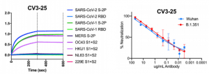
From 198 antibodies isolated from four COVID-19+ convalescent patients, 14 SARS-CoV-2 neutralizing antibodies were isolated. One targeted the N-terminal domain (NTD), one recognized an epitope in S2, and 11 bound the receptor-binding domain (RBD), and those IC50s ranged from 0.007 μg/ml to 15.1 μg/ml.

The S2 subunit contains at least one epitope that, although poorly immunogenic, is present on four of five human beta coronaviruses SARS-CoV-1, SARS-CoV-2, OC43, HKU1). That epitope, as defined by its recognition by CV3-25, is a valid candidate for the development of a global coronavirus vaccine. IC50 of C3-25 against SARS-CoV-2 was 0.34 μg/ml.

Man-specific lectins showing anti-viral properties against SARS-CoV-2, SARS-CoV, and MARS-Cov: Legume lectins for SARS-CoV-2

A group from Université Paul Sabatier, Toulouse, France, etc. has reported on Man-specific lectins showing anti-viral properties against SARS-CoV-2, SARS-CoV, and MARS-Cov.
https://www.mdpi.com/2073-4409/10/7/1619/htm

Man-specific lectins from plants, algae, fungi, and bacteria, have been largely studied with respect to their anti-viral properties against different types of enveloped viruses, including HIV-1, papilloma virus, herpes virus, hepatitis C virus, and Ebola virus. In this respect, the algal lectin griffithsin, the cyanobacteria lectins cyanovirin, actinohivin, and microvirin, and various GNA-related lectins like NPA and ASA have been particularly well documented. Most of these Man-specific lectins prevent the virus replication, at least under in vitro conditions, by interfering with the Man-containing N-glycans present on the cell surface of the virion envelope.

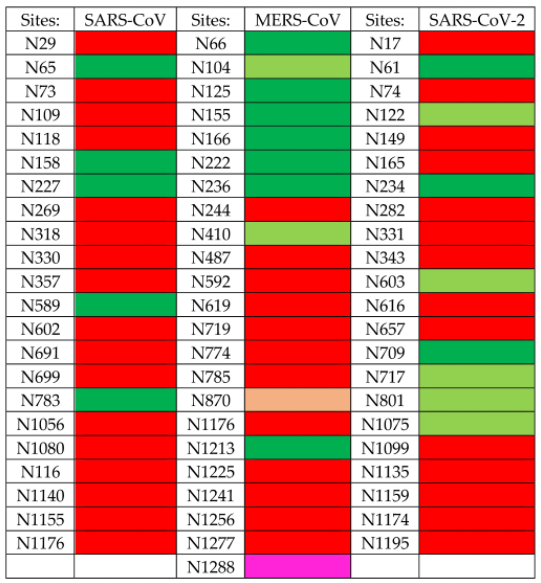
Glycans of the S-glycoproteins forming the spikes of SARS-CoV, MERS-CoV and SARSCoV-2, consist of high-mannose glycans and often sialylated N-glycans that predominantly occupy their N-glycosylation sites. However, depending on the coronaviruses, some discrepancies occur between the distribution of the two types of glycans on the surface of the virion, which introduces some diversity in the glycan shield covering the coronavirus spikes.


Figure explanation: Sites containing (almost) exclusively complex glycans (colored red), high-mannose glycans (colored green), and hybrid glycans (colored magenta). Sites harboring a mixture of complex glycans, high-mannose glycans, and a few hybrid glycans (pink), predominant high-mannose glycans (pale green) and predominant complex glycans (orange). In the case of SARS-CoV-2, high-mannose glycans consist essentially of tri-antennary glycans GlcNAc2-Man5–9, but the major high-mannose structure seems to be GlcNAc2Man5.

Accordingly, Man-specific lectins from plants, algae, fungi, and bacteria, which differ slightly due to their fine sugar-binding specificities, offer a vast panel of glycan probes more or less adapted to the specific recognition of the different coronaviruses. In this respect, GNA-related lectins together with Man-specific lectins from algae and cyanobacteria, appear as glycan probes nicely adapted to the recognition of the high-mannose shield which predominates at the top of the MERS-CoV spike. Otherwise, legume lectins with a higher affinity for N-glycans possessing the trimannoside Manα1,3Manα1,6Man core, seem better adapted to the recognition of the N-glycans distributed predominantly at the top of the glycan shield from SARS-CoV and SARS-CoV-2. 

Characteristics of antibodies (targeting RBD, NTD, and S2) induced by two times Pfizer mRNA vaccination: Effectiveness against current SARS-CoV-2 variants 

A group from Icahn School of Medicine at Mount Sinai, New York, USA, etc. has reported on characteristics of Pfizer mRNA vaccinated SARS-CoV-2 antibodies.
https://www.ncbi.nlm.nih.gov/pmc/articles/PMC8185186/

The antibody response peaked 1 week after the second vaccine dose, followed by a decline in titers over the following weeks as expected from an antibody response to vaccination. Interestingly, titers against the spike proteins of β-coronaviruses OC43 and HKU1 increased substantially after vaccination.

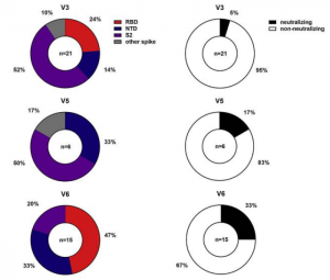
RBD and NTD were co-dominant epitopes of the mRNA vaccine-induced antibodies, and the majority of remaining epitopes was mapped to S2. All antibodies were tested for neutralizing activity against the USA-WA1/2020 strain of SARS-CoV-2. Only a minority of the binding antibodies showed neutralizing activity (see figure below, obtained from three individuals: V1, V5, V6).

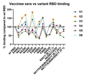
For sera from the six vaccinated individuals, binding between vaccinee sera and variant RBDs were evaluated. The highest reduction was only 2-fold for E406Q, N440K, E484K, and F490K (see figure below, six vaccinated individuals: V1-V6).
This would be a kind of good news showing that Pfizer mRNA vaccine would be effective against current SARS-CoV-2 variants.

losatran, ACE2 inhibitor, did not show any therapeutic effects on against SARS-CoV-2 in Phase II clinical trial 

A group from Department of Emergency Medicine, University of Minnesota, etc. has indicated that losatran, ACE2 inhibitor, did not show any therapeutic effects on against SARS-CoV-2.
https://www.ncbi.nlm.nih.gov/pmc/articles/PMC8225661/

This was a multicenter prospective double blind randomized placebo-controlled trial for the treatment of symptomatic outpatients with confirmed COVID-19 conducted across 3 hospital systems in Minnesota, United States between April and November 2020.

The intervention was losartan 25 mg versus equally appearing placebo. Participants self-administered study drug orally twice daily for 10 days for participants with eGFR >60 mL/min/1.73 m2, and once daily for those with eGFR 30–60 mL/min/1.73 m2. The threshold for angiotensin receptor blockade is 20 mg daily, with twice daily dosing more effective than once daily due to a 6–9 h half-life of the active metabolite. This dose was chosen in lieu of a 50 mg twice daily (maximum) dose after the FDA raised safety concerns regarding the higher dose regimen. This dose is expected to provide 37% inhibition of the angiotensin receptor.

The primary outcome did not differ significantly, and also viral loads were not statistically different between treatment groups at any time point. This clinical trial was terminated.

Fc-mediated effector functions and Antibody-Dependent Enhancement (ADE): a potent SARS-CoV-2 neutralizing antibody

A group from Biological Defense Program, DSO National Laboratories, Singapore, etc. has reported on Fc-mediated effector functions of a potent SARS-CoV-2 neutralizing antibody.
https://journals.plos.org/plosone/article?id=10.1371/journal.pone.0253487

Antibody-Dependent Enhancement (ADE) of disease remains a major concern for the use of anti-SARS-CoV-2 antibodies as therapeutics. ADE can occur if Fcγ Receptor (FcγR) engagement mediates an increase in the infection of phagocytic cells. Due to the potential of ADE, several ongoing SARS-CoV-2 antibody programs have chosen to use Fc isotypes that do not engage FcγR, like the IgG4 isotype, and engineered variants such as those carrying FcγR-null LALA variant. However, these may be counterproductive because the signaling mechanisms underpinning the efficacies of these antibodies, particularly the ability of FcγR engagement to induce other antiviral responses such as ADCC will be killed. To address these questions, authors isolated and characterized a RBD-binding neutralizing IgG1 antibody, named SC31, from an early convalescent patient. Authors assessed the impact of Fc functionality on its therapeutic efficacy by comparing SC31 with its LALA variant and demonstrated that the engagement of Fc receptors by SC31 triggers additional IFN-γ-mediated antiviral responses but importantly do not induce ADE.

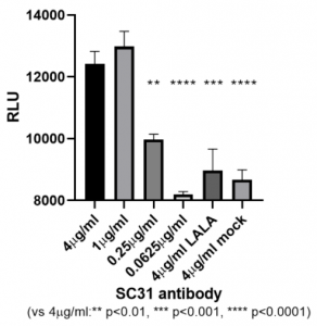
To determine the role of Fc-mediated effector functions in the therapeutic efficacy of SC31, the abilities of SC31 and its LALA variant were compared. The upstream activation of the FcγRIIIa ADCC signalling pathway was evaluated using a Jurkat reporter cell line expressing FcγRIIIa and with ADCC reporter assay after co-culture with target HEK293 cells expressing membrane-bound SARS-CoV-2 Spike protein. In contrast to its LALA variant, SC31 was confirmed to induce a dose-dependent activation of ADCC signaling.

Galectin-3 could be a good marker for Abdominal Aortic Aneurysm

A group from Taipei Medical University, Taiwan, etc. has reported that Galectin-3 could be a good marker for Abdominal Aortic Aneurysm (AAA).
https://www.ncbi.nlm.nih.gov/pmc/articles/PMC8200414/

Although ultrasound is the gold standard for the diagnosis and surveillance of AAAs with high sensitivity and specificity, the frequency of ultrasound surveillance varies with aneurysm diameter. Furthermore, ultrasound is not recommended for patients with subaneurysmal aortic dilatation. Therefore, circulating biomarkers of inflammation, which reflect the aneurysmal size, can assist in the detection and prognosis of AAA.

A cross-sectional study was performed to analyze plasma Gal-3 and IL-6 levels as circulating biomarkers in both control patients (n = 195) and patients with AAA (n = 151). Plasma Gal-3 levels were significantly higher in patients with AAA than in control patients (96.9 ± 4.5L vs. 76.5 ± 1.9 ng/mL), and the levels of IL-6 were also higher in AAA samples than in healthy control samples (92.8 ± 5.2 pg/mL vs. 72.5 ± 3.0 pg/mL). The diagnostic performance of Gal-3 and IL-6 were evaluated using ROC analysis. The results were that Gal-3 levels predicted AAA presence (AUC=0.91) significantly more accurately than did IL-6 levels (AUC=0.72).

Gal-3 is likely a chemotactic molecule for macrophages. Thus, its expression could be associated with various cardiovascular diseases. The increased risk of AAA observed in patients with higher Gal-3 levels may reflect the recruitment of inflammatory cells, including activated macrophages, in the arterial system and the subsequent secretion of Gal-3.


Binding specificities of C-type lectins, MBP, Langerin, and Dectin-2, as a summary 

A group from Tohoku Medical and Pharmaceutical University has reviewed 11 kinds of mammalian lectins responsible for pathogen recognition.
https://www.ncbi.nlm.nih.gov/pmc/articles/PMC8185196/

From this review, three C-type lectins were highlighted here for your information.
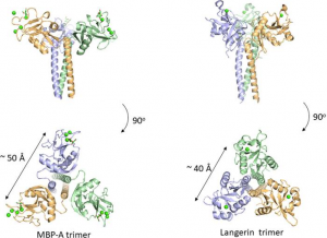
Many C-type lectins show selectivity toward mannose residues and this mannose binding is utilized for microbe sensing. One of the most studied examples is mannose-binding protein, MBP, which is known to activate the complement lectin pathway. Why does mannose recognition play this role in detecting non-self when mannose residues occur frequently in mammalian N-glycans, e.g., high-mannose glycan. The likely explanation is in the higher spatial density of mannose residues on bacteria relative to that of mammalian glycans. The C-type lectin domain has a Ca2+ ion coordinated with the OH3 and OH4 of the mannose. The affinity of 1:1 binding is weak with a dissociation constant of roughly 1 mM, however, the presentation of trimeric binding sites in MBP domains could interact with the multiple terminal mannose residues presented on microbes. The spacing between the mannose binding sites is around 50 Å, and is eminently suitable for binding packed terminal mannose residues with high affinity, but not single endogenous high mannose glycans. Langerin is also a C-type lectin with a coiled-coil region and a neck region in a trimeric structure, and has a distance of roughly 40 Å between binding sites. A trimeric oligosaccharide ligand with appropriate linker length for the 40 Å distance between each binding site has been reported for Langerin, with 1,000-fold higher affinity over the monomeric ligand.

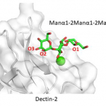
As for Dectin-2, the binding site accommodates internally positioned Manα1-2Man of mannans and other polysaccharides, whereas other C-type lectins like DC-SIGN and langerin bind only terminal Manα1-2Man structures. Recognition of internal mannose residue is advantageous in that multiple binding sites are presented toward lectin receptor. Dectin-2 is thus suitable for binding to longer mannan polysaccharides.