Differences in SARS-CoV-2 Spike glycosylation between CHO and HEK cells: Oligomannose increases in HEK cells

A group from Center for Biologics Evaluation and Research, Food and Drug Administration, Baltimore, MD, USA, etc. has reported on differences in SARS-CoV-2 Spike glycosylation between CHO and HEK cells.
https://www.ncbi.nlm.nih.gov/pmc/articles/PMC8497748/pdf/fchem-09-735558.pdf

CHO-Spike exhibits more complex (mainly bi-antennary) and higher sialylation (α2,3-linked), while HEK-Spike exhibits more high-mannose and a small amount of α2,3-and α2,6-linked sialic acids as follows.

Interactions between Bacillus Spp. and Pseudomonas Spp. promotes plant growth: Importance of pathogen metabolic pathways

A group from University of Moncton, Moncton, NB, Canada, etc. has reported that interactions between Bacillus Spp. and Pseudomonas Spp. promotes plant growth.
https://www.ncbi.nlm.nih.gov/pmc/articles/PMC8488376/

In this work, Pseudomonas spp. and Bacillus spp. isolates obtained from Canadian soils have been utilized to assess their ability to promote the growth of Cannabis sativa when inoculated alone or in combination treatments in different commercial soil substrates (Promix and Canna coco) commonly used to grow cannabis.

It was demonstrated that single treatments do not stimulate growth promotion under the conditions tested, but combinatorial treatments (Bacillus and Pseudomonas) have a significant positive effect on plant growth.

Here, 223, 825 = Pseudomonas, 979, 1082, 279 = Bacillus. Promix and Canna coco are different types of commercial soil substrates.

Five metabolic pathways could be identified as differently abundant in Canna coco, all of which were also found in Promix:

  • oxidative glucose degradation
  • creatinine degradation I
  • L-lysine biosynthesis II
  • S-methyl-5-thio-α-D-ribose 1-phosphate degradation I
  • L-methionine salvage cycle III)

These five pathways might be the background of plant growth enhancement, and could easily be linked back to either Pseudomonas spp. or Bacillus spp. metabolism.

Phospholipase metabolic pathway identified in the study is also an important secondary messenger pathway in plants, regulating response to phytohormones and pathogen elicitation. Secreted bacterial phospholipases have the capacity to liberate phosphorus from phospholipids, which the plant could assimilate, potentially augmenting plant growth. The phospholipase pathway together with sugar metabolism in Pseudomonas spp. might significantly increase the availability of essential soil nutrients for the plant and promote plant growth.

Syncytium formation (cell-to-cell fusion) could contribute to efficient spread of the SARS-CoV-2 delta variant: 2.5 times more effective as compared to WT

A group from Georg-August-University Göttingen, Wilhelmsplatz 1, 37073 Göttingen, Germany, etc. has reported that syncytium formation (cell-to-cell fusion) could contribute to efficient spread of the SARS-CoV-2 delta variant.
https://www.ncbi.nlm.nih.gov/pmc/articles/PMC8487035/

Vero, 293T, Caco-2, and Calu-3, these cell lines express endogenous ACE2 and Vero, Caco-2, and Calu-3 cells are often used for infection studies with authentic SARS-CoV-2. The delta variant S protein mediated entry into 293T and Vero cells with the same efficiency as the wild-type (WT) S protein, while entry into Caco-2 and Calu-3 cells was enhanced by 1.5 times and 2.0 times comparing with wild type. However, any increased ACE2 binding of the delta variant S protein was not observed, suggesting that increased entry into Caco-2 and Calu-3 cells was not due to augmented ACE2 binding.

SARS-CoV-2 S protein-driven syncytium formation is believed to contribute to the increased infectivity of the delta variant. To confirm syncytium formation drove by the delta variant, human lung cell line A549 expressing high level of ACE2 was adopted. As expected, WT S led to the formation of syncytia, while syncytia formation was not observed when cells were transfected with empty expression plasmid. Strikingly, the delta variant S protein caused more and larger syncytia, and quantification of cell-to-cell fusion revealed that fusion by the delta variant S protein was ∼2.5-fold more effective as compared to the WT S protein.

In conclusion, syncytium formation (cell-to-cell fusion) could contribute to efficient spread of the delta variant.

SARS-CoV-2 viral replication in human macrophages enhances an inflammatory cascade

A group from Department of Immunobiology, Yale University School of Medicine, New Haven, CT, USA, etc. has reported that SARS-CoV-2 viral replication in human macrophages enhances an inflammatory cascade.
https://www.biorxiv.org/content/10.1101/2021.09.27.461948v1

In order to identify SARS-CoV-2 infected cells, authors used MISTRG6-hACE2 mice (humanized-ACE2 mice). As expected, SARS-CoV-2 viral RNA was detected in epithelial cells, the main targets of SARS-CoV-2 infections. Surprisingly, however, human immune cells also had similar levels of viral RNA. To better visualize and characterize these cells, MISTRG6-hACE2 mice were infected with a reporter strain of virus, SARS-CoV-2-mNG19. The majority of epithelial cells (EPCAM+) in bronchioalveolar lavage (BAL) but only a small proportion of total lung epithelial cells were infected with SARS-CoV-2 as measured by mNG expressing cells. Human immune cells showed a clear mNG signal, and mNG+ human immune cells were predominantly human macrophages.

Since macrophages are phagocytic, it is needed to address whether the SARS-CoV-2 viral RNA in these cells replicates in these cells or is simply acquired by phagocytosis of infected cells or debris. To achieve this, we first characterized the mNG signal in human macrophages from MISTRG6 mice that were not transduced with hACE2. In these mice, epithelial cells were not infected with SARS-CoV-2. These mice had however similar levels of mNG signal in the human macrophages of AAV-ACE2 mice, arguing that viral uptake by human macrophages is independent of infected epithelial cells. To determine whether SARS-CoV-2 replicates in human macrophages, the key characteristics of viral replication, viral replication products, subgenomic RNA, double stranded RNA (dsRNA), and the key replicative enzyme RNA dependent RNA polymerase (RdRp) were evaluated.
(1) Quantified genomic and subgenomic viral RNA in mNG+ vs mNG- epithelial or human immune cells at 4dpi or 14dpi. Only mNG+ epithelial cells and mNG+ human immune cells showed detectable subgenomic viral RNA. The levels of subgenomic RNA in human immune cells and epithelial cells were similar, suggesting a similar extent of viral replication in human immune cells and epithelial cells.
(2) Stained for dsRNA in mNG+ cells to determine whether the mNG signal colocalized with dsRNA (an exclusive product of viral replication). As expected, mNG and dsRNA were detectable and colocalized in both epithelial cells and in macrophages.
(3) Visualized viral RdRp in human macrophages.

In addition, the existence of antibody mediated viral uptake by macrophages was confirmed by treating infected mice with monoclonal antibodies against Spike protein of SARS-CoV-2, starting at 35 hours post infection (hpi). It was clearly shown that monoclonal antibody treatment increased the percentage of mNG+ macrophages in lungs.

SARS-CoV-2 is rapidly inactivated by titanium dioxide (TiO2) surface coating

A group from Cambridge Institute of Therapeutic Immunology & Infectious Disease (CITIID), University of Cambridge, Cambridge, UK, etc. has reported SARS-CoV-2 is rapidly inactivated by titanium dioxide (TiO2) surface coating
https://www.ncbi.nlm.nih.gov/pmc/articles/PMC8450774/

For live SARS-CoV-2, virus was already significantly inactivated on the TiO2 surfaces after 20 min illumination.
After 5 h no detectable active virus remained.
Significantly, SARS-CoV-2 on the untreated surface was still fully infectious at 5 h post-addition of virus.

The coatings would need only periodic re-application depending on the amount of wear, for example 6-12 monthly, and would be widely available in the future.

Effects of Bacillus amyloliquefaciens QSB-6 strain on apple tree roots and disease

A group from National Key Laboratory of Crop Biology, College of Horticulture Science and Engineering, Shandong Agricultural University, Shandong, China has reported on Bacillus amyloliquefaciens QSB-6 strain isolated from apple orchard soils and its effect on apple tree roots and disease.
https://www.frontiersin.org/articles/10.3389/fmicb.2021.746799/full

Authors compared effects of 4 types of soils on apple tree roots.

  • untreated soil from a 31-year-old orchard (CK1)
  • the same soil fumigated with methyl bromide (CK2)
  • the same soil treated with the manure carrier only (T1)
  • the same soil treated with Bacillus amyloliquefaciens strain QSB-6 manure treatment (T2)

Interestingly, the CK2, T2, and T1 treatments significantly promoted the growth of apple tree roots, and the relative treatment effects were ranked from high to low: CK2 > T2 > T1 > CK1.

The number of rhizobacteria increased significantly after T2 treatment by 9.64 times higher in T2 than in CK1, and soil fungal numbers were reduced significantly by 85.58% (in CK2) and 81.74% (in T2) compared with CK1.
Extracellular metabolites from strain QSB-6 showed a strong inhibitory effect on Fusarium hyphal growth and spore germination as shown below.

(A) Fusarium proliferatum, (B) Fusarium solani, (C) Fusarium verticillioides, (D) Fusarium oxysporum

In summary, B. amyloliquefaciens QSB-6 has a good inhibitory effect on Fusarium in the soil and can significantly promote apple tree root growth. It has great potential as a biological control agent against apple disease.

Bacillus velezensis recruited Pseudomonas stutzeri for plant through metabolic cross-feeding

A group from Technical University of Denmark, Kongens Lyngby, Denmark, etc. has reported that Bacillus velezensis recruited Pseudomonas stutzeri for plant through metabolic cross-feeding
https://www.ncbi.nlm.nih.gov/pmc/articles/PMC8483172/

Generally, Bacillus spp. and Pseudomonas spp. are the most extensively studied beneficial microorganisms in the rhizosphere. Plant beneficial Bacillus, including B. velezensis SQR9 are known for plant growth promotion, disease suppression, and enhanced salt stress tolerance. Root colonization and plant growth-promoting properties require efficient biofilm formation (including exopolysaccharide EPS and TasA protein fibers) on the roots.

To explore the effects of B. velezensis SQR9 on rhizosphere microbiota, two-weeks-old cucumber seedlings were inoculated with strain SQR9 and the rhizosphere soil samples were collected after sixteen days. It was found that members of the genera Pseudomonas, Vogesella, Pseudoxanthomonas, Chryseobacterium, Pseudoduganella, Lysobacter, Klebsiella, and Cellvibrio were increased after SQR9 application. Remarkably, 38% of increased rhizobacteria mapped to the Pseudomonas genus, suggesting that B. velezensis SQR9 may recruit and then synergistically interact with Pseudomonas spp.

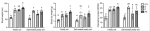
The rhizobacteria consortium had a stronger promoting effect in paddy soil, as the dual-species (Bacillus and Pseudomonas) consortium significantly increased the shoot height, shoot dry weight, and chlorophyll content of plants in comparison to plant inoculated with one species. In the case of salt treated paddy soil, Pseudomonas stutzeri XL272 protects the plant against salt stress at the most comparing with non-inoculated control plants. The protective effect of the rhizobacteria consortium was higher than that of P. stutzeri XL272 alone in comparison to control plants. These results support the suggestion that the PGPR can induce the assemblage of the indigenous beneficial microbiome, leading to the promotion of plant health and resistance to salt stress.

A big question is how B. velezensis SQR9 recruited Pseudomonas spp. etc.? Authors suggested that the metabolic cross-feeding such as branched-chain amino acids would be a key to understand the mechanism through analysis transcriptional alterations of bacillus and pseudomonas.
The image is like this; B. velezensis SQR9 is attracted by root exudates and colonizes the rhizosphere. After establishing biofilm on plant roots, it secretes metabolites that increase the abundance of indigenous plant beneficial genera (such as Pseudomonas spp.). Forming tightly associated biofilm, they share extracellular matrix and essential metabolites that increase their fitness in the rhizosphere, and help plant growth and increase resistant to salt stress.

Characteristics of a new lectin, GYL, extracted from bivalve Glycymeris yessoensis: immune defense activity

A group from Far Eastern Branch of Russian Academy of Sciences, Vladivostok, Russia has reported on a new lectin, GYL, extracted from bivalve Glycymeris yessoensis.
https://www.ncbi.nlm.nih.gov/pmc/articles/PMC8466245/

A lectin named GYL is a dimeric protein with a molecular mass of 36 kDa, as established by SDS-PAGE and MALDI-TOF analysis, consisting of 18 kDa subunits linked by a disulfide bridge.

The peptide sequence of GYL showed no significant homology with other lectins listed in the BLAST, however, the National Center for Biotechnology Information (NCBI) Conserved Domain Search program identified conserved EPN (Glu-Pro-Asn) and WND (Trp- Asn- Asp) motifs in the GYL peptide sequence, which are characteristic of the CRD of C-type lectins (CTL).

GYL was widely expressed throughout the tissues of healthy clams, however, levels of the lectin in the hemolymph and mantle were found to be 3.5- and 2.4-fold greater, respectively, than that found in the gonads.

GYL agglutinated all erythrocytes tested, suggesting that this activity is not highly specific. Although the detailed glycan binding specificity is not clearly shown, it was found that GYL preferentially bound peptidoglycan (PGN) and LPS expressed on bacteria cell walls, but had little binding activity toward β-1,3-glucan and mannan, which are usual components of the yeast cell wall.

Taking these things into consideration, it was suggested that GYL plays an important role in the immune defense of the clam against pathogenic microorganism infections. Temporal expression levels of GYL in the hemolymph following V. proteolyticus stimulation and diesel fuel exposure were investigated. Amazingly, the expression levels of lectin increased about 25-fold at 48 h post-injection, suggesting that bacterial infection and anthropogenic factors had a significant effect on the clam, causing them to increase the synthesis of GYL as a protective molecule.

A highly potent and broad neutralization antibody against SARS-CoV-2 was screened

A group from Vanderbilt University Medical Center, Nashville, USA, etc. has reported that a highly potent and broad neutralization antibody against SARS-CoV-2 was screened using a LIBRA-seq technology.
https://pubmed.ncbi.nlm.nih.gov/34592170/

To identify SARS-CoV-2 S-directed antibodies, authors utilized LIBRA-seq, a technology that enables high-throughput simultaneous determination of B cell receptor sequence and antigen reactivity at the single-cell level, expediting the process of lead candidate selection and characterization. Antigen-specific B cells were isolated from a donor with potently neutralizing antibodies in serum 3 months after infection confirmed by nasal swab RT-PCR testing for SARS-CoV-2.

Of the 73 IgG+ B cells with high LIBRA-seq scores for SARS-CoV-2 Spike, nine lead candidates were selected for characterization as recombinant monoclonal antibodies.
From VSV assays, antibody named 54042-4 showed the best potency, at a IC50 of 9 ng/mL, and broad neutralization against existing SARS-CoV-2 variants.
It was al so found that the 54042-4 heavy chain binds to RBD residues 443–447 and the 54042-4 light chain contacts RBD residues 498–500 from cryo-EM structure analysis.

Antibody-Dependent Enhancement of SARS-CVoV-2 infection is real

A group from The Institute of Medical Science, The University of Tokyo, etc. has reported that Antibody-Dependent Enhancement of SARS-CVoV-2 infection to immune cells is real.
https://journals.asm.org/doi/10.1128/mBio.01987-21

Viruses infect cells mainly via specific receptors at the cell surface. Antibody-dependent enhancement (ADE) of infection is an alternative mechanism of infection for viruses to infect immune cells that is mediated by antibodies and IgG receptors (FcγRs).

Authors generated BHK cells stably expressing human FcγRs (FcγRIA, FcγRIIA, or FcγRIIIA) or human angiotensin-converting enzyme 2 (hACE2) (the entry receptor for SARS-CoV-2). Wild-type BHK cells lack human ACE2 expression and are not susceptible to SARS-CoV-2.

BHK cells were infected with a firefly luciferase-expressing vesicular stomatitis virus (VSV) lacking the VSV-G gene and pseudotyped with SARS-CoV-2 spike (VSV-SARS2-S). Although BHK-hACE2 cells were susceptible to VSV-SARS2-S, the BHK-FcγRIA, BHK-FcγRIIA, and BHK-FcγRIIIA cells were not susceptible due to the lack of hACE2. However, using convalescent-phase plasma from COVID-19 patients and incubated with VSV-SARS2-S, it was found that two types of FcγRs, FcγRIIA and FcγRIIIA, mediate ADE of SARS-CoV-2 infection in the presence of ACE2.

It was also confirmed that SARS-CoV-2 infection is enhanced by convalescent-phase plasma in primary macrophages.