Colorectal Cancer and Tn-Antigen

A group from Fukushima Medical University has discussed about a key-point on colorectal cancer immunotherapy based on colorectal cancer and Tn-antigen.
https://www.mdpi.com/1422-0067/21/23/9081

In cancer, mutations are accumulated in the genome and epigenome. In colorectal cancer, 85% are derived from chromosomal instability and 15% from deficient mismatch-repair (dMMR).
It is also well known that in cancer, aberrant glycans are expressed on the cell surface, and O-type glycans are truncated, and resulting Tn-antigens are highly expressed. Tn-antigen binds to macrophage MGL and promotes il-10 secretion and acts immuno-suppressively, as well as inducing T-cell apoptosis. As a result, cancer cells evade immunity attacks.

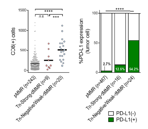
Cancer cells with deficient mismatch-repair have significantly higher expression of Tn-antigen than those that are not. In addition, those types of cancer cells tend to have less infiltration of CD8+ T-cells and lower expression of PD-L1 as an immune checkpoint molecule. Therefore, they have concluded that immunotherapy targeting on Tn-antigens and inhibition of immune checkpoint molecules may be effective for colorectal cancer with a strong deficient mismatch-repair.

The effects of ACE2 inhibitors in the new coronavirus (COVID-19), Dangerous in Diabetes Mellitus

The new coronavirus (COVID-19) is known to cause serious diseases with underlying disease such as acute kidney disease, diabetes, immunodeficiency, and obstructive airway disease.
Acute renal disease (HR = 3.23, 95% CI: 2.01 to 5.19),
Diabetes (HR = 2.07, 95% CI: 1.32 to 3.26),
Immunodeficiency (HR = 2.33, 95% CI: 1.29 to4.2),
Obstructive airway disease (HR = 2.13, 95 % CI: 1.06 -4.3),

Since ACE2 is the main infection receptor for SARS-CoV-2, the following group has performed statistical analysis based on data obtained from 617 patients on side effects of ACE inhibitors.
http://www.ijkd.org/index.php/ijkd/article/view/5920/1222

Side effects of ACE inhibitors are particularly severe in diabetes (HR = 3.51, 95% CI: 1.59 to 7.75).
In the figure below, DM=Diabetes Mellitus, ACE=0 means no ACE inhibitors, and ACE=1 means with ACE inhibitors.

CD147 is also suspected as a route of infection in the new coronavirus (SARS-CoV-2)

In envelope-having viruses, the binding of receptors and envelope proteins on the cell membrane surface of the host cell initiates the infection. In the new coronavirus (SARS-CoV-2), it is generally recognized that ACE2 is its receptor. ACE2 is expressed in the liver, lungs, stomach, kidneys, and large intestine, but ACE2 expression in the lungs is rather low, and it is suspected that other infection routes exist in view of the severity of COVID-19. C-type lectins, which are widely expressed in immune cells, are also candidates, but the following groups indicate that CD147 can be an infection pathway.
https://www.nature.com/articles/s41392-020-00426-x

The interaction between CD147 and S-protein has been verified by SPR and ELISA methods, and actual infection experiments using VeroE6 and BEAS-2B cells have also been conducted.
Infection through CD147 is said to be endocytosis, not cell membrane fusion, as an infection mechanism.

Obesity is dangerous in covid-19

A group of Jinyun People’s Hospital, China has reported a relationship between covid-19 and obesity.
https://eurjmedres.biomedcentral.com/articles/10.1186/s40001-020-00464-9

Statistical analysis through 12,591 COVID-19 patients showed that the BODY Mass Index (BMI Index) in humans infected with covid-19 was higher than that of mild individuals (MD (average difference in BMI) of 2.48 kg/m2, 95% [2.00 to 2.96 kg/m2]CI). In addition, obesity was shown to be more likely to develop severer disease in the pathology of COVID-19, for example, ICU treatment (OR = 1.57, 95% [1.18–2.09]CI, ECMO use (OR = 2.13, 95% [1.10–4.14]CI).

Excessive infiltration of naïve B cells triggers severe coronavirus (COVID-19)

In patients with the new coronavirus (COVID-19), excessive infiltration of CD4+/CD8+ cells, plasma cells, and macrophages into lung tissues is common, which is believed to lead to severe damages to lung tissues and pulmonary fibrosis.
A group of the National Defense Medical Center, Taipei has reported that excessive infiltration of naïve B cells triggers  observed pathophysiology of COVID-19.
https://journals.plos.org/plosone/article?id=10.1371/journal.pone.0242900

Possible mechanisms include:
COVID-19 causes the accumulation and activation of naïve B-cells in the mediastinal lymph nodes through activation of β2-integrin and α4β1 integrin. Excessive infiltration of naïve B-cells activated by SARS-CoV-2 S-proteins secretes large amounts of IgM and activates the humoral immune
response. This recruits a large amount of monocytes into lung tissues, and the monocytes differentiate to macrophages. The secreted IgM simultaneously activates the complement system, Fc receptors on dendritic cells, and macrophages, increasing the antigen presentation of SARS-CoV-2 and enhancing phagocytosis. In addition, an increase in antigen presentation of SARS-CoV-2 accelerates the extrafollicular response through stimulation of IL-12-dependent plasma cell differentiation in naïve B cell to produce more IgM , suppressing formation of germinal centers and B-cell proliferation.

In conclusion, the importance of therapies targeting naïve B-cells has been proposed.

Depletion in IgM Memory B cells correlates with severity in the new coronavirus (COVID-19)

In the new coronavirus (COVID-19), it is generally considered that impaired immune responses triggers unfavourable outcomes.
A group of Univ. of Pavia, Italy, suggests that the depletion of IgM Memory B cells is related to severe disease.
https://www.nature.com/articles/s41598-020-77945-8

A median value of IgM Memory B cell numbers was 65.0/uL (IQR: 51.0 – 85.0/uL) in healthy people, whereas a median vale was 5.9/uL  (IQR: 2.1 – 13.9/uL) in COVID-19 patients. Although the number of COVID-19 patients assessed was as small as 66, but the depletion in IgM Memory B cells was correlated with severe disease.
There was no correlation between age, sex, and the depletion in IgM Memory B cells.

A potential glycan marker for hepatocellular carcinoma derived from HCV infection: bi-antennary N-type glycan with Core Fucose and Bi-secting GlcNAc

There is a research report on biomarkers of hepatocellular carcinoma with cirrhosis derived from HCV infection.
https://www.mdpi.com/1422-0067/21/23/8913

It has been concluded that A2G1(6)FB (bi-antennary N-type glycan with Core Fucose and Bisecting GlcNAc) could be an excellent glycan marker.


In the figure below, TNM stands for Tumor-node-metastasis, and TNM=1, TNM=2, TNM=3, and TNM=4 represent the stage of hepatocellular carcinoma. The existing markers, AFP, PIVKA-II, do not show a clear correlation with A2G1(6)FB, and show excellent potency as an HCC marker.

A good Indicator for determining covid-19 and influenza A/B

The initial symptoms of covid-19 and influenza are very similar.
Is RT-PCR or antibody testing the only way to distinguish between these two infections?

Sapienza Univ. of Rome’s group has reported that the absolute count of monocytes, a type of leukocyte, is a good discriminating index between the two.
https://www.infezmed.it/media/journal/Vol_28_4_2020_9.pdf

If the absolute count of monocytes is 0.35×10^3/mL or more, COVID-19 can be distinguished from influenza A/B. AUC=0.68 (sensitivity=0.992, specificity=0.368)

This strongly suggests that covid-19 and influenza A/B have different immune responses.

S-protein or RBD-specific IgG is a key to recovering from covid-19 and completely eliminating viruses

From the laboratory tests of 1850 hospitalized COVID-19 patients in Wuhan, the following group discussed the dynamic changes in IgG to SARS-CoV-2. If the IgG level is low, the complete elimination of the virus becomes difficult, and thereby there is a chance that RT-PCR SARS-CoV-2 virus testing after recovery (i.e., after discharge) becomes positive.
https://www.nature.com/articles/s41467-020-19943-y

Dynamic changes in IgG include:

  • In severe cases, IgG level tends to be delayed by nearly a week compared to those with mild cases.
  • In severe cases, the level of IgG is significantly higher, and even in the elderly, the level of IgG tends to be higher.
  • It should be noted that patients with low levels of IgG at the time of hospital discharge may positive with RT-PCR reteaching of SARS-CoV-2.

Identification of immune-related biomarkers associated with novel coronavirus (COVID-19) mortality

It has been established that cytokine storms are occurring in the severe case of covid-19, and there are many research studies about such changes in blood cytokine concentrations.
Some of the most noteworthy studies have already been introduced in this blog site.

This article introduces the results obtained from covid-19 patients in Italy.
https://insight.jci.org/articles/view/144455

Clinical parameters indicate that the increase of the neutrophil/lymphocyte ratio, LDH, CRP, and D-dimer correlates with mortality.

In this study, 66 types of cytokines in the blood were evaluated, suggesting that MCP-1/CCL2, sTNFRSF1A, MMP-9, NGAL, S100A9, sST2, IL-10, and IL-15 were deeply related to the serious disease of COVID-19.
As a site note,
MCP-1/CCP, sTNFRSF1A are NK-kB-dependent markers,
MMP-9, NGAL, S100A9 are derived from neutrophils,
sST2 is a septic marker,
IL-15 is associated with NK-cell activation and function,
IL-10 is a counteraction of severe pneumonia,
has been discussed as such.

As the mechanism of serious disease becomes more concrete, effective therapeutic drugs will be narrowed down.

Powered by WordPress |Copyright © 2020 Emukk. All rights reserved