Identification of immune-related biomarkers associated with novel coronavirus (COVID-19) mortality

It has been established that cytokine storms are occurring in the severe case of covid-19, and there are many research studies about such changes in blood cytokine concentrations.
Some of the most noteworthy studies have already been introduced in this blog site.

This article introduces the results obtained from covid-19 patients in Italy.
https://insight.jci.org/articles/view/144455

Clinical parameters indicate that the increase of the neutrophil/lymphocyte ratio, LDH, CRP, and D-dimer correlates with mortality.

In this study, 66 types of cytokines in the blood were evaluated, suggesting that MCP-1/CCL2, sTNFRSF1A, MMP-9, NGAL, S100A9, sST2, IL-10, and IL-15 were deeply related to the serious disease of COVID-19.
As a site note,
MCP-1/CCP, sTNFRSF1A are NK-kB-dependent markers,
MMP-9, NGAL, S100A9 are derived from neutrophils,
sST2 is a septic marker,
IL-15 is associated with NK-cell activation and function,
IL-10 is a counteraction of severe pneumonia,
has been discussed as such.

As the mechanism of serious disease becomes more concrete, effective therapeutic drugs will be narrowed down.

In the new coronavirus (COVID-19), the role of immunonutrition is attracting attention as it becomes more severe in people over 65 years of age.

In the new coronavirus (COVID-19), immunonutrition is attracting attention so much, as it takes time to confirm the safety and efficacy of vaccines, and also because there are still no effective therapeutic drugs for COVID-19. In particular, the importance and urgency of immunonutrition research is increasing due to the high rate of severity in older people aged 65 and over.
https://www.ncbi.nlm.nih.gov/pmc/articles/PMC7211076/

The case fatality rate of COVID-19 increases with age. Reported rates are 0.2% for those aged 50-59, 0.4% for those aged 60-69, 1.3% for those aged 70-79, and 3.6% for those aged 80 and over.

Vitamin C, vitamin D, and zinc are the most important immunonutrition agents. The following is typical findings on each agent.

Role of vitamin C
Vitamin C has been confirmed to be effective in the treatment of pneumonia. Vitamin C is involved in maintaining and enhancing the activity of neutrophils, which are one of lymphocytes responsible for immune function, so it has the effect of enhancing immune function and resistance to viruses. In terms of potential mechanisms, it is well recognized that infectious diseases increase oxidative stress. Infections usually activate phagocytes that release active oxygen, which is an oxidising agent. Vitamin C becomes a well-known antioxidant that can cancel out these effects.

Role of vitamin D
Vitamin D receptors are expressed in many immune cells, and vitamin D is converted to 25 (OH) D3 in the liver and into an active form of 1,25 (OH) 2D3 in the function of immune cells. Vitamin D deficiency increases the risk of acute respiratory infections, and vitamin D metabolites are known to modulate the expression and secretion of inflammatory cytokines such as interferon IFN-γ, CXCL8, CXCL10 and chemokines, IL-6, and the tumor necrosis factor TNF-α.

The role of zinc
Zinc deficiency is known to cause impaired cellular immunity, including thymus atrophy with cd4+, CD8+ T-cell reduction, reduced peripheral blood lymphocyte count, reduced cytokine production from helper T-cells, reduced cytotoxic activity of NK cells, and inhibition of the formation of neutrophil extracellular traps (networks that bind to pathogens).

IL-10 may be an important target for reducing covid-19 mortality

The severity of covid-19 is due to very severe pneumonia and multiple organ failure. This is believed to be the result of an induced cytokine storm as a result of a SARS-CoV-2 virus infection. To suppress cytokine storms, Inhibitors of cytokines such as IL-6, IL-1, and GM-CSF are being considered, but further improvements are needed to reduce mortality.
The following group has proposed the importance of IL-10 in addition to the current strategies.
https://www.cell.com/action/showPdf?pii=S1471-4906%2820%2930256-8

IL-10 is generally recognized as an anti-inflammatory cytokine. However, IL-10 function seems to be multifaceted, and there have been some reports of IL-10 often acting as an inflammatory cytokine in the treatment of melanoma, renal cell carcinoma, and non-small cell lung cancer. In COVID-19, IL-6 and IL-10 are very highly expressed among various cytokines as infected with viruses and become more severe. So clinically speaking, IL-6 and IL-10 are identified as covariates that accurately predicted disease severity.
In the early stage of infection, IL-10 might be acting as an anti-inflammatory cytokine as a balancer as inflammation progresses, but it might be acting as an inflammatory cytokine when it becomes more severe. Therefore, the timing of IL-10 blocking is critical, but it may be effective to use IL-10 inhibitors together with IL-6 inhibitors.

Experimental validation will be a task of the future.

The third wave of the new coronavirus (covid-19) in Europe contains a mutation called A20.EU1 in the S-protein of SARS-CoV-2

The spread of the second wave of the new coronavirus (covid-19) is reported to be due to a mutation called D614G in the S-protein of SARS-CoV-2. It is said that the mutation of D614G increased the probability of open conformation of RBD for ACE2, boosting the infection rate.
https://www.cell.com/cell/fulltext/S0092-8674(20)31229-0

Although it seems that the novel coronavirus has now entered the third wave, there are reports that a mutation called A222V has been added to the NTD of the S-protein in this spread of infection. It is still unclear whether this mutation is a direct cause of the third wave of infection spread. In France, the percentage of strains that also contain mutations of S477N in the S-protein NTD is high compared to other European countries.
https://www.medrxiv.org/content/10.1101/2020.10.25.20219063v1

Multisystem Infectious Syndrome in Children in the new coronavirus (COVID-19) will be due to bacterial super antigens present in S-protein of SARS-CoV-2

An inflammatory disease similar to Kawasaki disease has been reported in children infected with SARS-CoV-2, and WHO has named the disease “Multisystem Infectious Syndrome in Children: MIS-C.”
The fact that the MIS-C shows clinical symptoms similar to toxic shock syndrome (TSS) hints at the presence of bacterial super antigens in SARS-CoV-2.
https://www.biorxiv.org/content/10.1101/2020.11.09.372169v1

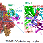
In this report, it was confirmed that the severity of MIS-C and cytokine storms are strongly correlated with the expansion of T-cell Receptor Beta Variable gene (TRBV) 11-2. Molecular dynamics studies have also confirmed that a T-cell receptor (containing TRBV11-2) and MHCII form a complex strongly coupled with a bacterial super-antigen-like structure (region from E661 to R685 around the polybasic insert PRRA) presents in SARS-CoV-2 S-protein. This will activate a number of T-cells nonspecifically and release a large amount of cytokines.

Infection with the new coronavirus (SARS-CoV-2) affects many immunoinhibitory receptors

There are reports that many immunoinhibitory receptors are affected when infected with the new coronavirus (SARS-CoV-2).
https://pubmed.ncbi.nlm.nih.gov/33200082/

Compared to SARS-CoV-1, Influenza A virus, and Respiratory synergy virus, SARS-CoV-2 has significantly increased the expression of the following immunoinhibotory receptors:
CEACAM1
SIGLECS
LILRB3
LILRB4
LILRB5
CLEAN12A
It concluded that these inhibitors could also be effective candidates for treatment of COVID-19.

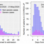
Differences between the first and second waves of the new coronavirus (COVID-19) in Japan

There is a report of investigating the difference between the first wave and the second wave of the new coronavirus in Japan.
https://www.journalofinfection.com/article/S0163-4453(20)30693-9/fulltext

The first wave peaked in April 2020, and the second wave peaked in August of the same year. In the second wave, it is said that the mutation of D614G has increased infectivity, but it is clearly shown that the severity rate is down. However, in the second wave, there are more young people infected than in the first wave, which may be causing the severity rate to be lowered.


It is said that it is already in the third wave now, and what kind of change is happening to the infectivity and the severity rate is paid attention further.

Severity rate in pregnant women is considerably higher: new coronavirus (COVID-19)

In the new coronavirus, the risk of severe COVID-19 is originally lower in women than in men, but it has been reported that pregnant women have a much higher rate of severity.
https://www.cdc.gov/mmwr/volumes/69/wr/mm6944e3.htm?s_cid=mm6944e3_w

Pregnant women between the ages of 15 and 24 have tripled the risk of those who must receive invasive ventilation (95% CI: 1.6 – 5.7),
and in pregnant women between the ages of 35 and 44, increase by 3.6 times (95% CI: 2.4 – 5.4).

Severity of the new coronavirus (COVID-19) and the changes of various immune cells (CD4+ Tcell, CD8+ Tcell, NK Cell, etc.)

When infected with the new coronavirus (SARS-CoV-2) and the severity of COVID-19 progresses, the following changes can be seen in various immune cells
https://www.ncbi.nlm.nih.gov/pmc/articles/PMC7591707/

Compared to healthy people,
Leukocytes and neutrophils increase significantly,
Conversely, lymphocytes, phosphates, T cells, and NK cells decrease.

In addition, interesting changes appear in a subset of T cells and NK cells,
the ratio of CD4+/CD8+ increases significantly,
PD-1 NK cells, CD244 NK cells, PD-1 CD4+ T cells, PD-1 CD8+ T cells, CD244 CD8+ T cells increase,
Conversely, CD27 NK cells and CD27 CD8+ T cells are reduced.

Thus, changes in immune cells and their subsets would be related to the mechanism of severity of COVID-19, and are clinically useful for screening of critical illness.

Up regulation of high mannose is behind the relationship between severity and age in the N1H1 influenza virus: a ferret model

In the 2009 N1H1 influenza pandemic, young people, like the new coronavirus, tended to be mild and became more severe as they aged.

The following group, using ferrets as a model, has investigated if any change in glycosylation might be involved in the background relationship between the severity of N1H1 influenza and age? They used lectin microarrays for comparative glycan profiling analysis with using Ferret lungs as samples.
https://pubs.acs.org/doi/10.1021/acs.jproteome.0c00455

As a result
In aged ferrets, high mannose was highly expressed as they became more severe.
In newly weaned ferrets, however, the expression of high mannose increased from 3 to 5 days after the infection, but since then, glycan modification has recovered to the same level as in healthy states, and stayed in mild and recovered.
It can be seen that glycan modification is deeply related to severity of influenza.