SARS-CoV-2 Omicron showed higher infectivity in human nasal epithelial cells, but the reason is not only due to strong binding to hACE2

A group from Department of Infectious Disease, Imperial College London, UK, etc. has reported about SARS-CoV-2 Omicron’s higher infectivity in human nasal epithelial cells (hNECs) and its possible reasons.
https://www.biorxiv.org/content/10.1101/2021.12.31.474653v1

SARS-CoV-2 Omicron showed a large early replication advantage in human nasal epithelial cells (hNECs), yielding viral titres ~100-fold higher than Delta by 24 hours post-infection. At 48 and 72 hours post-infection, viral titres were lower compared to Delta and at 72 hours the RNA collected from Omicron infected wells was fewer. In Vero-AT cells, replication of the two variants was equal. In Calu-3 cells, the viral yields of Omicron were lower than for Delta across all time points.

Previous SARS-CoV-2 variants, such as Delta, only enter cells efficiently by binding ACE2 and activating fusion via cell-surface protease TMPRSS2. Omicron, conversely, is able to enter cells in both a TMPRSS2-dependent and –independent manner, having evolved the ability to avoid endosomal restriction. This allows Omicron to infect any ACE2-expressing cell in the airway instead of relying solely on double ACE+ TMPRSS2+ cells.

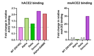
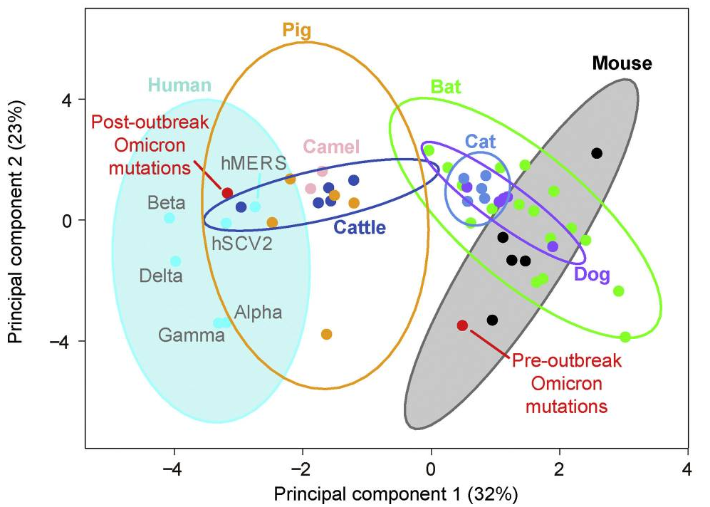
It was also found omicron Spike bound better to mouse ACE2 than any previous variant, as several others have reported. This suggests a possibility that omicron jumped from humans to mice, rapidly accumulated mutations conducive to infecting that host, then jumped back into humans.

The progenitor of SARS-CoV-2 Omicron did not evolve in human, but in mice

A group from University of Chinese Academy of Sciences, Beijing 100049, China has proposed a hypothesis that the progenitor of Omicron evolved in mice.
https://www.ncbi.nlm.nih.gov/pmc/articles/PMC8702434/

Generally speaking, the molecular spectrum of mutations that accumulate in a viral genome reflects a host-specific cellular environment.

The molecular spectrum of the pre-outbreak Omicron mutations was significantly different from the “standard” molecular spectrum for SARS-CoV-2 variants known to have evolved strictly in humans (hSCV2 spectrum), and interestingly, the molecular spectrum of the post-outbreak Omicron mutations was not significantly different from the hSCV2 spectrum. From a statistical point of view, these molecular spectrum analyses revealed that pre-outbreak Omicron mutations were unlikely to have been acquired in humans.

The molecular spectrum of post-outbreak Omicron mutations (which are known to have accumulated in humans) was located within the human 95% confidence ellipse. In contrast, the molecular spectrum of pre-outbreak Omicron mutations was within the mouse ellipse, suggesting that the pre-outbreak mutations accumulated in a rodent (in particular a mouse) host.

It was also confirmed that the pre-outbreak Omicron mutations in the RBD showed the greatest enhanced binding affinity for mouse ACE2 among 32 mammals with molecular docking analysis.

Some types of HCoV-specific CD8+ T cells cross-react against SARS-CoV-2

A group from Laboratory for Immunotherapy, RIKEN Center for Integrative Medical Science (IMS), Yokohama, Japan, has reported that some types of HCoV-specific CD8+ T cells may be cross-reacting against SARS-CoV-2.
https://www.ncbi.nlm.nih.gov/pmc/articles/PMC8640030/

Recently, the relationship between Human Leukocyte Antigens (HLA alleles) and COVID-19 is attracting attentions.

Authors assessed the interaction between HLA-A*24:02, as one of the major HLAs, and CD8+ T cells cross-reactive for SARS-CoV-2. In-vitro expansion assays can be used to detect pre-existing CD8+ T cells against cancer or cross-reactive CD8+ T cells to SARS-CoV-2.
cited from https://www.jstage.jst.go.jp/article/mhc/23/2/23_115/_pdf/-char/ja

PBMCs were cultured in the presence of selected each peptide, and the frequencies of epitope-specific IFN-γ-producing CD8+ T cells were assessed on day 21. Although selected six peptides from SARS-CoV-2 Spike protein were expected to bind to HLA-A*24:02 with high affinity, five of these did not induce a T-cell response, and only Pep#3 (Amino acid sequence = QYIKWPWYI, SARS-CoV-2 Spike position = 1208-1216)-specific CD8+ T cell counts were elevated in cells derived from all unexposed healthy donors (UHDs). Intriguingly, Pep#3 exhibited high sequence homology with other coronaviruses, including four types of seasonal coronaviruses (HCoV). Although all the six peptides of S protein were predicted to exhibit high affinity to HLA-A*24:02, five peptides were not homologous to other seasonal coronaviruses.
These results suggest that HCoV-specific CD8+ T cells may be cross-reacting against SARS-CoV-2, and that pre-existing memory CD8+ T cells may be responsible for the immune response.

To understand why CD8+ T cells exhibit multiple responses to various coronaviruses and the underlying mechanisms, we studied the cross-reactivity of CD8+ T cells at the single TCR level. After restimulation with Pep#3, CD8+ T cells from Pep#3-specific CD8+ T cell lines were isolated by gating CD107a+CD8+ cells and performed single-cell analysis of TCR repertoires. To analyze the clonalities of Pep#3-reactive CD8+ T cells, a total of 227 T cells were screened, and 44 TCR clonotypes were identified from five UHDs and one patient with hematological malignancies (HM). Furthermore, we focused on the dominant TCR types from among the clonotypes and identified four types of TCRα and TCRβ pairs from four donors (three UHDs and one HM). To assess the specificity and functions of cloned TCRs, we transduced the TCRα and TCRβ genes into the SKW3-CD8AB (human T-ALL) cell line and demonstrated their peptide-specific response. Intriguingly, the four types of TCR repertoires varied from each other in their epitope recognition. TCR-T (TCR-T-1) cells from UHD2 responded well to all the epitopes derived from coronaviruses, whereas the TCR-T (TCR-T-2) cells from UHD8 responded only to SARS-CoV-1 and SARS-CoV-2.

Trichoderma strains are promising biocontrol agents for plants: Chitinase and β-1,3-glucanase are the keys secreted from trichoderma

A group from College of Plant Protection, Hainan University, Haikou, Hainan, China, etc. has reported that the 13 trichoderma strains are promising biocontrol agents and could be developed as biofertilizers and biological pesticides for agricultural applications.
https://www.ncbi.nlm.nih.gov/pmc/articles/PMC8714372/

The 13 Trichoderma strains that significantly promoted the seed germination were further tested for their effect on root growth of watermelon, chili, eggplant, and tomato seedlings. In greenhouse experiment, all strains showed a promotion effect on root growth of chili seedlings, and among them, HL100 strain showed maximum increase 12.17% in root length compared to control. Three strains (GZ070, HL100, and HN059) significantly promoted the root growth of watermelon seedlings. HN059 strain showed maximum increase of 18.81% in root length compared to control. Trichoderma strains GZ070, HL100, HN059, JX013, XJ087, and NX043 strains and HL119, HN059, SC012, XJ035, SC098, and SC101 were found to promote the root growth of eggplant and tomato seedlings, respectively. The highest increase in root length of eggplant (40.99%) and tomato plants (34.68%) was recorded by GZ070 and SC098 strains, respectively.

Trichoderma grows rapidly and can quickly occupy the growth space of pathogenic fungi, which is one of the important mechanisms of their antimicrobial effect.
The production of cell wall-degrading enzymes (CWDEs) and volatile antibiotics is key parameters for Trichoderma as a biocontrol agent. It was found that 13 Trichoderma strains (PI > 85%) with excellent antagonism to pathogens could secrete chitinase and β-1,3-glucanase, which were closely related to the cell wall composition of pathogens. Therefore, Trichoderma can not only compete with pathogens for space and nutrition but also degrade the cell walls of pathogens, deform, or even digest the hyphae, and inhibit the growth of pathogens.

As references, some reports suggested that root colonization by Trichoderma strains could increase levels of defense-related plant enzymes, including various peroxidases, chitinases, β-1,3-glucanases, and the lipoxygenase-hydroperoxide lyase pathway. In cucumber, root colonization by strain T-203 causes an increase in phenolic glucoside levels in leaves; their aglycones (which are phenolic glucosides with the carbohydrate moieties removed) are strongly inhibitory to a range of bacteria and fungi.

Serum glycobiomarkers for colorectal carcinoma using reverse-phase Lectin Microarray: The combination of PHAE and HL sel was the best

A group from Institute of Chemistry, Slovak Academy of Sciences, Bratislava, Slovakia, etc. has reported that the combination of lectins (PHAE + HL sel) can detect only glycosylation changes associated with colorectal cancer (CRC) and not with age.
https://www.ncbi.nlm.nih.gov/pmc/articles/PMC8695905/

Here, reverse-phase lectin microarrays were used for the identification of changes in the serum glycome as potential CRC biomarkers.

What is the reverse-phase lectin microarrays:
Serum samples diluted 50× were spotted in different wells in triplicates using a microarray spotter on epoxide-coated slides. Subsequently, after spotting and blocking the slides, 70 μl of biotinylated lectins (5 μg/ml in PBS) was added and incubated at RT for 1 h. The slides were washed gently three times with PBS, and then 70 μl of streptavidin-fluorescent material conjugate (0.1 μg/ml in PBS) was added for 15 min. After a washing step and additional wash with deionized water, fluorescence intensity was read at 635 nm using a microarray reader.

Lectins used in this experiment:
AAL, RPL-Fuc1, PHAE, PHAL, ConA, DBA, WFL, WGA, RCAI, MAA, P sel, RPL-Sia2, SNAI, HPyL, HE sel, and HL sel

The lectin combination PHAE (specific to N-ghlycans with outer Gal and bisecting GlcNAc) + HL sel (specific to 6-O-Su sLex) provided the highest discrimination accuracy based on the AUC value (0.989).