Antibody response induced by a 3rd boost SARS-CoV-2 vaccination: after 8 months from the 2nd vaccination

A group from The Institute of Medical Biology, Chinese Academy of Medical Sciences and Peking Union Medical College, Kunming, Yunnan, China, etc. has reported on antibody response induced by a third boost dose of inactivated SARS-CoV-2 vaccine.
https://pubmed.ncbi.nlm.nih.gov/34666622/

In this study, 53 volunteers, who joined in the development and production of inactivated COVID-19 vaccines (it is not sure what company developed the relevant ones, though), received two doses (at 0 and 28 days) of the vaccines in 2020, and they received a 3rd dose 8 months after the 2nd dose recently.

At 0 days, 5 days, 7 days, and 14 days after the 3rd dose, blood was collected from 6 volunteers for the evaluation. It was found that both the anti-S antibody and neutralizing antibody against the original Wuhan strain gradually increased after 5 days, and the positive conversion rate of antibodies reached 100% at 14 days. Interestingly, the memory of IFN-γ-T cells against S, N, M, O antigens of SARS-CoV-2 can be quickly awakened after the 3rd dose.

These results indicate that although the neutralizing antibodies gradually decrease after two doses of inactivated vaccines, the antibody response could be awakened quickly by the 3rd vaccination and the T cell immune memory is still active even after 8 months from the 2nd vaccination.

SARS-CoV-2 specific antibody transfer to breastmilk is dominated by IgA and IgM

A group from Department of Biological Engineering, Massachusetts Institute of Technology, Cambridge, MA, USA, etc. has reported that SARS-CoV-2 specific antibody transfer to breastmilk is dominated by IgA and IgM.
https://www.ncbi.nlm.nih.gov/pmc/articles/PMC8531199/

Previous studies clearly illustrated the evolution and transfer of Fc-effector function in pregnant women via placenta to their infants, but less is known about the transfer of Fc-effector function across breastmilk.

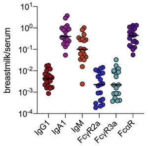
As expected, mothers infected with SARS-CoV-2 possessed SARS-CoV-2-specific antibodies in serum and breastmilk that were not present in SARS-CoV-2-uninfected specimens. But, interestingly, robust transfer of IgA and IgM was observed in breastmilk, but the transfer of IgG1 was more limited as shown below.

Importance of surfactin-type lipopeptide: Bacillus velezensis recognizes plant root cell pectin and thereby improve symbiotic relation

A group from University of Liège‐Gembloux Agro‐Bio Tech, Gembloux, Belgium, etc. has reported Bacillus velezensis recognizes plant root cell pectin and thereby improve symbiotic relation.
https://pubmed.ncbi.nlm.nih.gov/34724831/

Molecular interactions driving the early steps of partnership establishment between plant roots and rhizobacteria are not yet fully understood. Authors have found that Bacillus velezensis, one of beneficial bacteria, recognizes plant root cell wall pectin in synergy with soluble root exudates, and thereby the bacterium stimulates the production of a specific surfactin-type lipopeptide as key components of its secretome to improve the partnership in the course of early colonization.

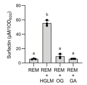
Actually, an 8-fold increase of the surfactin production was detected at the early exponential growth phase with homogalacturonan low methylated (HGLM) as shown below, however, the bacteria seem not to recognize oligomers with a lower polymerization degree (DP) than HGLM, as is clear from the fact that oligogalacturonides (OGs) did not stimulate surfactin biosynthesis. This might mean that longer polymers could indicate a healthy host suitable for bacterial colonization, while the monomers or low DP oligomers may reflect a dead or infected plant that is unable to adequately provide resources to the bacteria.


homogalacturonan low methylated (HGLM), DP of >150; oligogalacturonides (OG), DP of 15; galacturonic acid (GA), DP of 1; where REM means a root exudates mimicking medium.

But, there is no information about a lectin-like protein on the bacteria which can recognize the pectin backbone as a plant molecular pattern was not mentioned here anything.

Development of mannosylated macromolecules specific to each human mannose-binding lectins such as DC-SIGN, Dectin-2, and DEC-205

A group from Department of Chemistry, Massachusetts Institute of Technology, MS, USA, etc. has developed mannosylated macromolecules specific to each human mannose-binding lectins.
https://www.ncbi.nlm.nih.gov/pmc/articles/PMC8549053/

Carbohydrate-binding proteins (lectins) play vital roles in cell recognition and signaling, including pathogen binding and innate immunity. Thus, targeting lectins, especially those on the surface of immune cells, could advance immunology and drug discovery. Lectins are typically oligomeric; therefore, many of the most potent ligands are multivalent. An effective strategy for lectin targeting is to display multiple copies of a single glycan epitope on a polymer backbone; however, a drawback to such multivalent ligands is they cannot distinguish between lectins that share monosaccharide binding selectivity (e.g., mannose-binding lectins such as DC-SIGN, DC-SIGNR, MBL, SP-D, langerin, dectin-2, mincle, and DEC-205) as they often lack molecular precision.

Authors developed β-mannosylated glyco-IEGmers specific to each lectins with mannose binding specificity.

To generate the target mannosylated glycomacromolecules, iterative exponential growth (IEG) cycles beginning from (R)- or (S)-glycidyl propargyl ether (GPE, > 99% ee) were conducted to yield oligo/polytriazoles with precisely 8, 16, or 32 allyl side chains. Macromolecules were produced with three different overall absolute configurations: all (R) (“isotactic”), all (S) (“isotactic”), and alternating (R-alt-S) (“syndiotactic”). To append the mannose residues, these IEGmers were exposed to pure β-thiomannose sodium salt under UV light (λ = 365 nm).

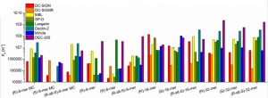
The binding affinity changed from sample to sample over several orders of magnitudes. For example, KA values for DC-SIGN ranged from approximately 1 × 10⁴ for (S)-8mer to about 1 × 10⁸ for (R)-16mer. For dectin-2, it also ranged from ∼1 × 10⁴ to ∼1 × 10⁸. DEC-205 bound all of the glyco-IEGmers with high KA values in the range of ∼1 × 10⁶ to 1 × 10⁹, with the highest KA of all pairwise interactions tested (2.2 × 10⁹).

This wide range in affinity is impressive as the mannose binding epitopes presented was identical among these lectins.

Snake venom (Dimeric PLA2s) from Vipera nikolskii showed high antiviral activity against SARS-CoV-2

A group from Department of Molecular Neuroimmune Signalling, Shemyakin-Ovchinnikov Institute of Bioorganic Chemistry, Russian Academy of Sciences, Moscow, Russia, has reported that Snake venom (Dimeric PLA2) from Vipera nikolskii showed high antiviral activity against SARS-CoV-2.
https://pubmed.ncbi.nlm.nih.gov/34714362/

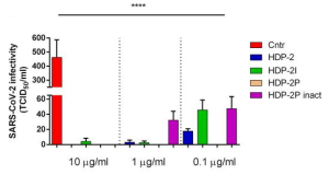
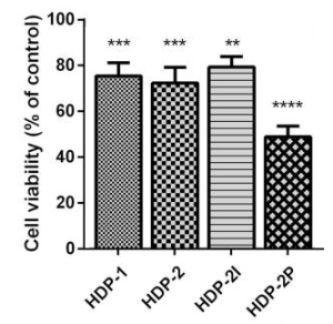
Two types of Snake venom (Dimeric PLA2) from Vipera nikolskii (HDP-1, HDP-2, and its subunits, HDP-2I and HDP-2P) were evaluated as natural antiviral products.
Complete suppression of the infectivity of SARS-CoV-2 was observed when the viral stock was treated with HDP-2P even at 0.1 µg/ml on Vero E6 cells.
On the other hand, the highest but moderate cytotoxicity was manifested by HDP-2P which at 100 μg/ml reduced cell viability on average by 51%.

These data highlight the potential of PLA2s as a natural product that could prove to be fruitful as the starting point for the development of antiviral drugs.

Saponin treatments could control rhizobacteria

A group from Research Institute for Sustainable Humanosphere, Kyoto University, Japan, etc. has reported that saponin treatments could control rhizobacteria.
https://www.ncbi.nlm.nih.gov/pmc/articles/PMC8538258/

Plant specialized metabolites (PSMs) secreted from roots are called root exudates, and these account for up to 40% of the carbon fixed during photosynthesis. Rhizosphere microbiomes assembled by root exudates promote plant growth and help the host plants overcome biotic and abiotic stresses. However, the association between PSMs and microbiota is not well characterized.
Saponins are a group of PSMs widely distributed in angiosperm plants. They exhibit biological and pharmacological activities, including antibacterial, antifungal, hemolytic, and cytotoxic properties.

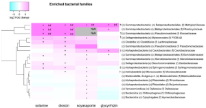
Burkholderiaceae, Methylophilaceae, Rhodocyclaceae, Moraxellaceae, Pseudomonadaceae, P3OB-42, Caulobacteraceae, Steroidobacteraceae, Geobacteraceae, and Sphingomonadaceae were enriched in saponin (α-Solanin, dioscin, soyasaponins, and glycyrrhizin) treatments as shown below.

For instance, members of the family Burkholderiaceae are reportedly involved in plant–pathogen suppression via the upregulation of induced systemic resistance-associated genes and the production of sulfurous volatile compounds and siderophores, and members of Sphingomonadaceae have been found to promote plant growth via phytohormone production, alleviation of heavy metal toxicity and drought stress, and pathogen suppression.

Therefore, it is plausible that saponin-producing plants may benefit from attracting those bacterial families to their rhizospheres and roots.